
mujhe Pap staining ki procedure btao details me or har step k baare me kyu ki jati hai or cells kesi dikhegi
Papanicolaou Pap staining procedure steps fixation hematoxylin OG EA stain cytology
https://pmc.ncbi.nlm.nih.gov/articles/PMC9345133
🔬 Papanicolaou (Pap) Staining - Poori Procedure
Pap Stain Kya Hai?
- Hematoxylin - nucleus stain karta hai
- Orange G (OG-6) - keratin/mature cells stain karta hai
- Eosin Azure (EA-50/EA-65) - cytoplasm ko differentiate karta hai
📋 STEP-BY-STEP PROCEDURE
🔷 STEP 1: FIXATION
- Cells ko preserve karta hai - proteins denature hoke cellular structure lock ho jati hai
- Autolysis (self-digestion) rokta hai
- Cells ki morphology maintain rehti hai
- Wet fixation zaroori hai - agar slide sookh gayi to artifacts aa jaate hain
⚠️ Yahi Pap stain ki sabse critical step hai. Agar fixation galat ho to baaki sab bekar.
🔷 STEP 2: HYDRATION (Rehydration)
- 80% alcohol - 10 dips
- 70% alcohol - 10 dips
- 50% alcohol - 10 dips
- Distilled water - 10 dips
- Alcohol-fixed slide ko water-based hematoxylin stain receive karne ke liye prepare karta hai
- Gradual hydration se cells distort nahi hoti
- Direct alcohol se water mein jaane se cells shrink ho sakti hain
🔷 STEP 3: NUCLEAR STAIN - HEMATOXYLIN
- Hematoxylin ek basic dye hai jo nucleus ke acidic components (DNA, RNA, chromatin) se bind karta hai
- Nucleus ko blue-purple (basophilic) color deta hai
- Chromatin pattern clearly dikhai deta hai - normal vs. malignant cells distinguish karne mein madad karta hai
- Regressive method (Harris Hematoxylin): Overstain karo, phir acid-alcohol se differentiate karo - better nuclear detail
- Progressive method (Gill's Hematoxylin): Exact time tak stain karo - easier, commonly used
🔷 STEP 4: WASHING
🔷 STEP 5: ACID ALCOHOL DIFFERENTIATION (Regressive method mein)
- Non-specific staining remove karta hai
- Cytoplasm se extra hematoxylin nikalta hai
- Sirf nucleus ka stain retain hota hai
- Extremely critical step - zyada time = nucleus ka stain bhi nikal jaata hai
🔷 STEP 6: BLUING
- Acid treatment ke baad hematoxylin red/brown ho jaata hai
- Alkaline environment (tap water pH ~7-8) mein blue-purple ho jaata hai
- Ye chemical reaction: Red soluble salt → Blue insoluble lake (aluminum-hematein complex)
🔷 STEP 7: DEHYDRATION (Pre-counterstain)
- 50% alcohol - 10 dips
- 70% alcohol - 10 dips
- 95% alcohol - 10 dips
- Counterstains alcohol-based hain, isliye water remove karna padega
- Agar paani raha to OG aur EA properly bind nahi karenge
🔷 STEP 8: ORANGE G (OG-6) STAINING
- OG ek acid dye hai jo cytoplasmic basic proteins (keratin) se bind karta hai
- Mature squamous cells aur keratinized cells ko orange-red color deta hai
- RBCs aur superficial cells strongly stain hoti hain
- OG-6 mein phosphotungstic acid hota hai jo non-specific staining rokta hai
🔷 STEP 9: EA STAINING (Eosin Azure - EA-50 ya EA-65)
- EA ek polychromatic mixture hai jisme hota hai:
- Eosin Y (acid dye) - cytoplasmic proteins, nucleoli, cilia ko pink-red stain karta hai
- Light Green - immature/active cells ke cytoplasm ko green-blue stain karta hai
- Bismarck Brown (EA-50 mein) - minimal role
- Cytoplasmic differentiation possible hoti hai - mature vs immature cells alag dikhte hain
| Feature | EA-50 | EA-65 |
|---|---|---|
| Use | Gynecological (cervical) samples | Non-gynecological (sputum, urine, FNA) |
| Eosin concentration | Kam | Zyada |
🔷 STEP 10: FINAL DEHYDRATION
- 95% alcohol (x2) - 10 dips each
- Absolute alcohol (100%) - 10 dips
- Mounting medium (DPX/Canada balsam) water-based nahi hota
- Pura paani remove karna padta hai warna slide cloudy ho jaati hai
🔷 STEP 11: CLEARING
- Alcohol ko replace karta hai
- Xylene ka refractive index glass aur mounting medium ke barabar hai
- Slide transparent (clear) ho jaati hai
- Mounting medium ke saath compatible hai
🔷 STEP 12: MOUNTING
- Cells preserve rehti hain permanently
- Refraction improve hoti hai - microscopy better hoti hai
- Physical damage se bachata hai
🎨 CELLS KESI DIKHEGI? (Microscopic Appearance)
| Structure | Color | Reason |
|---|---|---|
| Nucleus | Blue-Purple (Basophilic) | Hematoxylin + acidic DNA/chromatin |
| Chromatin | Dark blue, well-defined | Hematoxylin nuclear staining |
| Mature squamous cells cytoplasm | Orange / Pink-orange | Orange G binds keratin |
| Intermediate cells cytoplasm | Light green / Blue-green | Light green in EA stain |
| Parabasal cells cytoplasm | Dark green / Blue | Immature cells, more RNA |
| Nucleoli | Pink-red | Eosin in EA stain |
| Cilia | Pink | Eosin Y |
| RBCs | Orange-red | OG strongly stains them |
| Neutrophils | Small blue nuclei | Hematoxylin |
| Mucus | Pale pink/grey | Non-specific eosin |
🔍 NORMAL vs. ABNORMAL CELLS
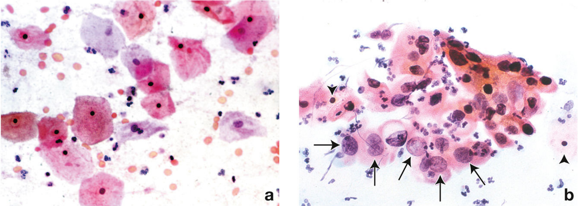
- Large, flat squamous cells with abundant pink-orange cytoplasm
- Small, dark (pyknotic) nuclei - condensed, inactive
- Cells ek dusre se alag, flat dikhti hain
- Large, irregular nuclei - koi pyknosis nahi
- High N:C ratio (nucleus bada, cytoplasm kam)
- Nuclei deeply stained, irregular chromatin
- Cells cluster mein hongi
- Kuch cells orange (keratinizing) dikhte hain
📊 Summary Table - Quick Reference
| Step | Time | Purpose |
|---|---|---|
| 95% Alcohol fixation | 15-20 min | Cell preservation |
| Hydration (graded alcohol) | 10 dips each | Water-based stain ke liye prepare |
| Gill's Hematoxylin | 10-15 min | Nucleus blue-purple |
| Wash + Acid alcohol | 1 dip | Differentiation |
| Bluing (tap water) | 3-10 min | Red → Blue conversion |
| Dehydration | 10 dips each | Counterstain ke liye |
| Orange G (OG-6) | 1-2 min | Keratin orange |
| EA-50/65 | 3-5 min | Cytoplasm pink/green |
| Absolute alcohol | 10 dips | Complete dehydration |
| Xylene | 3-5 min | Clearing |
| DPX mounting | - | Permanent preservation |
isi tarah H&E staining Gimsa staining or AFB staining leisman staining k baare me bhi btao
Leishman stain blood smear procedure steps methanol fixation malaria cells appearance
Giemsa staining procedure steps dilution buffer blood film malaria parasite appearance WBC colors
🔬 FOUR IMPORTANT STAINS - Complete Guide
═══════════════════════════════════
1️⃣ H&E STAINING (Hematoxylin & Eosin)
═══════════════════════════════════
Kya hai?
📋 H&E Procedure - Step by Step
STEP 1: FIXATION
- Cell metabolism band karta hai
- Autolysis (self-digestion) rokta hai
- Proteins cross-link hoke structure preserve hoti hai
- Formaldehyde amino groups ke saath react karta hai (mainly lysine residues)
- Pathogenic organisms kill hote hain
⚠️ Formalin lipids ke saath react nahi karta - isliye lipid membranes ke liye poor fixative hai.
STEP 2: PROCESSING (Tissue Processing)
- Washing - formalin hata
- Dehydration - graded alcohol (70% → 80% → 95% → 100%) - paani nikalo
- Clearing - Xylene/Xylol se alcohol replace karo
- Infiltration - Molten paraffin tissue mein ghusta hai
STEP 3: EMBEDDING & SECTIONING
STEP 4: DEPARAFFINIZATION
STEP 5: REHYDRATION
- 100% → 95% → 80% → 70% → Distilled water
STEP 6: HEMATOXYLIN STAINING
- Hematoxylin ek cationic (positive charge) dye hai
- Nucleus mein DNA/RNA aur chromatin negatively charged hain (anionic)
- Electrostatic attraction se bind karta hai
- Mordant (Alum/Aluminum salt) zaroori hai - dye ko tissue se permanently bind karta hai
- Nuclear detail: chromatin pattern, nucleoli, mitoses clearly dikhte hain
STEP 7: WASHING & BLUEING
STEP 8: DEHYDRATION (Partial)
STEP 9: EOSIN STAINING
- Eosin anionic (negative charge) dye hai
- Cytoplasmic proteins positively charged hain
- Eosin bind hota hai aur pink/red color deta hai
- Collagen, muscle, RBCs bhi pink ho jaate hain
STEP 10: DEHYDRATION → CLEARING → MOUNTING
- 95% → 100% alcohol (dehydration)
- Xylene × 2-3 (clearing)
- DPX/Canada balsam se coverslip
🎨 H&E mein Cells Kaisi Dikhti Hain?
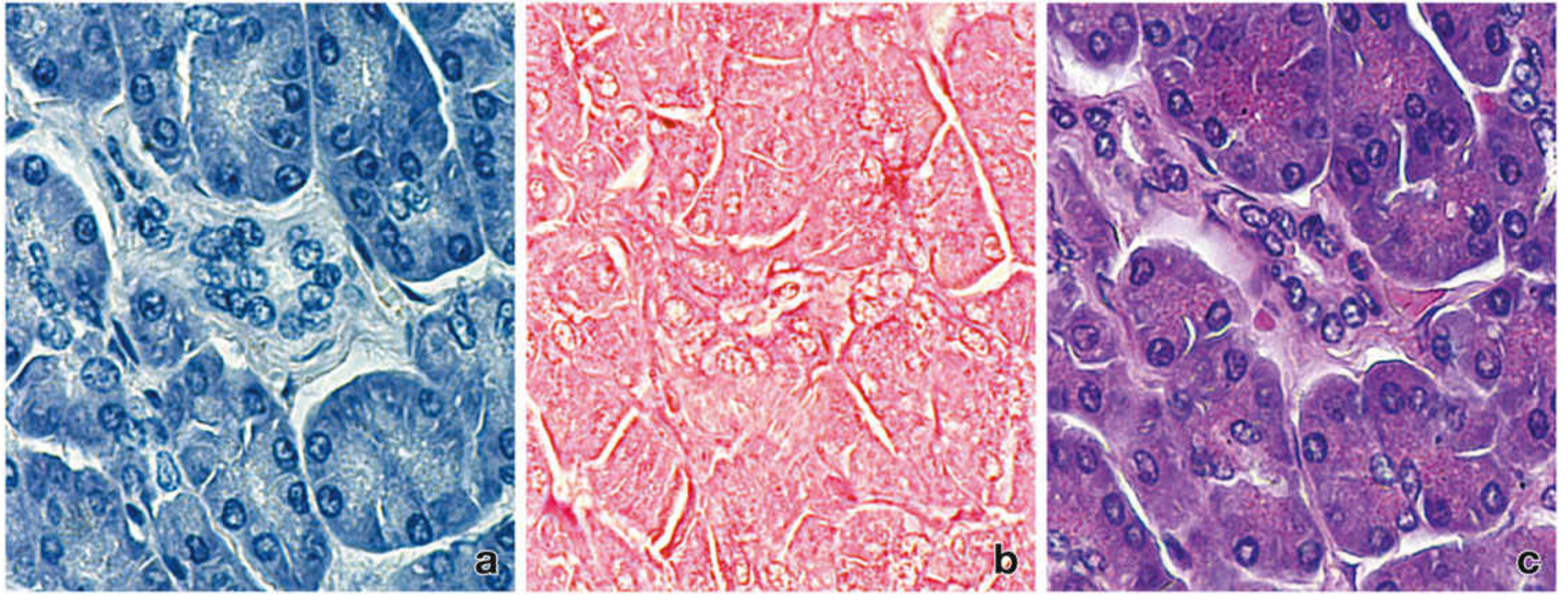
- Image (a) - Sirf hematoxylin: sab kuch blue
- Image (b) - Sirf eosin: sab kuch pink
- Image (c) - H&E combined: nuclei blue-purple, cytoplasm pink - perfect contrast!
| Structure | H&E Color | Reason |
|---|---|---|
| Nucleus / Chromatin | Blue-Purple (Basophilic) | Hematoxylin + DNA/RNA |
| Nucleolus | Dark blue/purple | RNA-rich |
| Cytoplasm (most cells) | Pink (Eosinophilic) | Eosin + cytoplasmic proteins |
| RBCs | Bright pink/orange-red | Hemoglobin eosinophilic |
| Collagen fibers | Pale pink | Eosin |
| Muscle | Deep pink/red | Myosin eosinophilic |
| Cartilage matrix | Blue-purple | Proteoglycans basophilic |
| Fat cells (adipocytes) | Empty white vacuoles | Lipid dissolved by processing |
| Calcification | Dark blue/purple | Calcium basophilic |
| Mitotic figures | Dark purple | Condensed chromosomes |
═══════════════════════════════════
2️⃣ GIEMSA STAINING
═══════════════════════════════════
Kya hai?
📋 Giemsa Procedure - Step by Step
STEP 1: SMEAR PREPARATION
- Thin film - individual cell morphology detail ke liye, species identification
- Thick film - parasite detection sensitivity ke liye (zyada blood, WBCs lyse ho jaati hain)
STEP 2: FIXATION (Thin film only!)
- Cells morphology preserve karta hai
- Slide se cells ko adhere karta hai
- THICK FILM FIX NAHI KARTE - kyunki RBCs ko lyse hona hai taaki parasites dikh sakein
STEP 3: GIEMSA WORKING SOLUTION BANANA
- Stock solution (3.8g Giemsa powder + 250ml methanol + 250ml glycerin)
- Working solution: 1:20 (thin film) ya 1:50 (thick film) dilution phosphate buffer (pH 7.2) mein
- Alkaline pH (>7.4) = blue dominant, parasite detail kho jaata hai
- Acidic pH (<7.0) = eosin dominant, RBCs too pink, Schuffner's dots nahi dikhte
STEP 4: STAINING
- Thin film: Giemsa (1:20) se 20-30 minutes
- Thick film: Giemsa (1:50) se 45-60 minutes (ya 10% solution for 8-10 min)
- Azure dyes negatively charged chromatin se bind karte hain (blue-purple)
- Eosin positively charged cytoplasm se bind karta hai (pink-red)
- Longer time = better penetration
STEP 5: WASHING
- Excess stain remove karo
- Iridescent "scum" float off karo
- Zyada wash mat karo - decolorization hogi
STEP 6: AIR DRY & EXAMINE
🎨 Giemsa mein Cells Kaisi Dikhti Hain?
| Structure | Giemsa Color | Reason |
|---|---|---|
| Leukocyte nucleus | Purple/Dark blue | Azure dyes + DNA |
| Neutrophil granules | Neutral pink-purple | Both acidic + basic dyes |
| Eosinophil granules | Bright red-orange | Eosin strongly binds |
| Basophil granules | Deep purple/black | Azure strongly binds |
| Monocyte cytoplasm | Grey-blue | Azure dyes |
| Lymphocyte cytoplasm | Sky blue | Scant, azure-stained |
| RBCs (normal) | Pale pink | Hemoglobin, eosinophilic |
| Platelets | Purple granules | Azure |
| Malaria parasite nucleus | Red/Magenta dot | Eosin + DNA |
| Malaria cytoplasm | Blue | Azure |
| Schuffner's dots (P. vivax) | Pink stippling | In infected RBC cytoplasm |
═══════════════════════════════════
3️⃣ AFB STAINING (Ziehl-Neelsen / Acid Fast Bacilli)
═══════════════════════════════════
Kya hai?
🧬 Principle (Kyun Kaam Karta Hai?)
- Normal dyes ko block karti hai
- Lekin Carbol Fuchsin (phenol + fuchsin) heat ke saath penetrate kar leta hai
- Ek baar andar gaya to acid-alcohol se nahi nikalta = "Acid Fast"
- Isliye inhe Acid-Fast Bacilli (AFB) kehte hain
📋 ZN Staining Procedure
STEP 1: SMEAR PREPARATION & FIXATION
STEP 2: CARBOL FUCHSIN (Primary Stain)
- Carbol Fuchsin = Basic fuchsin + 5% phenol in water/alcohol
- Phenol cell wall lipid layer ko dissolve karta hai, dye andar ghusta hai
- Heat penetration further increase karta hai
- Phenol + heat ke bina dye mycolic acid layer cross nahi kar sakta
⚠️ Kinyoun cold stain mein heat step nahi hoti - kyunki phenol concentration zyada hoti hai (8%).
STEP 3: WASHING
STEP 4: DECOLORIZATION - ACID ALCOHOL
- Non-AFB organisms se fuchsin nikaal deta hai (wo acid-alcohol resistant nahi hain)
- AFB ki mycolic acid wall dye ko tightly hold karti hai - nahi nikalti
- Yahi stain ka "diagnostic key" hai
- Modified AFB (Nocardia ke liye): 0.5-1% H2SO4 (weaker acid) use karte hain
STEP 5: WASHING
STEP 6: COUNTERSTAIN - METHYLENE BLUE
- Background aur non-AFB organisms ko blue color deta hai
- AFB aur background mein contrast banata hai - AFB clearly red dikhte hain blue background par
STEP 7: WASH, DRY, EXAMINE
🎨 AFB Stain Mein Kya Dikhta Hai?
| Structure | ZN Color | Reason |
|---|---|---|
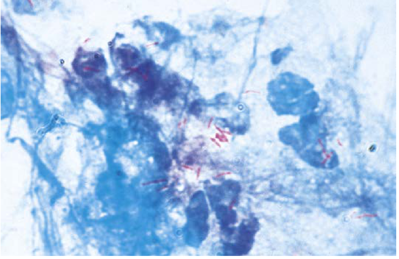
| AFB (M. tuberculosis) | Bright red/magenta rods | Carbol fuchsin retained (acid-fast) |
| Non-AFB bacteria | Blue | Counterstained with methylene blue |
| Neutrophils / Host cells | Blue | Non-acid-fast, counterstained |
| Background | Pale blue | Methylene blue |
| M. kansasii | Red, cross-barred "shepherd's crook" | Distinctive morphology |
| Nocardia | Weakly red (partially acid-fast) | Less mycolic acid, needs modified AFB |
| Bacilli per field | Report |
|---|---|
| 0 | No AFB seen |
| 1-9 per 100 fields | 1+ |
| 1-9 per 10 fields | 2+ |
| 1-9 per field | 3+ |
| >9 per field | 4+ |
═══════════════════════════════════
4️⃣ LEISHMAN STAINING
═══════════════════════════════════
Kya hai?
📋 Leishman Staining Procedure
STEP 1: BLOOD SMEAR BANAO
STEP 2: FIXATION + PRIMARY STAINING (Combined!)
- Leishman stain methanol mein dissolved hai
- Methanol hi fixative ka kaam karta hai + stain bhi deta hai - ek step mein dono!
- Pap stain se fark: Pap mein alag fixation step hoti hai, Leishman mein combined hai
- Methanol cells ko adhere karta hai, morphology preserve karta hai
⚠️ Bottle shake mat karo - precipitate suspend ho jaata hai aur slide par deposit hoke artifact banata hai.
STEP 3: BUFFER DILUTION
- Buffer paani provide karta hai - ionic dissociation se actual staining shuru hoti hai
- Romanowsky effect produce hota hai: methylene blue + eosin react karke purple color banate hain (yeh azure-eosin complex hai)
- pH 6.8 recommended - acidic pH eosinophilic structures (eosinophil granules, malaria parasites) better stain karta hai
- Metallic sheen confirm karta hai ki staining ho rahi hai
STEP 4: WASHING
- Excess stain hata
- Buffer remove karo
- Zyada wash = decolorization - colors fadenge
STEP 5: AIR DRY & EXAMINE
🎨 Leishman Stain Mein Cells Kaisi Dikhti Hain?
| Cell Type | Leishman Color | Why |
|---|---|---|
| RBCs | Pale pink/salmon | Hemoglobin eosinophilic |
| Neutrophil nucleus | Purple (multilobed) | Azure + DNA |
| Neutrophil granules | Neutral pink-lilac | Mixed charge, both dyes |
| Eosinophil nucleus | Purple (bilobed) | Azure |
| Eosinophil granules | Bright orange-red | Eosin strongly binds specific proteins |
| Basophil granules | Dark purple/violet | Azure strongly binds |
| Monocyte nucleus | Purple, kidney-shaped | Azure |
| Monocyte cytoplasm | Grey-blue, vacuolated | Azure |
| Lymphocyte nucleus | Deep purple | Azure, dense chromatin |
| Lymphocyte cytoplasm | Scant, sky blue | Azure |
| Platelets | Purple granules | Azurophilic |
| Malaria parasite (ring stage) | Blue cytoplasm + red nucleus dot | Azure + eosin |
| Malarial pigment (hemozoin) | Brown-black granules | Hematin byproduct |
🔄 Leishman vs Giemsa - Comparison
| Feature | Leishman | Giemsa |
|---|---|---|
| Fixation | Combined (methanol in stain) | Separate methanol step |
| Time | 5-8 min | 20-60 min |
| pH | 6.8-7.0 | 7.2 |
| Stability | Less stable (diluted = 3 hours) | More stable (glycerin present) |
| Malaria | Good for thin smears | WHO recommended, thick + thin |
| Main use | WBC differential (India/UK common) | Malaria, parasitology |
| Artifact risk | Higher (precipitation) | Lower |
📊 ALL FOUR STAINS - Master Comparison Table
| Feature | H&E | Giemsa | AFB (ZN) | Leishman |
|---|---|---|---|---|
| Main use | Histopathology (tissue biopsy) | Blood parasites, bone marrow | Mycobacteria (TB) | WBC differential, malaria |
| Specimen type | Paraffin tissue sections | Blood/bone marrow smear | Sputum, tissue | Blood smear |
| Fixative | 10% Formalin | Methanol | Heat fix | Methanol (in stain) |
| Primary stain | Hematoxylin (blue) | Azure B (blue-purple) | Carbol Fuchsin (red) | Leishman (methylene blue+eosin) |
| Counterstain | Eosin (pink) | Eosin (pink-red) | Methylene Blue (blue) | Buffer activates |
| Nucleus color | Blue-purple | Purple | Blue | Purple |
| Cytoplasm color | Pink | Pink-grey-blue | Blue | Pink-grey |
| Unique feature | Universal tissue stain | Schuffner's dots in malaria | AFB = bright RED rods | Combined fix+stain |
| Total time | ~2 hours | 30-60 min | 20-30 min | 5-8 min |
sabhi staining ki compete details ki pdf bnao
ls /home/daytona/workspace/ 2>/dev/null || echo "workspace not found"
mkdir -p /home/daytona/workspace/staining-guide && python3 -c "from reportlab.lib.pagesizes import A4; print('reportlab OK')" && python3 -c "from reportlab.platypus import SimpleDocTemplate; print('platypus OK')"
~/staining-guide/generate_pdf.py
from reportlab.lib.pagesizes import A4
from reportlab.lib import colors
from reportlab.lib.styles import getSampleStyleSheet, ParagraphStyle
from reportlab.lib.units import cm, mm
from reportlab.lib.enums import TA_LEFT, TA_CENTER, TA_JUSTIFY
from reportlab.platypus import (
SimpleDocTemplate, Paragraph, Spacer, Table, TableStyle,
PageBreak, HRFlowable, KeepTogether
)
from reportlab.pdfbase import pdfmetrics
from reportlab.pdfbase.ttfonts import TTFont
import os
OUTPUT_PATH = "/home/daytona/workspace/staining-guide/Histology_Staining_Complete_Guide.pdf"
# ── Color palette ──────────────────────────────────────────────
C_BLUE = colors.HexColor("#1a3a5c") # dark navy – headings
C_LIGHT_BLUE = colors.HexColor("#dce9f5") # light blue – header bg
C_ACCENT = colors.HexColor("#2e86de") # medium blue – sub-heads
C_PURPLE = colors.HexColor("#6c3483") # purple – H&E section
C_GREEN = colors.HexColor("#1e8449") # green – Giemsa section
C_RED = colors.HexColor("#c0392b") # red – AFB section
C_ORANGE = colors.HexColor("#d35400") # orange – Leishman section
C_PAP = colors.HexColor("#0e6655") # teal – Pap section
C_GRAY_LIGHT = colors.HexColor("#f4f6f7")
C_GRAY_MID = colors.HexColor("#aab7b8")
C_WHITE = colors.white
C_BLACK = colors.black
W, H = A4
MARGIN = 2*cm
doc = SimpleDocTemplate(
OUTPUT_PATH,
pagesize=A4,
rightMargin=MARGIN, leftMargin=MARGIN,
topMargin=2.5*cm, bottomMargin=2.5*cm,
title="Histology Staining – Complete Guide",
author="Orris Medical"
)
styles = getSampleStyleSheet()
# ── Custom paragraph styles ────────────────────────────────────
def make_style(name, parent="Normal", **kw):
s = ParagraphStyle(name, parent=styles[parent], **kw)
styles.add(s)
return s
title_style = make_style("MyTitle",
fontSize=26, leading=32, textColor=C_BLUE,
alignment=TA_CENTER, spaceAfter=6, fontName="Helvetica-Bold")
subtitle_style = make_style("MySubtitle",
fontSize=13, leading=18, textColor=C_ACCENT,
alignment=TA_CENTER, spaceAfter=4, fontName="Helvetica")
toc_head = make_style("TocHead",
fontSize=14, leading=18, textColor=C_BLUE,
fontName="Helvetica-Bold", spaceAfter=8)
toc_entry = make_style("TocEntry",
fontSize=11, leading=16, textColor=C_BLACK,
fontName="Helvetica", leftIndent=12)
sec_head = make_style("SecHead",
fontSize=16, leading=20, textColor=C_WHITE,
fontName="Helvetica-Bold", spaceAfter=2, spaceBefore=4,
backColor=C_BLUE, leftIndent=-5, borderPadding=(4,8,4,8))
sub_head = make_style("SubHead",
fontSize=13, leading=17, textColor=C_BLUE,
fontName="Helvetica-Bold", spaceBefore=10, spaceAfter=4)
step_head = make_style("StepHead",
fontSize=11, leading=15, textColor=C_WHITE,
fontName="Helvetica-Bold", spaceBefore=4, spaceAfter=2)
body = make_style("Body",
fontSize=10, leading=15, textColor=C_BLACK,
fontName="Helvetica", spaceAfter=4, alignment=TA_JUSTIFY)
bullet_style = make_style("Bullet",
fontSize=10, leading=15, textColor=C_BLACK,
fontName="Helvetica", spaceAfter=2,
leftIndent=18, bulletIndent=6)
note_style = make_style("Note",
fontSize=9.5, leading=14, textColor=colors.HexColor("#555555"),
fontName="Helvetica-Oblique", spaceAfter=4,
leftIndent=12, rightIndent=12,
backColor=colors.HexColor("#fdf6e3"),
borderPadding=(4,6,4,6))
caption_style = make_style("Caption",
fontSize=9, leading=12, textColor=colors.HexColor("#555555"),
fontName="Helvetica-Oblique", alignment=TA_CENTER, spaceAfter=6)
# ── Helper functions ───────────────────────────────────────────
def sp(h=0.3):
return Spacer(1, h*cm)
def hr(color=C_GRAY_MID, thickness=0.5):
return HRFlowable(width="100%", thickness=thickness, color=color,
spaceAfter=4, spaceBefore=4)
def colored_box_para(text, bg_color, text_color=C_WHITE, font_size=11, bold=True):
fn = "Helvetica-Bold" if bold else "Helvetica"
st = ParagraphStyle("_cb", fontSize=font_size, leading=font_size+5,
textColor=text_color, fontName=fn,
backColor=bg_color, borderPadding=(5,10,5,10),
spaceBefore=8, spaceAfter=4)
return Paragraph(text, st)
def step_box(num, title, color=C_ACCENT):
st = ParagraphStyle("_step", fontSize=11, leading=15,
textColor=C_WHITE, fontName="Helvetica-Bold",
backColor=color, borderPadding=(4,8,4,8),
spaceBefore=8, spaceAfter=2)
return Paragraph(f"● STEP {num}: {title}", st)
def bullet(text):
return Paragraph(f"• {text}", bullet_style)
def note(text):
return Paragraph(f"⚠ {text}", note_style)
def body_para(text):
return Paragraph(text, body)
def make_table(data, col_widths, header_bg=C_BLUE, header_fg=C_WHITE,
alt_bg=C_GRAY_LIGHT, font_size=9):
ts = TableStyle([
("BACKGROUND", (0,0), (-1,0), header_bg),
("TEXTCOLOR", (0,0), (-1,0), header_fg),
("FONTNAME", (0,0), (-1,0), "Helvetica-Bold"),
("FONTSIZE", (0,0), (-1,-1), font_size),
("LEADING", (0,0), (-1,-1), font_size+4),
("ROWBACKGROUNDS", (0,1), (-1,-1), [C_WHITE, alt_bg]),
("GRID", (0,0), (-1,-1), 0.4, C_GRAY_MID),
("VALIGN", (0,0), (-1,-1), "MIDDLE"),
("TOPPADDING", (0,0), (-1,-1), 5),
("BOTTOMPADDING", (0,0), (-1,-1), 5),
("LEFTPADDING", (0,0), (-1,-1), 7),
("RIGHTPADDING", (0,0), (-1,-1), 7),
("WORDWRAP", (0,0), (-1,-1), True),
("ALIGN", (0,0), (-1,-1), "LEFT"),
])
t = Table(data, colWidths=col_widths, repeatRows=1)
t.setStyle(ts)
return t
# ── Header/Footer ──────────────────────────────────────────────
def header_footer(canvas, doc):
canvas.saveState()
W_p, H_p = A4
# Header bar
canvas.setFillColor(C_BLUE)
canvas.rect(0, H_p - 1.5*cm, W_p, 1.5*cm, fill=1, stroke=0)
canvas.setFillColor(C_WHITE)
canvas.setFont("Helvetica-Bold", 11)
canvas.drawString(MARGIN, H_p - 1.0*cm, "Histology Staining – Complete Guide")
canvas.setFont("Helvetica", 9)
canvas.drawRightString(W_p - MARGIN, H_p - 1.0*cm, "Orris Medical Reference")
# Footer
canvas.setFillColor(C_BLUE)
canvas.rect(0, 0, W_p, 1.2*cm, fill=1, stroke=0)
canvas.setFillColor(C_WHITE)
canvas.setFont("Helvetica", 9)
canvas.drawString(MARGIN, 0.4*cm, "For educational purposes only")
canvas.drawRightString(W_p - MARGIN, 0.4*cm, f"Page {doc.page}")
canvas.restoreState()
# ══════════════════════════════════════════════════════════════════
# BUILD CONTENT
# ══════════════════════════════════════════════════════════════════
story = []
# ─── COVER PAGE ───────────────────────────────────────────────
story.append(sp(3))
# Big colored banner
banner_data = [["HISTOLOGY STAINING"]]
banner_t = Table(banner_data, colWidths=[W - 2*MARGIN])
banner_t.setStyle(TableStyle([
("BACKGROUND", (0,0), (-1,-1), C_BLUE),
("TEXTCOLOR", (0,0), (-1,-1), C_WHITE),
("FONTNAME", (0,0), (-1,-1), "Helvetica-Bold"),
("FONTSIZE", (0,0), (-1,-1), 32),
("ALIGN", (0,0), (-1,-1), "CENTER"),
("VALIGN", (0,0), (-1,-1), "MIDDLE"),
("TOPPADDING", (0,0), (-1,-1), 22),
("BOTTOMPADDING", (0,0), (-1,-1), 22),
]))
story.append(banner_t)
story.append(sp(0.5))
sub_banner_data = [["Complete Procedure Guide"]]
sub_t = Table(sub_banner_data, colWidths=[W - 2*MARGIN])
sub_t.setStyle(TableStyle([
("BACKGROUND", (0,0), (-1,-1), C_ACCENT),
("TEXTCOLOR", (0,0), (-1,-1), C_WHITE),
("FONTNAME", (0,0), (-1,-1), "Helvetica"),
("FONTSIZE", (0,0), (-1,-1), 16),
("ALIGN", (0,0), (-1,-1), "CENTER"),
("VALIGN", (0,0), (-1,-1), "MIDDLE"),
("TOPPADDING", (0,0), (-1,-1), 10),
("BOTTOMPADDING", (0,0), (-1,-1), 10),
]))
story.append(sub_t)
story.append(sp(1.5))
# 5 stain boxes on cover
stains = [
("PAP STAIN", "Cervical Cytology • FNA • Sputum", C_PAP),
("H&E STAIN", "Histopathology Tissue Sections", C_PURPLE),
("GIEMSA STAIN", "Blood Parasites • Malaria • Bone Marrow", C_GREEN),
("AFB STAIN (ZN)","Mycobacteria • Tuberculosis • Leprosy", C_RED),
("LEISHMAN STAIN","WBC Differential • Malaria • Blood Smear", C_ORANGE),
]
cover_data = []
for name, desc, clr in stains:
cover_data.append([
Paragraph(f"<b>{name}</b>", ParagraphStyle("_ct", fontSize=13, textColor=C_WHITE,
fontName="Helvetica-Bold", alignment=TA_CENTER, leading=17)),
Paragraph(desc, ParagraphStyle("_cd", fontSize=10, textColor=C_WHITE,
fontName="Helvetica", alignment=TA_LEFT, leading=14)),
])
ct = Table(cover_data, colWidths=[5.5*cm, W - 2*MARGIN - 5.5*cm])
row_styles = []
for i, (_, _, clr) in enumerate(stains):
row_styles += [
("BACKGROUND", (0,i), (-1,i), clr),
("ROWHEIGHT", (0,i), (-1,i), 1.1*cm),
]
ct.setStyle(TableStyle([
("VALIGN", (0,0), (-1,-1), "MIDDLE"),
("TOPPADDING", (0,0), (-1,-1), 8),
("BOTTOMPADDING",(0,0),(-1,-1), 8),
("LEFTPADDING", (0,0), (-1,-1), 12),
("RIGHTPADDING",(0,0), (-1,-1), 12),
("LINEABOVE", (0,0), (-1,0), 0, C_WHITE),
("LINEBELOW", (0,-1),(-1,-1), 0, C_WHITE),
("INNERGRID", (0,0), (-1,-1), 0.5, C_WHITE),
] + row_styles))
story.append(ct)
story.append(sp(1.5))
story.append(Paragraph("Complete Step-by-Step Procedures, Principles & Cell Appearances",
ParagraphStyle("_cov", fontSize=11, textColor=colors.HexColor("#555555"),
fontName="Helvetica-Oblique", alignment=TA_CENTER)))
story.append(Paragraph("Orris Medical Reference | July 2026",
ParagraphStyle("_cov2", fontSize=10, textColor=C_GRAY_MID,
fontName="Helvetica", alignment=TA_CENTER, spaceBefore=4)))
story.append(PageBreak())
# ─── TABLE OF CONTENTS ────────────────────────────────────────
story.append(colored_box_para("TABLE OF CONTENTS", C_BLUE, font_size=16))
story.append(sp(0.5))
toc_items = [
("1.", "PAP STAINING (Papanicolaou Stain)", "3"),
("2.", "H&E STAINING (Hematoxylin & Eosin)", "7"),
("3.", "GIEMSA STAINING", "11"),
("4.", "AFB STAINING (Ziehl-Neelsen)", "14"),
("5.", "LEISHMAN STAINING", "17"),
("6.", "Comparison Table – All 5 Stains", "20"),
]
toc_data = [[Paragraph(n, toc_entry), Paragraph(t, toc_entry),
Paragraph(p, ParagraphStyle("_pg", fontSize=11, fontName="Helvetica-Bold",
alignment=TA_CENTER, textColor=C_ACCENT))]
for n, t, p in toc_items]
toc_t = Table(toc_data, colWidths=[1*cm, 13*cm, 2*cm])
toc_t.setStyle(TableStyle([
("FONTSIZE", (0,0), (-1,-1), 11),
("LEADING", (0,0), (-1,-1), 18),
("LINEBELOW",(0,0), (-1,-1), 0.3, C_GRAY_MID),
("TOPPADDING",(0,0),(-1,-1), 6),
("BOTTOMPADDING",(0,0),(-1,-1), 6),
("ROWBACKGROUNDS",(0,0),(-1,-1),[C_WHITE, C_GRAY_LIGHT]),
]))
story.append(toc_t)
story.append(PageBreak())
# ══════════════════════════════════════════════════════════════════
# SECTION TEMPLATE HELPER
# ══════════════════════════════════════════════════════════════════
def section_header(num, title, subtitle, color):
data = [[
Paragraph(f"{num}", ParagraphStyle("_n", fontSize=28, fontName="Helvetica-Bold",
textColor=C_WHITE, alignment=TA_CENTER, leading=34)),
Paragraph(f"<b>{title}</b><br/><font size='11'>{subtitle}</font>",
ParagraphStyle("_t", fontSize=18, fontName="Helvetica-Bold",
textColor=C_WHITE, leading=24)),
]]
t = Table(data, colWidths=[1.8*cm, W - 2*MARGIN - 1.8*cm])
t.setStyle(TableStyle([
("BACKGROUND", (0,0), (-1,-1), color),
("VALIGN", (0,0), (-1,-1), "MIDDLE"),
("TOPPADDING", (0,0), (-1,-1), 12),
("BOTTOMPADDING",(0,0),(-1,-1), 12),
("LEFTPADDING",(0,0),(-1,-1), 10),
]))
return t
def step_row(num, title, step_color, content_paras):
"""Returns a KeepTogether block for a single step."""
elems = []
elems.append(sp(0.2))
data = [[
Paragraph(str(num), ParagraphStyle("_sn", fontSize=14, fontName="Helvetica-Bold",
textColor=C_WHITE, alignment=TA_CENTER, leading=18)),
Paragraph(f"<b>{title}</b>", ParagraphStyle("_st", fontSize=11,
fontName="Helvetica-Bold", textColor=C_WHITE, leading=15)),
]]
st = Table(data, colWidths=[1.0*cm, W - 2*MARGIN - 1.0*cm])
st.setStyle(TableStyle([
("BACKGROUND", (0,0), (-1,-1), step_color),
("VALIGN", (0,0), (-1,-1), "MIDDLE"),
("TOPPADDING", (0,0), (-1,-1), 5),
("BOTTOMPADDING",(0,0),(-1,-1), 5),
("LEFTPADDING",(0,0),(-1,-1), 8),
]))
elems.append(st)
for p in content_paras:
elems.append(p)
return KeepTogether(elems)
def app_color(sec_color):
"""Returns a lighter version for step backgrounds."""
return sec_color
# ══════════════════════════════════════════════════════════════════
# 1. PAP STAINING
# ══════════════════════════════════════════════════════════════════
story.append(section_header("1", "PAP STAINING", "Papanicolaou Stain – Cytology", C_PAP))
story.append(sp(0.4))
story.append(colored_box_para("Overview & Principle", C_PAP, font_size=12))
story.append(body_para(
"The Papanicolaou (Pap) stain is a <b>polychromatic cytological staining technique</b> used to examine "
"cells in cervical smears, sputum, urine, FNA, and other body fluids. It uses three dyes: "
"<b>Hematoxylin</b> (nuclear stain), <b>Orange G / OG-6</b> (keratin stain), and "
"<b>Eosin Azure / EA-50/65</b> (cytoplasmic counterstain). The combination produces excellent "
"nuclear detail and differential cytoplasmic staining."))
story.append(sp(0.2))
story.append(colored_box_para("Dyes Used", C_PAP, font_size=11))
dye_data = [
["Dye", "Type", "Target", "Color Produced"],
["Hematoxylin (Harris/Gill's)", "Basic (cationic)", "DNA, RNA, chromatin (acidic)", "Blue-Purple"],
["Orange G (OG-6)", "Acid (anionic)", "Keratin, basic proteins", "Orange"],
["Eosin Y (in EA)", "Acid (anionic)", "Cytoplasmic proteins, nucleoli", "Pink-Red"],
["Light Green (in EA)", "Acid (anionic)", "Immature cell cytoplasm", "Blue-Green"],
]
story.append(make_table(dye_data, [5*cm, 3*cm, 4.5*cm, 3*cm], header_bg=C_PAP))
story.append(sp(0.3))
story.append(colored_box_para("Step-by-Step Procedure", C_PAP, font_size=12))
pap_steps = [
("FIXATION", [
body_para("Immerse wet smear <b>immediately</b> in <b>95% Ethyl Alcohol for 15–20 minutes</b>."),
bullet("Preserves cell morphology by denaturing proteins"),
bullet("Prevents autolysis and drying artifacts"),
bullet("Wet fixation is critical — air drying causes cell shrinkage and distortion"),
note("Most critical step. If slide dries before fixation, results are unreliable."),
]),
("HYDRATION (Graded Alcohol)", [
body_para("Pass slide through descending alcohols: <b>80% → 70% → 50% → Distilled water</b> (10 dips each)."),
bullet("Removes alcohol to allow water-based hematoxylin to penetrate"),
bullet("Gradual hydration prevents cell distortion"),
]),
("HEMATOXYLIN (Nuclear Stain)", [
body_para("Stain in <b>Gill's or Harris Hematoxylin for 10–15 minutes</b>."),
bullet("Basic dye binds acidic nuclear components (DNA/RNA) via electrostatic attraction"),
bullet("Aluminium mordant forms lake complex with hematoxylin — permanent staining"),
bullet("Regressive method: overstain then differentiate | Progressive: exact timed staining"),
]),
("WASHING", [
body_para("Running tap water for <b>2–3 minutes</b>. Removes excess hematoxylin."),
]),
("ACID ALCOHOL DIFFERENTIATION", [
body_para("<b>0.5% acid alcohol — only 1 dip!</b> (Regressive method only)"),
bullet("Removes non-specific cytoplasmic hematoxylin staining"),
bullet("Nuclear stain is retained due to mordant binding"),
note("Over-differentiation removes nuclear stain. Exactly 1 dip — no more."),
]),
("BLUING", [
body_para("Running tap water or Scott's tap water substitute for <b>3–10 minutes</b>."),
bullet("Acid treatment turns hematoxylin brown-red → alkaline environment converts to blue-purple"),
bullet("Chemical shift: Red soluble salt → Blue insoluble aluminum-hematein lake"),
]),
("DEHYDRATION (Pre-counterstain)", [
body_para("Pass through <b>50% → 70% → 95% alcohol</b> (10 dips each)."),
bullet("Removes water — counterstains (OG, EA) are alcohol-based"),
bullet("Water presence prevents proper counterstain binding"),
]),
("ORANGE G (OG-6) STAINING", [
body_para("Dip in OG-6 solution for <b>1–2 minutes</b>. Then 95% alcohol 2–3 dips."),
bullet("Acid dye binds basic cytoplasmic proteins, especially keratin"),
bullet("Superficial squamous cells and RBCs stain bright orange"),
bullet("Phosphotungstic acid in OG-6 prevents non-specific staining"),
]),
("EA STAINING (Eosin Azure)", [
body_para("Stain in <b>EA-50 (gynecological) or EA-65 (non-GYN) for 3–5 minutes</b>."),
bullet("EA is polychromatic: Eosin Y stains nucleoli, cilia pink-red"),
bullet("Light Green stains immature/metabolically active cell cytoplasm blue-green"),
bullet("Differential cytoplasmic staining allows maturation assessment"),
]),
("FINAL DEHYDRATION → CLEARING → MOUNTING", [
body_para("95% alcohol × 2 → Absolute alcohol → Xylene × 3 → <b>DPX coverslip</b>."),
bullet("Complete water removal required for resinous mounting medium"),
bullet("Xylene clears tissue (same refractive index as glass/mountant)"),
bullet("DPX provides permanent preservation for archival storage"),
]),
]
for i, (title, content) in enumerate(pap_steps):
story.append(step_row(i+1, title, C_PAP, content))
story.append(sp(0.4))
story.append(colored_box_para("Cell Appearance in Pap Stain", C_PAP, font_size=12))
pap_result = [
["Structure", "Color", "Reason"],
["Nucleus / Chromatin", "Blue-Purple", "Hematoxylin + DNA/chromatin"],
["Nucleolus", "Dark blue-purple", "RNA-rich"],
["Superficial squamous cells", "Orange / Pink-orange", "Orange G binds keratin"],
["Intermediate cells", "Light green / Blue-green", "Light Green in EA"],
["Parabasal cells", "Dark green / Blue", "Immature, RNA-rich cytoplasm"],
["Cilia", "Pink-red", "Eosin Y"],
["Nucleoli", "Pink-red", "Eosin Y"],
["RBCs", "Orange-red", "OG strongly binds hemoglobin"],
["Neutrophils", "Small blue nuclei, pink cytoplasm", "Hematoxylin + Eosin"],
["Malignant cells", "Large dark nuclei, high N:C ratio", "Increased chromatin density"],
]
story.append(make_table(pap_result, [5.5*cm, 4*cm, 6*cm], header_bg=C_PAP))
story.append(sp(0.3))
story.append(note("Normal: Large cytoplasm, small pyknotic nucleus. Malignant: Large nucleus, scant cytoplasm, irregular chromatin. Bethesda system used for reporting."))
story.append(PageBreak())
# ══════════════════════════════════════════════════════════════════
# 2. H&E STAINING
# ══════════════════════════════════════════════════════════════════
story.append(section_header("2", "H&E STAINING", "Hematoxylin & Eosin – Histopathology Gold Standard", C_PURPLE))
story.append(sp(0.4))
story.append(colored_box_para("Overview & Principle", C_PURPLE, font_size=12))
story.append(body_para(
"Hematoxylin & Eosin (H&E) is the <b>most widely used stain in histopathology</b>. It is applied to "
"formalin-fixed, paraffin-embedded tissue sections. Hematoxylin (basic dye) stains nuclei blue-purple; "
"Eosin (acidic dye) stains cytoplasm and extracellular matrix pink. This combination gives superb "
"cellular detail for pathological diagnosis."))
story.append(colored_box_para("Dyes Used", C_PURPLE, font_size=11))
he_dye = [
["Dye", "Type", "Target", "Color Produced"],
["Hematoxylin + Al mordant", "Basic (cationic)", "DNA, RNA, chromatin", "Blue-Purple (Basophilic)"],
["Eosin Y (alcohol-based)", "Acid (anionic)", "Cytoplasmic proteins, collagen, muscle", "Pink-Red (Eosinophilic)"],
]
story.append(make_table(he_dye, [5*cm, 3*cm, 4.5*cm, 3*cm], header_bg=C_PURPLE))
story.append(sp(0.3))
story.append(colored_box_para("Step-by-Step Procedure", C_PURPLE, font_size=12))
he_steps = [
("FIXATION", [
body_para("Immerse tissue <b>immediately</b> in <b>10% Neutral Buffered Formalin (NBF) for 24–48 hours</b>."),
bullet("Terminates cell metabolism and enzyme activity"),
bullet("Prevents autolysis (self-digestion by intracellular enzymes)"),
bullet("Formaldehyde cross-links amino groups (mainly lysine) of proteins"),
bullet("Kills pathogens — biosafety for lab handling"),
note("Formalin does NOT fix lipids — fat cells appear as empty vacuoles after processing."),
]),
("TISSUE PROCESSING (Automated Processor)", [
body_para("Sequential steps in tissue processor: Washing → Dehydration → Clearing → Infiltration."),
bullet("Dehydration: Graded alcohol (70% → 80% → 95% → 100%) removes water"),
bullet("Clearing: Xylene/Xylol replaces alcohol (miscible with both alcohol & paraffin)"),
bullet("Paraffin infiltration: Molten paraffin (60°C) fills tissue spaces"),
bullet("Paraffin provides solid support for thin sectioning"),
]),
("EMBEDDING & SECTIONING", [
body_para("Tissue embedded in paraffin block. <b>Microtome</b> cuts <b>5–7 µm sections</b> mounted on glass slides."),
bullet("5–7 µm thickness = single cell layer — essential for light microscopy"),
bullet("Sections floated on warm water bath to flatten, then mounted on charged slides"),
]),
("DEPARAFFINIZATION", [
body_para("<b>Xylene × 2 changes, 5 minutes each</b>."),
bullet("Dissolves paraffin wax completely"),
bullet("Stains cannot penetrate wax — must be removed first"),
]),
("REHYDRATION", [
body_para("Descending alcohol series: <b>100% → 95% → 80% → 70% → Distilled water</b>."),
bullet("Hematoxylin is water-based — tissue must be rehydrated"),
bullet("Graded steps prevent osmotic shock to tissue"),
]),
("HEMATOXYLIN STAINING", [
body_para("Immerse in <b>Mayer's or Harris Hematoxylin for 3–10 minutes</b>."),
bullet("Hematoxylin oxidized to hematein (active form) binds aluminium mordant"),
bullet("Al-hematein complex (positively charged) binds negatively charged DNA/RNA"),
bullet("Nuclear chromatin, nucleoli, and mitotic figures stained blue-purple"),
]),
("WASHING & BLUEING", [
body_para("Running tap water or Scott's solution for <b>5–10 minutes</b>."),
bullet("Acid residues removed; alkaline water (pH 7-8) causes 'blueing' reaction"),
bullet("Hematoxylin shifts from brown-red to characteristic blue-purple (bathochromic shift)"),
]),
("EOSIN STAINING", [
body_para("Stain in <b>1% Eosin Y in 95% alcohol for 1–3 minutes</b>."),
bullet("Eosin (anionic dye) binds positively charged cytoplasmic proteins"),
bullet("Collagen, muscle fibers, RBCs all stain pink-red"),
bullet("Alcohol-based eosin gives crisper results than aqueous eosin"),
]),
("DEHYDRATION → CLEARING → MOUNTING", [
body_para("95% alcohol → 100% alcohol → <b>Xylene × 3 → DPX/Canada Balsam coverslip</b>."),
bullet("Complete dehydration required — water causes cloudiness in permanent mount"),
bullet("Xylene is transparent and miscible with mounting resin"),
bullet("DPX mount is permanent — slides usable for decades"),
]),
]
for i, (title, content) in enumerate(he_steps):
story.append(step_row(i+1, title, C_PURPLE, content))
story.append(sp(0.4))
story.append(colored_box_para("Cell Appearance in H&E", C_PURPLE, font_size=12))
he_result = [
["Structure", "Color", "Term"],
["Nucleus / Chromatin", "Blue-Purple", "Basophilic"],
["Nucleolus", "Dark blue/purple", "Basophilic (RNA)"],
["Cytoplasm (most cells)", "Pink", "Eosinophilic"],
["RBCs", "Bright pink/orange-red", "Eosinophilic (Hemoglobin)"],
["Collagen fibers", "Pale pink", "Eosinophilic"],
["Skeletal/cardiac muscle", "Deep pink-red", "Eosinophilic (Myosin)"],
["Cartilage matrix", "Blue-purple", "Basophilic (Proteoglycans)"],
["Fat cells (adipocytes)", "Empty white vacuoles", "Lipid dissolved in processing"],
["Calcification (calcium)", "Dark blue-purple", "Basophilic"],
["Mitotic figures", "Dark purple", "Condensed chromosomes"],
["Plasma cells", "Clockface purple nucleus, pink cytoplasm", "Eccentric nucleus, RER-rich"],
["Mast cell granules", "Purple/metachromatic", "Heparin/histamine granules"],
]
story.append(make_table(he_result, [5.5*cm, 4.5*cm, 5.5*cm], header_bg=C_PURPLE))
story.append(sp(0.3))
story.append(note("Basophilic = Blue-purple (DNA, RNA, GAGs, calcium). Eosinophilic = Pink-red (proteins, collagen, RBCs). These terms describe affinity for the respective dyes."))
story.append(PageBreak())
# ══════════════════════════════════════════════════════════════════
# 3. GIEMSA STAINING
# ══════════════════════════════════════════════════════════════════
story.append(section_header("3", "GIEMSA STAINING", "Romanowsky Stain – Blood Parasites & Haematology", C_GREEN))
story.append(sp(0.4))
story.append(colored_box_para("Overview & Principle", C_GREEN, font_size=12))
story.append(body_para(
"Giemsa stain is a <b>Romanowsky-type stain</b> used for blood smears, bone marrow, and parasite detection. "
"It is the <b>WHO-recommended stain for malaria diagnosis</b>. Composed of Azure B (polychromed methylene blue) "
"and Eosin Y dissolved in methanol and glycerol. The Romanowsky effect (azure-eosin complex) produces the "
"characteristic purple color of nuclei distinct from pink cytoplasm. pH 7.2 is critical for optimal results."))
story.append(colored_box_para("Composition", C_GREEN, font_size=11))
gi_comp = [
["Component", "Amount", "Role"],
["Giemsa powder (Azure B + Eosin Y)", "3.8 g", "Primary staining dyes"],
["Methanol (anhydrous)", "250 mL", "Solvent + fixative action"],
["Glycerol", "250 mL", "Stabilizer, extends shelf life"],
["Phosphate buffer pH 7.2", "As working diluent", "Critical for Romanowsky effect"],
]
story.append(make_table(gi_comp, [6*cm, 3*cm, 6.5*cm], header_bg=C_GREEN))
story.append(sp(0.3))
story.append(colored_box_para("Step-by-Step Procedure", C_GREEN, font_size=12))
gi_steps = [
("SMEAR PREPARATION", [
body_para("Make <b>thin film</b> (individual cell morphology) or <b>thick film</b> (parasite sensitivity) on clean glass slide. Air dry completely."),
bullet("Thin film: 1 drop of blood, spread at 45° angle — single cell layer"),
bullet("Thick film: 2-3 drops, spread in circular motion — 3x more sensitive for parasites"),
]),
("FIXATION (THIN FILM ONLY!)", [
body_para("<b>Absolute methanol — 2-3 dips (30 seconds)</b> in Coplin jar."),
bullet("Methanol denatures proteins and fixes cellular morphology"),
bullet("Adheres cells firmly to glass slide"),
note("THICK FILM IS NOT FIXED. RBCs must lyse during staining to expose parasites. Methanol prevents lysis."),
]),
("PREPARE GIEMSA WORKING SOLUTION", [
body_para("Dilute stock Giemsa in <b>phosphate buffer pH 7.2</b>:"),
bullet("Thin film: 1:20 dilution → 20-30 minutes staining"),
bullet("Thick film: 1:50 dilution → 45-60 minutes (or 10% solution for 8-10 min rapid)"),
note("pH 7.2 is critical. Alkaline pH (>7.4) = over-blue, obscures detail. Acidic pH (<7.0) = Schuffner's dots lost."),
]),
("STAINING", [
body_para("Immerse slides in working Giemsa solution for specified time (see above)."),
bullet("Azure dyes (negatively charged) bind positively charged nuclear components → purple"),
bullet("Eosin (negatively charged) binds positively charged cytoplasmic proteins → pink"),
bullet("Parasite nucleus stains red/magenta (Romanowsky effect = azure-eosin complex)"),
]),
("WASHING", [
body_para("Dip gently in <b>buffered water pH 7.2 (3-5 dips)</b>. Float off iridescent scum."),
note("Excessive washing decolorizes the film. 3-5 gentle dips only."),
]),
("AIR DRY & EXAMINE", [
body_para("Stand vertically to dry. Examine under oil immersion (100x)."),
bullet("Thin film: Examine first at 40x for overview, then 100x oil for detail"),
bullet("Thick film: Used for screening — quantify parasites per 1000 RBCs"),
]),
]
for i, (title, content) in enumerate(gi_steps):
story.append(step_row(i+1, title, C_GREEN, content))
story.append(sp(0.4))
story.append(colored_box_para("Cell Appearance in Giemsa", C_GREEN, font_size=12))
gi_result = [
["Cell / Structure", "Color", "Notes"],
["Leukocyte nucleus", "Purple/Dark blue", "All WBCs — Azure dyes + DNA"],
["Neutrophil granules", "Neutral pink-purple", "Mixed charge — both dyes bind"],
["Eosinophil granules", "Bright red-orange", "Eosin strongly binds specific proteins"],
["Basophil granules", "Deep purple/violet-black", "Azure strongly binds heparin"],
["Monocyte cytoplasm", "Grey-blue, vacuolated", "Azure dyes, phagocytic vacuoles"],
["Lymphocyte cytoplasm", "Scant, sky blue", "Azure, minimal cytoplasm"],
["RBCs (normal)", "Pale pink/salmon", "Hemoglobin, eosinophilic"],
["Platelets", "Purple granules", "Azurophilic granules"],
["Malaria parasite nucleus", "Red/Magenta dot", "Romanowsky effect, eosin-azure"],
["Malaria cytoplasm", "Blue ring", "Azure dyes bind RNA"],
["Schüffner's dots (P. vivax)", "Pink stippling in RBC", "Characteristic of P. vivax/ovale"],
["Malarial pigment (hemozoin)", "Brown-black granules", "Hematin byproduct in parasites"],
["Histoplasma in macrophages", "Blue intracellular yeasts", "Fungal organisms"],
]
story.append(make_table(gi_result, [5*cm, 4*cm, 6.5*cm], header_bg=C_GREEN))
story.append(PageBreak())
# ══════════════════════════════════════════════════════════════════
# 4. AFB STAINING (ZIEHL-NEELSEN)
# ══════════════════════════════════════════════════════════════════
story.append(section_header("4", "AFB STAINING", "Ziehl-Neelsen Stain – Mycobacteria / Tuberculosis", C_RED))
story.append(sp(0.4))
story.append(colored_box_para("Overview & Principle", C_RED, font_size=12))
story.append(body_para(
"The Ziehl-Neelsen (ZN) stain detects <b>Acid-Fast Bacilli (AFB)</b> such as <i>Mycobacterium tuberculosis</i>, "
"M. leprae, and other mycobacteria. The key principle: mycobacterial cell walls contain "
"<b>mycolic acids</b> (long-chain C60-C90 fatty acids), forming a waxy lipid layer. "
"Carbol fuchsin + heat penetrates this layer. Once inside, the dye CANNOT be removed "
"by acid-alcohol — hence 'acid-fast'. Non-AFB organisms are decolorized and "
"counterstained blue."))
story.append(colored_box_para("Reagents", C_RED, font_size=11))
afb_reagents = [
["Reagent", "Composition", "Role"],
["Carbol Fuchsin", "Basic fuchsin + 5% phenol in ethanol/water", "Primary dye — penetrates mycolic acid wall"],
["Acid Alcohol", "3% HCl in 95% ethanol", "Decolorizer — removes dye from non-AFB"],
["Methylene Blue (1%)", "Aqueous solution", "Counterstain — stains non-AFB blue"],
]
story.append(make_table(afb_reagents, [4*cm, 6*cm, 5.5*cm], header_bg=C_RED))
story.append(sp(0.3))
story.append(colored_box_para("Step-by-Step Procedure (Ziehl-Neelsen)", C_RED, font_size=12))
afb_steps = [
("SMEAR PREPARATION & FIXATION", [
body_para("Spread sputum/specimen on slide, air dry, then <b>heat fix</b> (pass over flame 3 times)."),
bullet("Heat fixation adheres cells to slide"),
bullet("Inactivates pathogenic organisms — biosafety for TB specimens"),
bullet("Specimen can also be fixed at 80°C for 15 minutes (safer method)"),
]),
("CARBOL FUCHSIN (Primary Stain)", [
body_para("Flood slide with <b>Carbol Fuchsin</b>. Apply <b>gentle heat until steaming</b>. Leave <b>5 minutes</b>. Cool."),
bullet("Phenol (5%) disrupts the lipid-rich mycolic acid cell wall"),
bullet("Heat further increases dye penetration into waxy wall"),
bullet("Basic fuchsin (positively charged) binds nucleic acids inside organism"),
note("Kinyoun modification: No heating — higher phenol (8%) compensates. Safer but slightly less sensitive."),
]),
("WASHING", [
body_para("Running water wash. Removes unbound surface carbol fuchsin."),
]),
("ACID ALCOHOL DECOLORIZATION", [
body_para("<b>3% HCl in 95% alcohol for 1–3 minutes</b> until no more red color runs off."),
bullet("Removes carbol fuchsin from NON-acid-fast organisms (their walls lack mycolic acids)"),
bullet("AFB RETAIN the dye — mycolic acids form insoluble dye-lipid complex"),
bullet("This is the diagnostic key: retained red = acid-fast = mycobacterium"),
note("Modified AFB (Nocardia, weakly acid-fast): Use 0.5% H₂SO₄ instead of acid-alcohol."),
]),
("WASHING", [
body_para("Running water wash. Prepares slide for counterstain."),
]),
("METHYLENE BLUE COUNTERSTAIN", [
body_para("<b>1% Methylene Blue for 1–2 minutes</b>."),
bullet("Stains decolorized background cells, neutrophils, and non-AFB bacteria blue"),
bullet("Creates color contrast: red AFB against blue background"),
bullet("Without counterstain, AFB would be invisible against colorless background"),
]),
("WASH, DRY & EXAMINE", [
body_para("Wash, air dry, examine under <b>oil immersion (1000x)</b>. Scan 100 fields minimum."),
bullet("Report as: No AFB / 1+ / 2+ / 3+ / 4+ per WHO grading"),
bullet("At least 300 fields should be examined before reporting negative"),
]),
]
for i, (title, content) in enumerate(afb_steps):
story.append(step_row(i+1, title, C_RED, content))
story.append(sp(0.4))
story.append(colored_box_para("Cell Appearance in AFB (ZN) Stain", C_RED, font_size=12))
afb_result = [
["Structure", "Color", "Notes"],
["Acid-Fast Bacilli (M. tuberculosis)", "Bright red/magenta rods", "Retained carbol fuchsin"],
["Non-AFB bacteria", "Blue", "Methylene blue counterstain"],
["Neutrophils / Host cells", "Blue background", "Non-acid-fast"],
["Background", "Pale blue", "Methylene blue"],
["M. kansasii", "Red, cross-barred 'shepherd's crook'", "Distinctive banded morphology"],
["Nocardia", "Weakly pink/red (partial)", "Weakly acid-fast — needs modified AFB"],
["M. leprae", "Red, globi in macrophages", "Modified Fite-Faraco stain preferred"],
]
story.append(make_table(afb_result, [5*cm, 4*cm, 6.5*cm], header_bg=C_RED))
story.append(sp(0.3))
afb_grade = [
["Grading (Carbol Fuchsin, 1000x)", "Report"],
["0 AFB in 300 fields", "No AFB seen"],
["1-2 per 300 fields", "Doubtful — repeat"],
["1-9 per 100 fields", "1+"],
["1-9 per 10 fields", "2+"],
["1-9 per field", "3+"],
[">9 per field", "4+"],
]
story.append(colored_box_para("AFB Grading (WHO/IUATLD)", C_RED, font_size=11))
story.append(make_table(afb_grade, [8*cm, 7.5*cm], header_bg=C_RED))
story.append(PageBreak())
# ══════════════════════════════════════════════════════════════════
# 5. LEISHMAN STAINING
# ══════════════════════════════════════════════════════════════════
story.append(section_header("5", "LEISHMAN STAINING", "Romanowsky Stain – WBC Differential & Malaria", C_ORANGE))
story.append(sp(0.4))
story.append(colored_box_para("Overview & Principle", C_ORANGE, font_size=12))
story.append(body_para(
"Leishman stain is a <b>Romanowsky-type stain</b> named after Scottish pathologist William Boog Leishman. "
"It is widely used for <b>WBC differential counts, malaria diagnosis, and trypanosomiasis</b>. "
"Composed of polychromed methylene blue (azure mixture) + Eosin Y dissolved in methanol. "
"Key advantage: <b>methanol in the stain acts as both fixative and solvent</b>, combining the "
"fixation and primary staining into one step. Similar to Giemsa but faster."))
story.append(colored_box_para("Composition", C_ORANGE, font_size=11))
lei_comp = [
["Component", "Role"],
["Polychromed Methylene Blue (Azure mixture)", "Nuclear stain — blue-purple; Romanowsky effect"],
["Eosin Y", "Cytoplasmic stain — pink-red; granule staining"],
["Methanol (anhydrous)", "Solvent + FIXATIVE — combined in one step"],
["Phosphate Buffer pH 6.8-7.0", "Dilutant — activates ionic dissociation for staining"],
]
story.append(make_table(lei_comp, [8*cm, 7.5*cm], header_bg=C_ORANGE))
story.append(sp(0.3))
story.append(colored_box_para("Step-by-Step Procedure", C_ORANGE, font_size=12))
lei_steps = [
("BLOOD SMEAR PREPARATION", [
body_para("Prepare <b>thin blood smear</b> from EDTA blood or finger prick. Spread evenly, <b>air dry completely</b>."),
bullet("Thin film: single cell layer for individual morphology assessment"),
bullet("Must be completely dry before staining — trace moisture causes artifacts"),
note("Do NOT shake stain bottle before use — precipitates cause artifacts mimicking platelets."),
]),
("FIXATION + PRIMARY STAINING (Combined Step!)", [
body_para("Flood slide with <b>undiluted Leishman stain for 1–2 minutes</b>. Do NOT rinse."),
bullet("Methanol in stain = FIXATIVE: preserves morphology, adheres cells to slide"),
bullet("Methanol also serves as solvent carrying the dye"),
bullet("This combined fixation-staining step distinguishes Leishman from Giemsa"),
note("This is Leishman's major advantage — no separate fixation step needed."),
]),
("BUFFER DILUTION (Critical Step)", [
body_para("WITHOUT washing, add <b>double volume of phosphate buffer pH 6.8</b>. Mix gently by rocking. Leave <b>3 minutes</b>. A greenish metallic sheen should appear on surface."),
bullet("Buffer provides water — essential for ionic dissociation of dyes"),
bullet("Romanowsky effect activated: azure-eosin complex forms → purple color for nuclei"),
bullet("pH 6.8: slightly acidic → better eosinophilic structures (eosinophil granules, malaria)"),
bullet("Metallic sheen on surface = positive quality control indicator"),
]),
("WASHING", [
body_para("Rinse slide with <b>deionized water for 10–15 seconds</b>. Drain."),
bullet("Removes excess stain and buffer"),
note("Excessive washing removes stain. 10-15 seconds only. If over-washed, restain."),
]),
("AIR DRY & EXAMINE", [
body_para("Stand slides upright to drain and <b>air dry away from dust</b>. Examine under oil immersion (100x)."),
bullet("Do not blot — causes artifacts"),
bullet("Total staining time: 5–8 minutes (much faster than Giemsa)"),
]),
]
for i, (title, content) in enumerate(lei_steps):
story.append(step_row(i+1, title, C_ORANGE, content))
story.append(sp(0.4))
story.append(colored_box_para("Cell Appearance in Leishman Stain", C_ORANGE, font_size=12))
lei_result = [
["Cell / Structure", "Color", "Notes"],
["RBCs", "Pale pink/salmon", "Hemoglobin eosinophilic"],
["Neutrophil nucleus", "Purple (multilobed)", "Azure + DNA"],
["Neutrophil granules", "Neutral pink-lilac", "Mixed charge, both dyes"],
["Eosinophil nucleus", "Purple (bilobed)", "Azure"],
["Eosinophil granules", "Bright orange-red", "Eosin strongly binds MBP"],
["Basophil granules", "Deep purple/violet-black", "Azure + heparin"],
["Monocyte nucleus", "Purple, kidney/horseshoe-shaped", "Azure"],
["Monocyte cytoplasm", "Grey-blue, vacuolated", "Azure, phagocytic vacuoles"],
["Lymphocyte nucleus", "Deep purple, dense", "Azure, condensed chromatin"],
["Lymphocyte cytoplasm", "Scant, sky blue", "Azure dyes"],
["Platelets", "Purple granules, pale blue area", "Azurophilic granules"],
["Malaria ring stage", "Blue cytoplasm + red nucleus dot", "Azure + eosin (Romanowsky)"],
["Malarial pigment (hemozoin)", "Brown-black coarse granules", "Hematin byproduct"],
["Trypanosoma", "Purple nucleus/kinetoplast, blue cytoplasm", "Blood parasites"],
["Leishman-Donovan bodies", "Blue oval amastigotes in macrophages", "Kala-azar diagnosis"],
]
story.append(make_table(lei_result, [5*cm, 4*cm, 6.5*cm], header_bg=C_ORANGE))
story.append(PageBreak())
# ══════════════════════════════════════════════════════════════════
# 6. MASTER COMPARISON TABLE
# ══════════════════════════════════════════════════════════════════
story.append(colored_box_para("MASTER COMPARISON – ALL 5 STAINS", C_BLUE, font_size=14))
story.append(sp(0.3))
comp_data = [
["Feature", "PAP", "H&E", "Giemsa", "AFB (ZN)", "Leishman"],
["Primary Use", "Cytology\n(cervical,\nFNA, sputum)", "Histopathology\ntissue biopsy", "Blood parasites\nmalaria\nbone marrow", "Mycobacteria\nTB, Leprosy\nNocardia", "WBC differential\nmalaria\nblood smear"],
["Specimen", "Cytology smear\n(wet)", "Paraffin tissue\nsections", "Blood/BM smear", "Sputum, tissue,\nBAL", "Blood smear"],
["Fixative", "95% Ethyl\nAlcohol (wet)", "10% NBF\n(formalin)", "Methanol\n(separate)", "Heat fixation", "Methanol\n(in stain!)"],
["Primary Stain", "Hematoxylin\n+ OG + EA", "Hematoxylin\n+ Eosin", "Azure B\n+ Eosin Y", "Carbol\nFuchsin (red)", "Polychromed\nMethylene Blue\n+ Eosin"],
["Counterstain", "None\n(EA acts as\ncounterstain)", "Eosin\ncounterstains\nhematoxylin", "None needed", "Methylene\nBlue (blue)", "Buffer activates\nRomanowsky\neffect"],
["Nucleus Color", "Blue-Purple", "Blue-Purple", "Purple", "Blue\n(methylene blue)", "Purple"],
["Cytoplasm", "Pink/Green\n(differential)", "Pink", "Pink-grey-blue", "Blue\n(background)", "Pink-grey"],
["Special Feature", "Diff cytoplasmic\nstaining by\nmaturation", "Universal tissue\nstain, gold\nstandard", "Schüffner's dots\nin P.vivax", "AFB = bright\nRED rods on\nblue background", "Combined\nfix+stain\nstep"],
["Total Time", "20–30 min", "~2 hours\n(with processing)", "30–60 min", "20–30 min", "5–8 min"],
["Reporting", "Bethesda\nsystem", "Histopathology\nreport", "Parasite count\nper 1000 RBCs", "WHO AFB\ngrading 1+–4+", "WBC\ndifferential %"],
]
col_w = [(W - 2*MARGIN) / 6] * 6
col_w[0] = 3*cm
remaining = W - 2*MARGIN - 3*cm
for i in range(1, 6):
col_w[i] = remaining / 5
comp_t = Table(comp_data, colWidths=col_w, repeatRows=1)
row_colors = [C_WHITE] * len(comp_data)
section_colors_map = [C_BLUE, C_PAP, C_PURPLE, C_GREEN, C_RED, C_ORANGE]
row_ts = [
("BACKGROUND", (0,0), (-1,0), C_BLUE),
("TEXTCOLOR", (0,0), (-1,0), C_WHITE),
("FONTNAME", (0,0), (-1,0), "Helvetica-Bold"),
("FONTSIZE", (0,0), (-1,-1), 8),
("LEADING", (0,0), (-1,-1), 11),
("GRID", (0,0), (-1,-1), 0.4, C_GRAY_MID),
("VALIGN", (0,0), (-1,-1), "MIDDLE"),
("TOPPADDING", (0,0), (-1,-1), 4),
("BOTTOMPADDING", (0,0), (-1,-1), 4),
("LEFTPADDING", (0,0), (-1,-1), 5),
("ROWBACKGROUNDS", (0,1), (-1,-1), [C_WHITE, C_GRAY_LIGHT]),
("FONTNAME", (0,1), (0,-1), "Helvetica-Bold"),
("TEXTCOLOR", (0,1), (0,-1), C_BLUE),
]
comp_t.setStyle(TableStyle(row_ts))
story.append(comp_t)
story.append(sp(0.5))
# Leishman vs Giemsa mini table
story.append(colored_box_para("Leishman vs Giemsa – Quick Comparison", C_GREEN, font_size=11))
lg_data = [
["Feature", "Leishman", "Giemsa"],
["Fixation", "Combined in stain (methanol)", "Separate methanol step"],
["Total time", "5–8 minutes", "30–60 minutes"],
["Buffer pH", "6.8–7.0", "7.2"],
["Stability (diluted)", "Unstable — use within 3 hours", "Stable — glycerol present"],
["Malaria diagnosis", "Good for thin smears", "WHO recommended (thin + thick)"],
["Common in", "India, UK haematology labs", "Parasitology, reference labs"],
["Artifact risk", "Higher (precipitation)", "Lower"],
]
story.append(make_table(lg_data, [4.5*cm, 7*cm, 4*cm], header_bg=C_GREEN))
story.append(sp(0.4))
story.append(colored_box_para("Romanowsky Effect – Explanation", C_BLUE, font_size=11))
story.append(body_para(
"The <b>Romanowsky effect</b> is seen in Giemsa, Leishman, Wright, and other polychromatic blood stains. "
"It produces a <b>purple color distinct from either blue or red alone</b>. The mechanism: "
"polychromed methylene blue (oxidized to Azure B, Azure A etc.) combines with eosin to form "
"an <b>azure-eosin complex</b>. This complex selectively stains certain structures (malaria parasite nucleus, "
"Schüffner's dots) a distinct purple-red (magenta) that neither dye alone can produce. "
"This is the basis for differentiating malaria parasite chromatin (red-magenta dot) from the blue cytoplasm."))
story.append(sp(0.3))
# References
story.append(hr(C_BLUE, 1))
story.append(colored_box_para("Sources & References", C_BLUE, font_size=11))
refs = [
"Histology: A Text and Atlas with Correlated Cell and Molecular Biology (Wolters Kluwer)",
"Junqueira's Basic Histology – Text and Atlas, 17th Edition",
"Henry's Clinical Diagnosis and Management by Laboratory Methods (Elsevier)",
"Tietz Textbook of Laboratory Medicine, 7th Edition",
"Robbins & Kumar Basic Pathology (Elsevier)",
"Jawetz, Melnick & Adelberg's Medical Microbiology, 28th Edition",
"Nuances of the Papanicolaou Stain – PMC/NIH (PMID: PMC9345133)",
"WHO Guidelines – Giemsa Staining of Malaria Blood Films (WHO WPRO SOP-07A)",
"Newcomer Supply – Papanicolaou Stain Procedure (Technical Reference)",
]
for r in refs:
story.append(bullet(r))
story.append(sp(0.3))
story.append(Paragraph(
"This document is for educational/reference purposes only. Always follow your institution's SOPs for diagnostic staining.",
ParagraphStyle("_disc", fontSize=8.5, textColor=colors.HexColor("#888888"),
fontName="Helvetica-Oblique", alignment=TA_CENTER)))
# ── BUILD PDF ──────────────────────────────────────────────────
doc.build(story, onFirstPage=header_footer, onLaterPages=header_footer)
print(f"PDF saved to: {OUTPUT_PATH}")
cd /home/daytona/workspace/staining-guide && python3 generate_pdf.py 2>&1
~/staining-guide/generate_pdf.py
Successfully replaced 1 instance(s) of the string in '/home/daytona/workspace/staining-guide/generate_pdf.py'
cd /home/daytona/workspace/staining-guide && python3 generate_pdf.py 2>&1
ls -lh /home/daytona/workspace/staining-guide/Histology_Staining_Complete_Guide.pdf
Histology Staining Complete Guide
PDF Document · PDF
PDF mein kya kya hai:
| Section | Color Theme | Content |
|---|---|---|
| 1. PAP Stain | Teal | 10 steps + dye table + cell appearance |
| 2. H&E Stain | Purple | 9 steps + what basophilic/eosinophilic means |
| 3. Giemsa Stain | Green | 6 steps + malaria cell appearance |
| 4. AFB (ZN) Stain | Red | 7 steps + WHO grading table |
| 5. Leishman Stain | Orange | 5 steps + WBC differential colors |