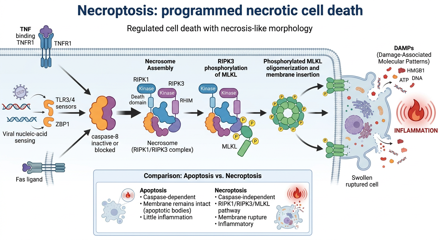
Infographic on necroptosis
necroptosis
necroptosis pathway RIPK1 RIPK3 MLKL review
Create an educational scientific infographic titled “Necroptosis: programmed necrotic cell death.” Show a clear left-to-right molecular pathway: stress signals (TNF binding TNFR1, viral nucleic-acid sensing, Fas ligand) lead to an intracellular decision point labeled “caspase-8 inactive or blocked”, then assembly of a “necrosome” containing RIPK1 and RIPK3, then RIPK3 phosphorylation of MLKL, then phosphorylated MLKL oligomerizes and inserts into the plasma membrane. End with a swollen ruptured cell releasing DAMPs and an inflammation icon. Include a small comparison box: apoptosis = caspase-dependent, membrane remains intact, little inflammation; necroptosis = caspase-independent, RIPK1/RIPK3/MLKL, membrane rupture, inflammatory. Include labels that it is regulated cell death with necrosis-like morphology. Use biologically accurate cell and protein diagrams, concise legible text, and a clean professional medical-textbook infographic style.