
Complete notes on juvenile idiopathic arthritis, starting from: - Etiology - Incidence - Pathophysiology - Clinical criterias for diagnosis - Investigations - Most relevant differential diagnosis for it What are the different types of juvenile idiopathic arthritis . how to approach a case of juvenile idiopathic arthritis involved for my FCPS Part II examination? What history and physical examination findings, with differential diagnosis in mind, should be taken from the patient?
"juvenile idiopathic arthritis"[MeSH Terms] AND classification
juvenile idiopathic arthritis types clinical features joint involvement
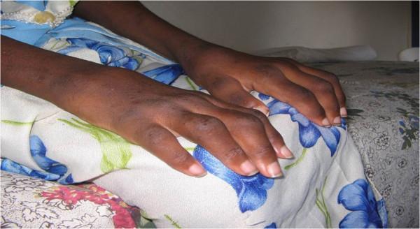
Clinical photograph demonstrating symmetrical polyarticular joint involvement in the hands of a pediatric patient, diagnostic of Rheumatoid Factor-negative (RF-ve) Polyarticular Juvenile Idiopathic Arthritis (JIA). The image shows bilateral, widespread swelling and structural deformities across multiple joint groups. Key anatomical regions involved include the wrist joints, metacarpophalangeal (MCP) joints, proximal interphalangeal (PIP) joints, and distal interphalangeal (DIP) joints. Visible features include significant soft tissue swelling and altered alignment of the digits, which are characteristic of chronic inflammatory arthritis. This visual provides an educational example of the polyarticular JIA subtype, highlighting the symmetrical distribution of disease in the upper limbs and the potential for multi-joint progression in untreated or late-presenting pediatric cases within a clinical rheumatology context.
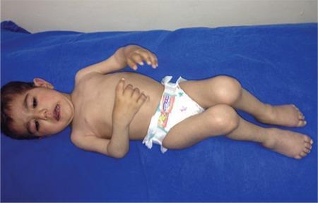
Clinical photograph of a young child demonstrating widespread joint involvement characteristic of polyarticular juvenile idiopathic arthritis (JIA). The patient is shown in a supine position with significant multi-joint deformities and contractures. The upper extremities exhibit symmetric arthritis of the small joints; both wrists and metacarpophalangeal (MCP) joints appear swollen with the hands held in a flexed, claw-like posture. Both arms are adducted across the torso, indicating restricted range of motion in the shoulders and elbows. The lower extremities show prominent involvement of the knees and ankles, which appear enlarged and are held in a flexed position. Significant swelling and deformity are visible in the metatarsophalangeal (MTP) joints of the feet. The overall clinical presentation illustrates the symmetric, polyarticular nature of the disease, involving five or more joints, often including both large and small joints of the upper and lower extremities. This visual serves as an educational example of the severe physical manifestations and functional limitations associated with advanced or inadequately controlled JIA.
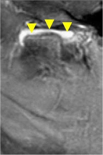
This diagnostic image is a post-contrast, fat-saturated sagittal T1-weighted MRI of the left temporomandibular joint (TMJ). The image demonstrates clinical features of active inflammatory arthritis, specifically Juvenile Idiopathic Arthritis (JIA). Three yellow arrowheads highlight intense contrast enhancement (CE) and synovial thickening within the superior joint space, located immediately above the mandibular condyle. The hyperintense (bright) signal in the synovium signifies increased vascularity and active inflammation. Structurally, there is an increase in soft tissue volume within the joint capsule, representing synovial proliferation. This imaging modality serves as a gold standard for detecting early signs of active TMJ involvement in pediatric rheumatology, distinguishing inflammatory changes like synovitis from later-stage arthritic sequelae such as bony erosions or condylar remodeling.
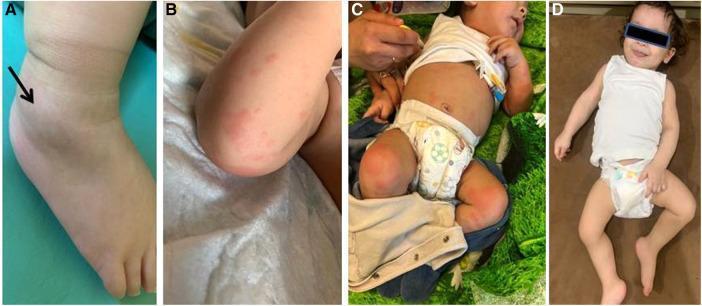
A four-panel clinical photograph collage documenting the clinical features and treatment response in a pediatric patient with systemic juvenile idiopathic arthritis (sJIA). (A) Close-up of the right ankle showing significant periarticular swelling (indicated by a black arrow), consistent with active arthritis. (B) High-magnification view of a characteristic maculopapular rash, appearing as salmon-pink, non-confluent macules on the extremity. (C) Clinical photograph of the infant prior to biologic therapy, demonstrating a widespread erythematous rash on the trunk and lower limbs, alongside a distressed general condition. (D) Comparative follow-up photograph 3 months after initiating canakinumab (an IL-1̢ inhibitor) treatment, showing complete resolution of the cutaneous rash, absence of visible joint swelling, and a marked improvement in the patient's overall clinical state and well-being. The set serves as an educational tool for identifying systemic features of pediatric autoinflammatory conditions and monitoring the efficacy of targeted biologic interventions.
systemic JIA quotidian fever salmon rash
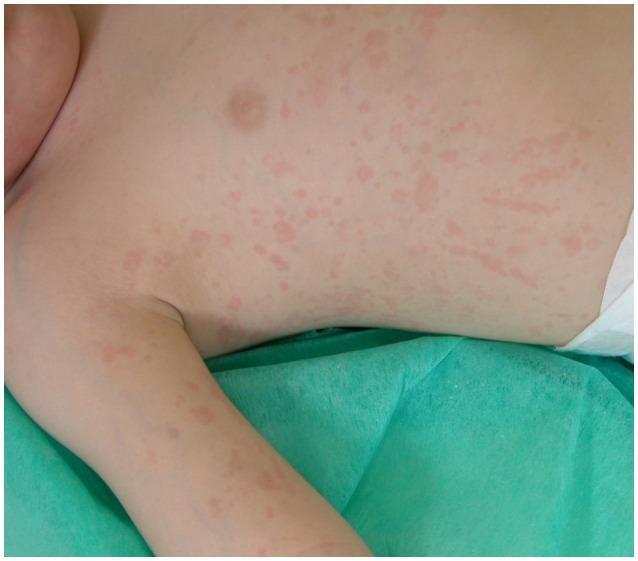
This clinical photograph displays a widespread cutaneous manifestation on a pediatric patient's torso and upper extremity. The presentation is characterized as a salmon-macular rash, featuring numerous discrete and confluent salmon-pink erythematous lesions. The lesions are primarily macular, appearing flat and flush with the skin surface, with irregular margins and varying sizes. Some areas exhibit linear or streaky patterns among the scattered spots. This specific visual finding is a hallmark clinical feature of systemic juvenile idiopathic arthritis (sJIA), often presenting during fever spikes. The image demonstrates the classic morphology and distinctive hue essential for differentiating sJIA from other pediatric exanthems or inflammatory conditions. The distribution includes the chest, abdomen, and the inner aspect of the arm, highlighting the diffuse and migratory nature of the rash in rheumatological contexts.
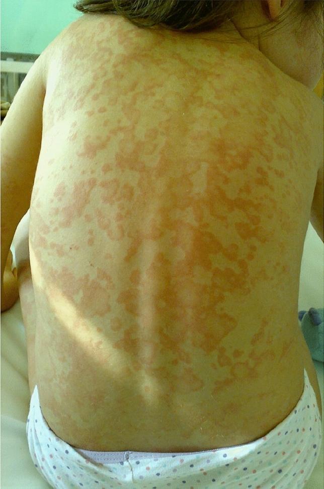
This clinical photograph shows the back of a pediatric patient exhibiting a widespread, salmon-pink erythematous rash. The lesions are distributed extensively across the upper and lower back and shoulders. Morphologically, the rash is composed of discrete macules and slightly raised papules that frequently coalesce into larger, irregularly shaped, nonfixed patches. The borders of the lesions are indistinct and poorly demarcated, blending into the surrounding fair skin. There is a notable absence of scaling, crusting, vesiculation, or blistering. Clinically, this type of evanescent rash is a hallmark cutaneous manifestation of systemic juvenile idiopathic arthritis (sJIA), typically characterized by its transient nature, often appearing during peaks of spiking fever and subsiding as the temperature normalizes. The image serves as a key educational example for diagnosing autoinflammatory disorders through cutaneous physical examination.
enthesitis related arthritis HLA-B27 sacroiliac
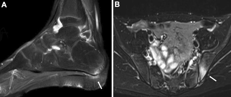
This composite diagnostic image consists of two fat-suppressed T2-weighted magnetic resonance images (MRI) illustrating classic manifestations of Enthesitis-Related Arthritis (ERA). Image A is a sagittal view of the foot and ankle, demonstrating enthesitis at the insertion of the plantar fascia into the calcaneus. A focal area of high signal intensity (arrow) at the calcaneal attachment signifies localized edema and active inflammation, contrasting with the low-signal cortical bone. Image B is an axial view of the pelvis, showing the sacroiliac (SI) joints. A distinct area of hyperintensity (arrow) is visible on the left side, indicating bone marrow edema and inflammation consistent with sacroiliitis. The surrounding pelvic musculature and soft tissues show intermediate signal intensities, while the fat suppression technique highlights the pathological fluid/edema. These findings are pedagogically significant for identifying the progression of spondyloarthropathies in HLA-B27 positive pediatric patients, transitioning from peripheral enthesitis to axial involvement.
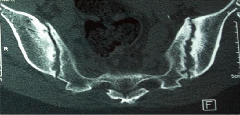
This diagnostic image is an axial computed tomography (CT) scan of the pelvis, specifically targeting the sacroiliac (SI) joints. The scan demonstrates bilateral Grade 3 sacroiliitis, a hallmark of axial spondyloarthritis. Key visual findings include significant subchondral sclerosis, visible as hyperdense (white) bone thickening on both the sacral and iliac sides of the joints. Multiple cortical erosions are present, creating an irregular, jagged appearance of the joint margins. There is also evident narrowing and blurring of the joint space. These features are characteristic of chronic inflammatory changes in conditions such as Enthesitis-Related Arthritis (ERA) or ankylosing spondylitis. The image serves as a high-quality educational example for identifying radiographic manifestations of chronic sacroiliitis in the context of HLA-B27-positive autoimmune disease.
Juvenile Idiopathic Arthritis (JIA) - Complete FCPS Part II Notes
1. DEFINITION
Previously called Juvenile Rheumatoid Arthritis (JRA) in the USA and Juvenile Chronic Arthritis (JCA) in Europe - both terms are now obsolete.
2. INCIDENCE & EPIDEMIOLOGY
| Parameter | Data |
|---|---|
| Most common rheumatic disease of childhood | Yes |
| Prevalence (global) | 1-4 per 1,000 children |
| Incidence | 6-19 per 100,000 per year |
| Age of onset | Any age < 16 years; bimodal peaks at 2-4 years and 9-14 years |
| Female predominance | Most subtypes (except systemic JIA and ERA) |
| Oligoarticular | 40-50% of all JIA |
| Polyarticular RF-negative | 20-35% |
| Systemic JIA | 5-15% |
| Enthesitis-related arthritis | 10-15% |
| Psoriatic JIA | 5-10% |
| Polyarticular RF-positive | <10% |
| Undifferentiated | ~5% |
3. ETIOLOGY
3a. Genetic Factors
- HLA region confers up to 13% of total genetic risk
- Each JIA subtype may have a distinct adult counterpart based on HLA associations:
- RF+ polyarticular JIA → HLA-DRB1 (mirrors adult seropositive RA)
- Oligoarticular + RF-negative polyarticular → shares HLA with adult seronegative RA
- Systemic JIA → genetically distinct from all other subtypes
- ERA → strongly associated with HLA-B27 (60-80%)
- GWAS has confirmed 22 additional susceptibility genes outside HLA, including IL-2, IL-2RA, PTPN2, RUNX1 (more prominent in JIA than in adult RA)
- JIA shares more genetic loci with Type 1 Diabetes than with RA, particularly via the IL-2 pathway
3b. Immunological Factors
- Aberrant T-cell activation (both CD4+ Th1 and Th17 pathways)
- Pro-inflammatory cytokines: IL-1, IL-6, IL-17, TNF-α
- Systemic JIA - primarily driven by IL-1 and IL-6 (more autoinflammatory than autoimmune)
- ANA positivity suggests B-cell dysregulation (relevant in oligoarticular JIA and uveitis risk)
3c. Environmental Triggers
- Infections (viral/bacterial) may act as triggers in genetically susceptible individuals
- Trauma has been reported as a precipitant
- No confirmed infectious agent consistently identified
4. PATHOPHYSIOLOGY
Synovial Joint Inflammation
- Initiating event: Unknown antigen triggers innate and adaptive immune response
- Synovial membrane becomes infiltrated with T lymphocytes, macrophages, and plasma cells
- Synovitis: Synoviocyte proliferation → pannus formation
- Pannus invades cartilage and subchondral bone → erosions (more prominent in RF+ polyarticular)
- Cytokine cascade: TNF-α, IL-1, IL-6 → amplify inflammation, cause systemic effects
- Chronic hyperemia from inflammation in children causes periarticular bony overgrowth (widening/squaring of tibial spines, carpal bones) - different from adult erosive pattern
Subtype-Specific Immunopathology
| Subtype | Dominant Mechanism |
|---|---|
| Oligoarticular | ANA-driven; T-cell dysregulation; strong uveitis link |
| Systemic JIA | IL-1 / IL-6 autoinflammation; macrophage activation; resembles periodic fever syndromes |
| RF+ Polyarticular | Anti-CCP antibodies; similar to adult RA; immune complex deposition |
| ERA | HLA-B27 related; enthesis-centered T-cell response; axial involvement |
Uveitis Pathogenesis
- Anterior uveitis (most common) is driven by local immune activation in the uveal tract
- Risk linked to ANA positivity and young age of onset
- Typically asymptomatic (insidious) in oligoarticular JIA - requires slit-lamp screening
5. ILAR CLASSIFICATION CRITERIA (7 Subtypes)
5a. Oligoarticular JIA (40-50%)
| Feature | Detail |
|---|---|
| Definition | 1-4 joints in first 6 months |
| Persistent subtype | ≤4 joints throughout entire disease course |
| Extended subtype | >4 joints after first 6 months |
| Demographics | F > M; peak onset 2-4 years |
| Joints | Typically lower limb large joints - knees, ankles |
| ANA | Positive in 60-70% |
| Uveitis | 30% overall; 16% persistent; 25% extended |
| Exclusions | RF+ at ≥2 tests; psoriasis; HLA-B27+ male >6 yrs; SpA features; systemic features |
5b. Polyarticular JIA - RF Negative (20-35%)
| Feature | Detail |
|---|---|
| Definition | ≥5 joints in first 6 months; RF negative on ≥2 tests ≥3 months apart |
| Demographics | F > M; bimodal peaks 2-4 and 10-14 years |
| Joints | Small and large joints; can be symmetric or asymmetric |
| ANA | 50% positive |
| HLA-B27 | 10% positive |
| Uveitis | 4% (usually asymptomatic anterior) |
5c. Polyarticular JIA - RF Positive (<10%)
| Feature | Detail |
|---|---|
| Definition | ≥5 joints; RF+ on ≥2 tests ≥3 months apart |
| Demographics | F >> M; peak onset 9-12 years |
| Joints | Symmetric polyarthritis including small joints of hands and feet |
| ANA | 40% positive |
| Anti-CCP | Often positive |
| Uveitis | 2% |
| Rheumatoid nodules | Present (nontender, on bony prominences/extensor surfaces) |
| Adult equivalent | Seropositive RA |
5d. Systemic JIA (5-15%)
| Feature | Detail |
|---|---|
| Definition | Arthritis + quotidian (daily) fever ≥2 weeks + ≥1 of: evanescent rash, lymphadenopathy, hepato/splenomegaly, serositis |
| Demographics | M = F; peak onset 1-5 years |
| Joints | Oligo or polyarticular; knees, wrists, ankles; cervical spine |
| Fever | Daily spiking (quotidian), typically ≥39°C, once or twice/day, returns to baseline |
| Rash | Salmon-pink evanescent macular rash - appears with fever spikes, disappears when afebrile |
| ANA | 20% |
| HLA-B27 | 5-10% |
| Uveitis | 1% (lowest risk) |
| Serious complication | Macrophage Activation Syndrome (MAS) |
5e. Enthesitis-Related Arthritis / ERA (10-15%)
| Feature | Detail |
|---|---|
| Definition | Arthritis + enthesitis, OR either + ≥2 of: sacroiliitis/axial involvement, HLA-B27+, male onset >6 years, symptomatic anterior uveitis, FDR with HLA-B27 disease |
| Demographics | M > F; peak onset 9-12 years |
| Joints | Lower extremity large joints; axial skeleton involvement |
| Enthesitis | Heel (Achilles insertion), plantar fascia, tibial tubercle, iliac crest |
| HLA-B27 | 60-80% positive |
| Uveitis | 7%; typically acute symptomatic (unlike other subtypes) |
| Adult equivalent | Ankylosing Spondylitis |
5f. Psoriatic JIA (5-10%)
| Feature | Detail |
|---|---|
| Definition | Arthritis + psoriasis, OR arthritis + ≥2 of: dactylitis, nail pitting/onycholysis, psoriasis in FDR |
| Demographics | F > M; bimodal peaks 2-4 and 9-11 years |
| Joints | Wrists + small joints of hands/feet initially; asymmetric; dactylitis ("sausage digits") |
| ANA | 40% |
| HLA-B27 | 20% |
| Uveitis | 10% |
5g. Undifferentiated JIA (~5%)
6. CLINICAL FEATURES - GENERAL
Articular Features (Common to All)
- Joint swelling - soft tissue (synovitis) ± effusion
- Morning stiffness (>45 min in children - they characteristically exhibit "gelling" after inactivity)
- Warmth and limited range of motion - tenderness less prominent than in adults
- Limp - often the presenting complaint in young children rather than pain complaint
- Preference to hold joints in flexion (position of comfort)
- Growth disturbances - leg length discrepancy (affected limb may be longer due to hyperemia early; shorter late); micrognathia from TMJ involvement
Extra-articular Features by System
| System | Feature |
|---|---|
| Eyes | Anterior uveitis (most common extra-articular feature; insidious in oligoarticular; symptomatic in ERA) |
| Skin | Salmon rash (systemic), psoriasis (PsA), rheumatoid nodules (RF+) |
| Fever | Quotidian fever in systemic JIA |
| Lymphoid | Lymphadenopathy, hepatosplenomegaly (systemic JIA) |
| Cardiovascular | Pericarditis, myocarditis (systemic JIA) |
| Growth | Short stature, osteopenia, micrognathia |
| TMJ | Mandibular hypoplasia, limited mouth opening |
7. INVESTIGATIONS
Laboratory Tests
| Investigation | Significance |
|---|---|
| CBC | Leukocytosis (systemic JIA); anemia of chronic disease; thrombocytosis (active disease marker) |
| ESR / CRP | Elevated in active disease; may be normal in oligoarticular |
| ANA | Positive in 60-70% oligoarticular; risk marker for uveitis (not diagnostic of JIA) |
| RF | Two positive tests ≥3 months apart defines RF+ polyarticular subtype; also rules in/out |
| Anti-CCP | More specific than RF; positive in RF+ polyarticular JIA |
| HLA-B27 | Positive in 60-80% ERA; supports that subtype |
| Ferritin | Markedly elevated (often >10,000 µg/L) in systemic JIA and MAS |
| LFTs + Fibrinogen | For MAS screening: elevated LFTs, decreased fibrinogen |
| Uric acid | To exclude gout |
| Blood culture | To exclude septic arthritis |
| LDH | Elevated - helps exclude leukemia |
| Synovial fluid analysis | Inflammatory pattern (WBC 5,000-80,000, predominantly neutrophils); sterile; exclude infection and crystals |
Imaging
| Modality | Findings |
|---|---|
| X-ray | Insensitive early; periarticular osteopenia, soft tissue swelling; later: joint space narrowing, erosions, bony overgrowth (widening/squaring of carpal bones, tibial spines) |
| Ultrasound | First-line for effusion detection, synovitis, enthesitis; guides aspiration |
| MRI | Gold standard for early synovitis, bone marrow edema (BME), cartilage loss; essential for TMJ, hip, and sacroiliac joint evaluation; contrast-enhanced for synovial enhancement |
| Bone scan | Rarely used; identifies polyarticular involvement |
Ophthalmological Screening
- Slit-lamp examination is mandatory for ALL JIA subtypes
- Frequency depends on subtype, ANA status, and age of onset (highest risk: ANA+ oligoarticular, age <6 years → every 3 months)
- Must be done even without symptoms (uveitis in oligoarticular JIA is asymptomatic)
8. DIAGNOSTIC CRITERIA SUMMARY (ILAR 2001)
- Age of onset < 16 years
- Arthritis (swelling, or ≥2 of: limited ROM, tenderness on motion, warmth) in one or more joints
- Duration ≥ 6 weeks
- Exclusion of all other causes
PRINTO 2019 Updated Criteria have been proposed as a revision to the ILAR criteria, reorganizing JIA into 6 categories with refined inclusion/exclusion criteria, though ILAR criteria remain most widely cited in examinations.
9. DIFFERENTIAL DIAGNOSIS
Most Relevant Differentials
| Diagnosis | Key Distinguishing Features |
|---|---|
| Septic arthritis | Acute onset, single hot joint, fever, child ill-appearing; synovial WBC >100,000; positive culture |
| Reactive arthritis | Post-infectious (GI/GU); transient; typically <6 weeks; preceding infection history; HLA-B27 may be + |
| Acute rheumatic fever | Migratory polyarthritis; carditis; erythema marginatum; elevated ASO; response to salicylates |
| Leukemia / lymphoma | Bone pain > joint pain (disproportionate); night sweats; anemia; thrombocytopenia; elevated LDH; bone marrow biopsy |
| Systemic Lupus Erythematosus | Malar rash; renal involvement; positive dsDNA and Anti-Sm; ANA >1:320; multi-system |
| Kawasaki Disease | Fever >5 days; rash; strawberry tongue; lymphadenopathy; conjunctivitis; coronary involvement (< 5 years) |
| Lyme arthritis | Geographic exposure; tick bite history; positive Borrelia serology; monoarticular knee |
| Transient synovitis (hip) | Child 3-10 years; unilateral hip pain; afebrile or low-grade; self-limiting |
| Osteomyelitis | Bone pain + point tenderness; periosteal elevation on X-ray; elevated CRP/ESR; positive bone scan/MRI |
| Inflammatory bowel disease arthropathy | GI symptoms; peripheral arthritis (parallels bowel disease) or axial arthritis (independent) |
| Psoriatic arthritis (adult) | Psoriatic plaques precede arthritis more reliably in adults vs children |
| Hemophilia | Hemarthrosis; bleeding history; clotting factor levels |
10. APPROACH TO JIA FOR FCPS PART II EXAMINATION
Structured Clinical Approach
HISTORY TAKING
A. Presenting Complaint
- Duration of joint symptoms (must be ≥6 weeks to qualify for JIA)
- Which joint(s) involved? How many? Which came first?
- Morning stiffness: how long? (>45 min is significant)
- Gelling phenomenon (stiffness after periods of rest)
- Limp - especially in young children who may not verbalize pain
B. Characterization of Joint Symptoms
- Swelling (soft vs fluctuant)
- Pain - note: JIA children often have less pain than expected given degree of swelling
- Warmth, erythema (redness suggests infection more than JIA)
- Range of motion restriction
- Functional impairment: dressing, climbing stairs, sports
C. Systemic Symptoms (Crucial for Subtype Identification)
| Symptom | Implication |
|---|---|
| Quotidian fever (≥39°C, once/twice daily, returns to baseline) | Systemic JIA |
| Salmon-pink rash with fever | Systemic JIA |
| Eye symptoms (redness, photophobia, blurred vision) | Uveitis (note: absence of symptoms does NOT rule out uveitis in oligoarticular) |
| Back/buttock pain worse in morning, relieved by activity | ERA / sacroiliitis |
| Heel pain | Enthesitis (ERA) |
| Skin rash (psoriatic plaques, nail changes, dactylitis) | Psoriatic JIA |
| Weight loss, night sweats, pallor | Malignancy (red flag) |
| Preceding infection (GI, UTI, throat) | Reactive arthritis |
| Oral ulcers | SLE; Behcet's |
| Malar rash, photosensitivity | SLE |
D. Growth and Development History
- Linear growth (JIA impairs growth)
- Jaw development (TMJ involvement → micrognathia)
- Pubertal development (chronic inflammation delays puberty)
E. Past Medical History
- Previous infections, vaccinations
- Previous episodes of joint swelling
- Eye examinations history
F. Family History
- Psoriasis (first-degree relative → supports psoriatic JIA)
- HLA-B27 associated diseases: ankylosing spondylitis, inflammatory bowel disease
- Autoimmune disease in family
- Any history of rheumatic diseases
G. Drug and Social History
- Current medications (including NSAIDs already trialed)
- School attendance and functional impairment
- Geographic exposure (Lyme disease endemic area?)
PHYSICAL EXAMINATION
A. General Examination
- Growth parameters (height, weight - plot on centile chart; short stature common)
- Temperature chart (quotidian pattern?)
- Facial appearance: micrognathia (TMJ involvement), bird facies
- Pallor (anemia of chronic disease or leukemia)
- Lymphadenopathy (generalized in systemic JIA vs local in infection/malignancy)
- Hepatosplenomegaly (systemic JIA, SLE, malignancy)
B. Skin Examination
- Salmon-pink evanescent macular rash - look during fever (systemic JIA); disappears between spikes
- Psoriatic plaques (elbows, knees, scalp, umbilicus, natal cleft)
- Nail pitting, onycholysis (psoriatic JIA)
- Erythema marginatum (ARF)
- Malar rash, discoid lesions (SLE)
- Rheumatoid nodules on extensor surfaces (RF+ polyarticular)
C. Joint Examination (CRITICAL)
| Step | What to Look For |
|---|---|
| Inspection | Swelling, deformity, muscle wasting, alignment |
| Palpation | Warmth, effusion (ballottement, bulge sign at knee), synovial thickening, tenderness |
| ROM | Active and passive; note restriction |
| Function | Gait, grip strength, dressing ability |
- Knees: Most commonly involved; bulge sign, patellar tap for effusion
- Ankles and subtalar joints
- Wrists and small joints of hands (MCPs, PIPs, DIPs)
- Cervical spine: rotation, flexion (C2-C3 involvement common in JIA)
- TMJ: mouth opening (<35 mm is abnormal); micrognathia
- Hips: log roll, FABER, Thomas test (often painful, limited internal rotation)
- Sacroiliac joints: Faber test, sacral compression (ERA)
- Entheses: Achilles insertion, plantar fascia, tibial tubercle, iliac crest (ERA)
D. Eye Examination
- Visual acuity
- Pupil shape (irregular pupil → posterior synechia from chronic uveitis)
- Band keratopathy (calcium deposits in cornea from chronic uveitis)
- Refer ALL JIA patients for formal slit-lamp examination regardless of symptoms
E. Cardiovascular
- Pericardial rub (systemic JIA)
- Heart murmur (ARF differential - mitral regurgitation)
PUTTING IT TOGETHER: DIFFERENTIAL DIAGNOSIS AT THE BEDSIDE
| Clinical Pattern | First Consider |
|---|---|
| Single warm swollen joint, fever, child ill-looking | Septic arthritis (EMERGENCY) |
| Migratory polyarthritis + carditis, fever, post-strep | Acute Rheumatic Fever |
| Quotidian fever + salmon rash + arthritis + hepatosplenomegaly | Systemic JIA |
| Young girl, 2-4 years, single knee swelling, no pain, no systemic features | Oligoarticular JIA |
| Older girl, symmetric small joint polyarthritis, RF+ | RF+ Polyarticular JIA (like adult RA) |
| Teenage boy, lower limb arthritis + heel pain + HLA-B27+ | ERA / early AS |
| Arthritis + dactylitis + nail pitting ± family psoriasis | Psoriatic JIA |
| Arthritis + malar rash + renal disease + high-titer ANA | SLE |
| Bone pain > joint pain + anemia + thrombocytopenia | Leukemia (MUST exclude before JIA label) |
| Post-GI/GU infection + oligoarthritis, <6 weeks | Reactive arthritis |
11. MACROPHAGE ACTIVATION SYNDROME (MAS) - CRITICAL COMPLICATION
- Occurs most commonly in systemic JIA
- A life-threatening hyperactivation of macrophages and T-cells
- Clinical features: persistent high fever (loss of quotidian pattern), hepatosplenomegaly, CNS dysfunction, bleeding
- Lab hallmarks: falling ESR (paradoxical), rising CRP, very high ferritin, pancytopenia, elevated LFTs, decreased fibrinogen, elevated D-dimer, elevated triglycerides
- Treatment: High-dose IV corticosteroids + IL-1 blockade (anakinra) + cyclosporine A
12. COMPLICATIONS
| Complication | Subtype Most at Risk |
|---|---|
| Chronic uveitis → blindness, band keratopathy, cataract, glaucoma | Oligoarticular (ANA+) |
| MAS | Systemic JIA |
| Growth retardation / short stature | All, especially systemic |
| Micrognathia (TMJ involvement) | Systemic, polyarticular |
| Amyloidosis | Long-standing disease (rare in modern therapy era) |
| Osteoporosis | Chronic disease + steroid use |
| Drug toxicity | Methotrexate (hepatotoxicity), biologics (infections) |
SUMMARY TABLE: ILAR JIA SUBTYPES AT A GLANCE
| Feature | Oligoarticular | RF-Poly | RF+Poly | Systemic | ERA | Psoriatic |
|---|---|---|---|---|---|---|
| % cases | 40-50 | 20-35 | <10 | 5-15 | 10-15 | 5-10 |
| Sex | F>>M | F>M | F>>M | M=F | M>>F | F>M |
| Age | 2-4 yr | 2-4, 10-14 yr | 9-12 yr | 1-5 yr | 9-12 yr | 2-4, 9-11 yr |
| Joints | ≤4 | ≥5 | ≥5 Symmetric | Oligo/poly | Lower limb + axial | Wrists/small |
| ANA | 60-70% | 50% | 40% | 20% | 20% | 40% |
| RF | - | - | + | - | - | - |
| HLA-B27 | - | 10% | 10-15% | 5-10% | 60-80% | 20% |
| Uveitis | 30% (silent) | 4% | 2% | 1% | 7% (acute) | 10% |
| Key feature | ANA, uveitis | Asymmetric poly | Nodules, RA-like | Quotidian fever, rash | Enthesitis, B27 | Dactylitis, psoriasis |
- The Harriet Lane Handbook, 23rd ed. (Johns Hopkins), Table 27.1 - JIA Classification
- Firestein & Kelley's Textbook of Rheumatology, 11th ed. - Chapter 24 (Genetics), Chapter 58 (Imaging in JIA)
- Rheumatology, 2-Volume Set (2022, Elsevier) - ILAR/PRINTO Classification Criteria for JIA
- Robbins & Kumar Basic Pathology - Pathophysiology of JIA
Alter the notes by adding mnemonics (mnemonics should be memorable, easily recallable, and especially relatable to the disease) for remembering the information, especially the incidents, different types, investigations, and treatment.
Juvenile Idiopathic Arthritis (JIA) - Complete FCPS Part II Notes with Mnemonics
1. DEFINITION
🧠 Mnemonic: "JIA = 6-16 Rule" 6 weeks minimum duration + onset before 16 years = JIA until proven otherwise
2. INCIDENCE & EPIDEMIOLOGY
| Letter | Subtype | Frequency |
|---|---|---|
| Old | Oligoarticular | 40-50% (most common) |
| Parrots | Polyarticular RF- | 20-35% |
| Prefer | Polyarticular RF+ | <10% |
| Singing | Systemic | 5-15% |
| Extremely | Enthesitis-Related (ERA) | 10-15% |
| Pretty | Psoriatic | 5-10% |
| Unes | Undifferentiated | ~5% |
Think: Oligoarticular is the oldest (most common) bird in the JIA flock - it dominates.
3. ETIOLOGY
- J - Junk HLA genes (HLA region = 13% of genetic risk)
- I - IL-2 pathway (more dominant in JIA than adult RA)
- A - Autoimmune triggers + environmental (viral/bacterial)
- H - HLA-B27 (especially ERA subtype, 60-80%)
- G - GWAS genes (22 confirmed outside HLA: PTPN2, RUNX1, IL-2RA)
- I - Innate dysregulation (systemic JIA - IL-1/IL-6 driven)
- E - Environmental triggers (infections, trauma as precipitants)
4. PATHOPHYSIOLOGY
| Letter | Step |
|---|---|
| Precipitant | Unknown antigen triggers immune response |
| Activation | T-cells, macrophages infiltrate synovium |
| Synovitis | Synoviocyte proliferation → pannus formation |
| Tearing | Pannus invades cartilage and subchondral bone |
| Amplification | TNF-α, IL-1, IL-6 cytokine cascade perpetuates damage |
In children specifically, chronic hyperemia causes bony OVERGROWTH (squaring/widening of carpal bones and tibial spines) - the opposite of adult erosive disease. Think: "Kids GROW, adults ERODE."
Cytokines by Subtype
- All other subtypes → TNF-α dominant (respond to anti-TNF therapy)
- Systemic JIA → IL-1 + IL-6 (respond to anakinra/canakinumab + tocilizumab, NOT anti-TNF primarily)
5. THE 7 SUBTYPES - ILAR CLASSIFICATION
Master Mnemonic: "OPPOSE U"
| Letter | Subtype |
|---|---|
| O | Oligoarticular |
| P | Polyarticular RF- |
| P | Polyarticular RF+ |
| O | sOmething systemic (systemic JIA) |
| S | Spondylitis-like (ERA) |
| E | Extra-skin (Psoriatic) |
| U | Undifferentiated |
5a. Oligoarticular JIA (40-50%)
- Only = 1-4 joints only
- Little = youngest age (peak 2-4 years)
- Innocent = least systemic features; "silent" uveitis is the hidden danger
- Girls = F >> M
- Oligoarticular = the most common subtype
- "Persistent" = Polite (stays ≤4 joints forever; 16% uveitis)
- "Extended" = Expanding (grows beyond 4 joints after 6 months; 25% uveitis)
| Feature | Detail |
|---|---|
| Joints | 1-4; lower limb large joints (knees, ankles) |
| ANA | 60-70% positive |
| Uveitis | 30% overall - SILENT (no symptoms!) |
| Exclusions | RF+; psoriasis; HLA-B27+ male >6 yrs; systemic features |
5b. Polyarticular RF-Negative (20-35%)
"RF-Negative Poly = Free to be Bimodal and Asymmetric"
- Five or more joints
- RF negative
- Bimodal peaks (2-4 AND 10-14 years)
- Asymmetric or symmetric involvement
| Feature | Detail |
|---|---|
| Definition | ≥5 joints, RF- × 2 tests ≥3 months apart |
| ANA | 50% |
| Uveitis | 4% |
5c. Polyarticular RF-Positive (<10%)
- RF + Anti-CCP positive
- Older onset (9-12 years)
- Symmetric small joint polyarthritis
- Equivalent to adult RA (adult counterpart)
| Feature | Detail |
|---|---|
| Unique features | Rheumatoid nodules (bony prominences, extensor surfaces) |
| Uveitis | Lowest of all poly types: 2% |
| Adult equivalent | Seropositive RA |
5d. Systemic JIA (5-15%)
| Letter | Feature |
|---|---|
| Spike | Quotidian fever (daily ≥39°C, returns to baseline) |
| Arthritis | Oligo or polyarticular (knees, wrists, ankles) |
| Inflamed organs | Hepatosplenomegaly + lymphadenopathy + serositis |
| Luminous rash | Salmon-pink evanescent rash (appears WITH fever, disappears without it) |
| Snag | MAS = Macrophage Activation Syndrome (the deadly snag) |
🌅 Visual: Imagine the salmon-pink rash as the COLOR OF A SUNSET - it appears dramatically then fades. The fever is the tide - rash rides the tide.
| Diagnostic Requirement | ≥2 weeks quotidian fever + arthritis + ≥1 of: rash, lymphadenopathy, hepatosplenomegaly, serositis |
|---|---|
| M = F | Equal sex distribution (unique!) |
| ANA | Only 20% positive |
| Uveitis | 1% - lowest risk of all subtypes |
5e. Enthesitis-Related Arthritis / ERA (10-15%)
- Enthesis + arthritis (definition)
- Real males (M >> F predominance; teenage boys)
- Axial skeleton involvement (sacroiliitis, spine)
- Plus: HLA-B27+ in 60-80%, heel/Achilles pain, future ankylosing spondylitis
| Letter | Criterion |
|---|---|
| Axial | Axial spine / sacroiliac involvement |
| Symptom | Symptomatic anterior uveitis (acute, painful - unlike other types) |
| HLA-B27 | HLA-B27 positive |
| Eight-plus | Age >6 years + Male sex |
| Sib/parent | First-degree relative with HLA-B27 disease |
ERA = arthritis OR enthesitis + ≥2 ASHES criteria
5f. Psoriatic JIA (5-10%)
- Dactylitis (sausage digits)
- Arthritis (wrists + small hand/foot joints; asymmetric)
- Nail changes (pitting, onycholysis)
- Arthritis + psoriasis → definite
- Arthritis + 2 of (Dactylitis, Nail changes, Family history of psoriasis) → probable
5g. Undifferentiated (~5%)
SUBTYPE MEMORY TABLE
| Subtype | ANA% | Uveitis% | HLA-B27% |
|---|---|---|---|
| Oligoarticular | 60-70 | 30 (silent) | Low |
| RF- Poly | 50 | 4 | 10 |
| RF+ Poly | 40 | 2 | 10-15 |
| Systemic | 20 | 1 (lowest) | 5-10 |
| ERA | 20 | 7 (acute) | 60-80 |
| Psoriatic | 40 | 10 | 20 |
Quick memory rule for uveitis: "Oligo sees the most (30%), Systemic sees the least (1%). ERA's uveitis is the LOUDEST (symptomatic), Oligo's is the QUIETEST (silent)."
6. CLINICAL FEATURES
- Swelling (synovial thickening ± effusion)
- Warmth (less redness than infection)
- Activity-limiting stiffness (morning stiffness >45 min; "gelling")
- Restricted range of motion
- Muscle wasting (periarticular, disuse)
🧒 Pearl: Young children present with LIMP, not pain complaint. "JIA children limp before they complain."
7. INVESTIGATIONS
| Letter | Test | Purpose |
|---|---|---|
| Leuco | CBC (leukocytosis/anemia/thrombocytosis) | Activity; exclude leukemia |
| ANA | ANA | Subtype + uveitis risk |
| Blood | Blood culture | Exclude septic arthritis |
| WBC synovial | Synovial fluid analysis | Inflammatory vs septic vs crystal |
| Optics | Slit-lamp (ophthalmology) | Silent uveitis screening |
| RF | RF + Anti-CCP | Subtype classification |
| King | ESR / CRP | Inflammatory activity |
| Complete | HLA-B27 | ERA classification |
| Liver enzymes | LFTs + ferritin | MAS screening |
| Ultrasound | Ultrasound joints | Effusion, synovitis, enthesitis |
| Erosion check | X-ray | Structural damage (late) |
| Scan MRI | MRI | Early synovitis, TMJ, hips, SIJ |
Key Lab Patterns by Scenario
- Ferritin - massively elevated (>10,000 µg/L) ← the cardinal marker
- ALT/AST - elevated
- Leucocytes - FALL (pancytopenia, paradoxical)
- LDH - elevated
- Fibrinogen - FALLS (coagulopathy)
- Anemia - worsening
- Serum ferritin - rises while ESR paradoxically FALLS
- Triglycerides - elevated
🚨 Paradox pearl: In MAS complicating systemic JIA, the ESR falls despite severe systemic disease (because fibrinogen falls). Rising CRP + falling ESR + skyrocketing ferritin = MAS until proven otherwise.
8. DIAGNOSTIC CRITERIA (ILAR 2001)
- Age < 16 years ✓
- Arthritis ≥ 6 weeks ✓
- Exclusion of other diagnoses ✓
9. DIFFERENTIAL DIAGNOSIS
| Letter | Diagnosis | Key Distinguishing Clue |
|---|---|---|
| Septic | Septic arthritis | Single hot joint, child toxic, WBC >100,000 in synovial fluid |
| Hematology | Hematological malignancy (leukemia) | Bone pain >> joint pain; anemia; high LDH; blasts on smear |
| Acute rheumatic | Acute Rheumatic Fever | Migratory arthritis; carditis; elevated ASO; post-strep |
| Reactive | Reactive arthritis | Post-infection (GI/GU); self-limiting (<6 weeks) |
| Pain syndromes | Fibromyalgia / pain amplification | Normal labs; trigger points; no synovitis |
| Lyme | Lyme arthritis | Tick exposure; Borrelia serology; monoarthritis (knee) |
| Orthopaedic | Osteomyelitis / transient synovitis | Point tenderness on bone; febrile; hip on X-ray/USS |
| Onco/heme | Other vasculitis (Kawasaki disease) | Fever >5 days; coronary aneurysm; age <5; conjunctivitis |
| Kollegen | Connective tissue disease (SLE) | Malar rash; renal disease; dsDNA+; multi-system |
| Spondyloarthropathy | Adult-type spondyloarthritis | Age/sex/HLA-B27 overlap with ERA; IBD arthritis |
🚨 ALWAYS exclude leukemia FIRST before labeling childhood arthritis as JIA. Rule: "No blasts, no malignancy → then think JIA."
10. TREATMENT
STAIR 5 → BIOLOGICS (biologic DMARDs)
STAIR 4 → csDMARDs (Methotrexate = first-line DMARD)
STAIR 3 → Intraarticular corticosteroids
STAIR 2 → Short-course systemic corticosteroids (if needed)
STAIR 1 → NSAIDs (foundation of the staircase)
Step 1 - NSAIDs (The Floor)
- Naproxen, Ibuprofen, Indomethacin
- First-line for symptom control
- Not disease-modifying
Step 2 - Corticosteroids
- Intra-articular triamcinolone - most effective for oligoarticular JIA (single joint)
- Systemic steroids - bridging therapy in systemic JIA, severe polyarticular; minimize long-term use
Step 3/4 - Conventional DMARDs (csDMARDs)
| Drug | Use |
|---|---|
| Methotrexate (MTX) | First-line DMARD; weekly; most evidence in JIA |
| Sulfasalazine | ERA/spondyloarthritis subtype preferred |
| Leflunomide | Alternative to MTX; similar efficacy |
| Hydroxychloroquine | Mild disease, psoriatic JIA adjunct |
🧠 MTX memory: "MTX = Monday Tablet eXact" - give once weekly (not daily!) + folic acid supplement to reduce side effects (mucositis, hepatotoxicity)
Step 5 - Biologics
| Target | Drug | Best Subtype |
|---|---|---|
| TNF-α | Etanercept, Adalimumab, Infliximab | Polyarticular, ERA, Psoriatic |
| IL-6 | Tocilizumab | Systemic JIA (+ polyarticular) |
| IL-1 | Anakinra, Canakinumab | Systemic JIA (first-line biologic) |
| T-cell co-stimulation | Abatacept (CTLA-4-Ig) | RF+ polyarticular, refractory |
| IL-17 | Secukinumab | ERA (axial disease) |
- IL-1 blockers (anakinra/canakinumab) + IL-6 blockers (tocilizumab) = the two pillars
- Anti-TNF is less effective in systemic JIA
- "Systemic JIA hates TNF blockers, loves IL-1/IL-6 blockers"
Uveitis Treatment
- Topical steroids + mydriatics (first-line; eye drops)
- Methotrexate oral (systemic DMARD for refractory)
- Adalimumab IV/SC (biologic of choice for JIA uveitis - best evidence)
MAS Treatment
- Pulse IV corticosteroids (high dose methylprednisolone)
- Anakinra (IL-1 blockade, rapid acting)
- Cyclosporine A (if refractory)
- Killing JAK pathway (JAK inhibitors - ruxolitinib for refractory MAS)
11. OPHTHALMOLOGY SCREENING PROTOCOL
- ANA positive
- Not old - young age of onset (<6 years)
- Arthritis subtype = oligoarticular or psoriatic
| Risk Category | Screening Frequency |
|---|---|
| High risk (ANA+, oligo, <6 yr onset) | Every 3 months |
| Moderate risk | Every 6 months |
| Low risk (systemic, ERA) | Every 12 months |
Note: ERA uveitis is symptomatic (red painful eye) - patients self-present. Oligoarticular uveitis is silent - regular screening is the only way to detect it.
12. COMPLICATIONS SUMMARY
| Letter | Complication |
|---|---|
| Growth | Short stature, delayed puberty, micrognathia |
| Uveitis | Blindness, band keratopathy, cataract, glaucoma |
| Amyloidosis | Long-standing uncontrolled disease (rare now) |
| MAS | Macrophage Activation Syndrome (most feared acute complication) |
13. FCPS PART II HISTORY & EXAMINATION - QUICK RECALL
History Mnemonic: "MAST JIA" (imagine a ship's mast with JIA sails)
| Letter | History Domain |
|---|---|
| Morning stiffness | Duration (>45 min?); gelling? |
| Affected joints | Number, which joints, symmetric/asymmetric |
| Systemic symptoms | Fever pattern (quotidian?), rash, organomegaly |
| Time | Duration ≥6 weeks? (diagnostic requirement) |
| Joints elsewhere | TMJ, spine, sacroiliac joints (neck pain, back pain, heel pain) |
| Infection/eye | Any preceding infection (reactive); eye symptoms |
| Associated features | Psoriasis, family history, HLA-B27 diseases in family |
Examination Mnemonic: "SWIRL"
| Letter | Examination Step |
|---|---|
| Skin | Salmon rash, psoriatic plaques, rheumatoid nodules, nail changes |
| Weight/growth | Plot centiles; short stature is a sign of chronic disease |
| Inflamed joints | Systematic joint exam: swelling, warmth, effusion, ROM, tenderness |
| Rare joints** | TMJ (mouth opening), cervical spine, hips (log roll), entheses (heel) |
| Lymph/organs | Lymphadenopathy, hepatosplenomegaly, eye examination |
MASTER ONE-PAGE RECALL: THE JIA HOUSE
        🏠 JIA HOUSE
   ________________________
  |   ROOF = BIOLOGICS     |  ← step 5 (targeted: anti-TNF/IL-1/IL-6)
  |   FLOOR 2 = MTX/DMARDs |  ← step 4 (methotrexate = king DMARD)
  |   FLOOR 1 = Steroids   |  ← step 3 (intraarticular or short-course)
  |   GROUND = NSAIDs      |  ← step 1-2 (foundation)
  |________________________|
        |         |
    LEFT PILLAR   RIGHT PILLAR
   (ANA + uveitis (HLA-B27 + ERA
    screening)     = axial disease)

  BASEMENT = DIAGNOSIS (6 weeks, <16 yrs, exclude others)
  BOILER ROOM = MAS (systemic JIA emergency, treat with PACK)
- The Harriet Lane Handbook, 23rd ed. (Johns Hopkins), Table 27.1
- Firestein & Kelley's Textbook of Rheumatology, 11th ed.
- Rheumatology, 2-Volume Set (2022, Elsevier) - ILAR/PRINTO Classification Criteria