
Connections and functions of prefrontal cortex
Reading File
Finding Sources
Finding Sources
Reading File
Reading File
Finding Sources
Reading File
Reading File
Loading Image
Reading File
Loading Image
I now have comprehensive, high-quality content from multiple authoritative textbooks. Here is the full answer:
Prefrontal Cortex: Connections and Functions
The prefrontal cortex (PFC) is the largest subdivision of the frontal lobes and consists of higher-order heteromodal association cortex - the region anterior to the motor, premotor, and limbic areas. It is the part of the brain most commonly implicated in "frontal lobe" disorders.
Anatomical Subdivisions
The PFC is not a single area. Its major subdivisions are:
| Subdivision | Location | Key Role |
|---|---|---|
| Dorsolateral PFC | Lateral convexity | Working memory, cognitive set, executive control |
| Orbitofrontal cortex | Undersurface / orbital | Emotional regulation, social behavior, reward |
| Medial PFC / anterior cingulate | Medial surface | Motivation, initiative, affect |
| Ventromedial PFC | Medial-inferior | Decision-making, emotional weighting |
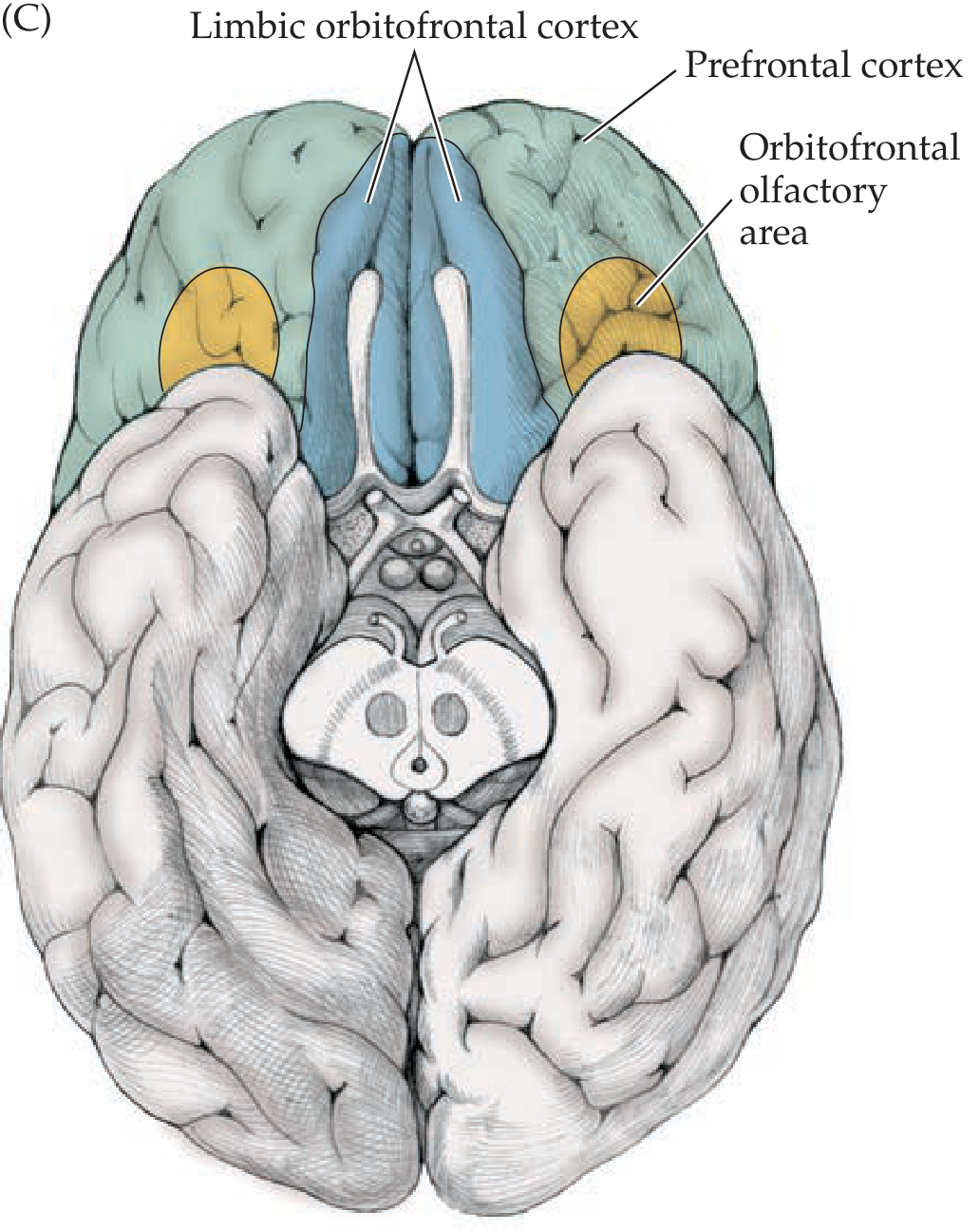
The medial surface and orbitofrontal surface, shown below (medial view showing prefrontal cortex in teal and limbic anterior cingulate/orbitofrontal cortex in blue):
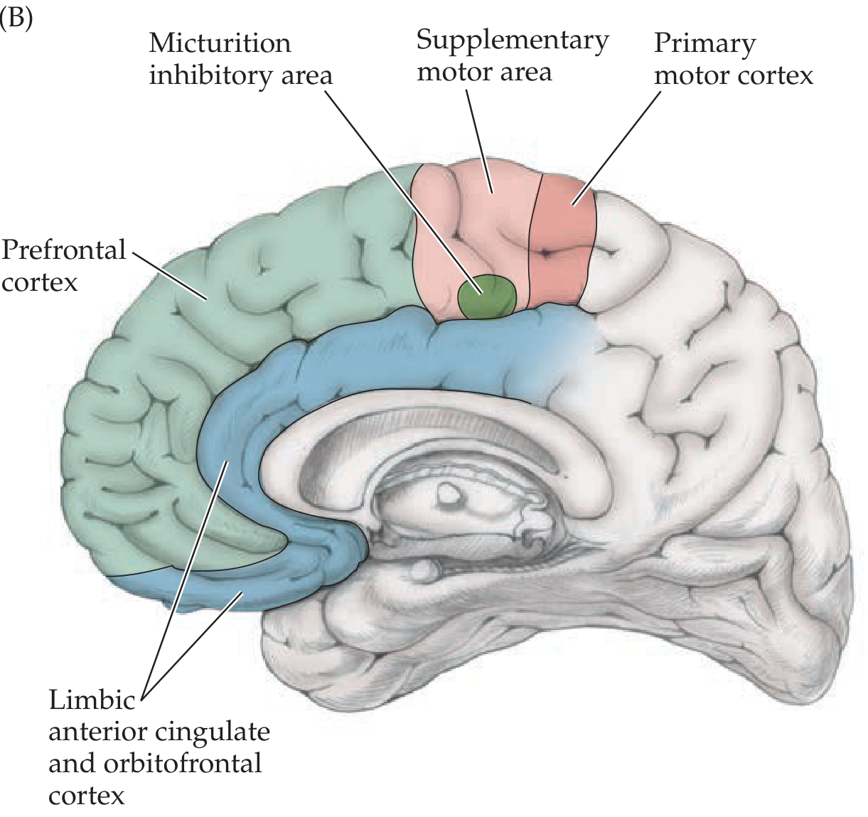
And the orbitofrontal/basal view:
Connections of the Prefrontal Cortex
The PFC has numerous, mostly bidirectional cortical and subcortical connections. The anatomical pattern allows it to integrate multimodal sensory, motor, and limbic information - the structural basis for its role in higher-order cognition.
A. Cortical Connections
| Target | Detail |
|---|---|
| Parietal, occipital, and temporal association cortex | Both unimodal sensory association cortex and heteromodal association cortex |
| Motor association cortex (premotor, SMA) | Frontal-to-frontal within the same lobe |
| Anterior cingulate gyrus | Major limbic cortical connection |
| Posteromedial orbitofrontal cortex | Limbic-prefrontal integration |
| Anteromedial temporal cortex | Via the uncinate fasciculus |
| Hippocampal formation | Indirectly via cingulate gyrus and parahippocampal gyrus |
B. Subcortical Connections
| Structure | Pathway / Detail |
|---|---|
| Mediodorsal thalamic nucleus | The most important thalamic relay to and from PFC (bidirectional) |
| Medial pulvinar & intralaminar nuclei | Additional thalamic connections |
| Amygdala | Via the uncinate fasciculus - specifically to orbital and medial frontal regions |
| Basal ganglia | PFC projects mainly via the head of the caudate nucleus |
| Hypothalamus | Autonomic and endocrine regulation |
| Septal region & subthalamic region | Limbic modulation |
| Cerebellum | Via thalamic relays (fronto-cerebellar circuit) |
| Midbrain | Descending projections |
C. Neuromodulatory (Brainstem) Inputs
Like all cortical areas, the PFC receives ascending modulatory inputs from:
- Dopamine (VTA - mesocortical pathway)
- Norepinephrine (locus coeruleus)
- Serotonin (raphe nuclei)
- Acetylcholine (nucleus basalis of Meynert)
- Histamine (tuberomammillary nucleus)
- Orexin (lateral hypothalamus)
Key fact: The dorsolateral PFC is the primary target of the mesocortical dopamine pathway from the VTA. D1 receptor stimulation here supports working memory and executive function. The orbital/ventromedial PFC receives limbic inputs especially via the uncinate fasciculus (amygdala).
- Neuroanatomy through Clinical Cases 3rd Edition, p. 941
Functions of the Prefrontal Cortex
A useful mnemonic is RIO:
R - Restraint (inhibition of inappropriate behaviors) I - Initiative (motivation to pursue productive activities) O - Order (sequencing, cognitive organization)
1. Executive Functions
The PFC integrates information from across the brain to execute goal-directed behavior:
- Planning and sequencing complex actions
- Cognitive flexibility (mental set-shifting)
- Selective attention amid distractors
- Prognostication - anticipating consequences of actions
- Solving complex mathematical, legal, and philosophical problems
- Controlling behavior according to moral norms
2. Working Memory
Dorsolateral PFC is the critical substrate for working memory - the ability to hold and manipulate limited information in an immediately available store while performing cognitive operations (analogous to "carrying" in arithmetic). Different subregions store different types of temporary information (shape, form, movement). This "brain's working memory" underlies much of what we call higher intelligence.
3. Decision Making
The orbitofrontal and ventromedial PFC integrate information from limbic areas (emotional) and heteromodal cortex (abstract/rational) to produce emotionally weighted decisions - enabling "intuitive" judgments when time or information is limited. Patients with orbitofrontal damage (classically, Phineas Gage) make poor social and economic decisions despite intact intelligence tests.
4. Learning and Memory Encoding
The dorsolateral PFC works in concert with the medial temporal lobes during new learning:
- Left frontal + medial temporal lobes: activated during encoding of new verbal information
- Right frontal + medial temporal lobes: activated during encoding of new nonverbal information
5. Social and Emotional Regulation
Via limbic connections (orbitofrontal, anterior cingulate), the PFC:
- Inhibits inappropriate social behaviors and aggression
- Regulates emotional responses
- Supports moral reasoning
6. Motivation and Initiative
The medial PFC and anterior cingulate provide the motivational drive to initiate and sustain goal-directed activity.
Effects of Prefrontal Damage (Clinical Correlates)
Prefrontal damage produces a characteristic syndrome that may spare routine intelligence testing yet severely impair real-world functioning:
| Loss | Clinical Manifestation |
|---|---|
| Working memory | Cannot string together sequential tasks toward complex goals |
| Executive planning | Cannot solve complex problems; easily distracted |
| Cognitive flexibility | Failure on Wisconsin Card Sorting Test |
| Motivation | Decreased initiative, amotivation (medial lesions) |
| Impulse control | Disinhibition, inappropriate social behavior (orbital lesions) |
| Emotional regulation | Rapid mood swings, loss of social judgment |
| Parallel tasking | Cannot perform multiple tasks simultaneously |
- Guyton and Hall Textbook of Medical Physiology, p. 721
Neurotransmitter Relevance
| NT | Relevance to PFC |
|---|---|
| Dopamine (D1) | Working memory and executive function (mesocortical pathway); hypo-dopaminergia in PFC hypothesized in schizophrenia negative/cognitive symptoms |
| Norepinephrine | Enhances prefrontal signal-to-noise; NET blockade raises both NE and DA in PFC |
| GABA | GAD67 (GABA synthesis enzyme) is reduced in PFC in schizophrenia |
| Glutamate | NMDA/AMPA receptor changes implicated in PFC dysfunction |
- Stahl's Essential Psychopharmacology; Kaplan & Sadock's Comprehensive Textbook of Psychiatry, p. 4535-4536
Summary: The PFC is positioned as the brain's "executive hub" - anatomically receiving from every sensory modality, limbic system, and subcortical structure, while projecting back to regulate all of them. Its functions span working memory, planning, decision-making, impulse control, social cognition, and motivational drive. Disruption of any of its major connections (thalamo-prefrontal, amygdalo-prefrontal, basal ganglia-prefrontal) produces characteristic and clinically recognizable deficits.
Differentiate between sympathetic nervous system and parasympathetic nervous system in physiology for 8 marks mbbs refer books and give me pls
Reading File
Finding Sources
Finding Sources
Reading File
Reading File
Reading File
Finding Sources
Reading File
Reading File
Finding Sources
I now have all the content needed from Guyton, Ganong's, Costanzo, and Lippincott Pharmacology. Let me compile the comprehensive 8-mark MBBS answer.
Differences Between Sympathetic and Parasympathetic Nervous System
(8-mark MBBS Answer - Physiology)
Introduction
The Autonomic Nervous System (ANS) is the part of the peripheral nervous system that controls involuntary visceral functions. It is divided into two major functional divisions:
- Sympathetic Nervous System (SNS) - "Fight or Flight"
- Parasympathetic Nervous System (PNS) - "Rest and Digest"
Both consist of a two-neuron chain: a preganglionic neuron (CNS to ganglion) and a postganglionic neuron (ganglion to effector).
Differences: Structural / Anatomical
| Feature | Sympathetic | Parasympathetic |
|---|---|---|
| Origin (Outflow) | Thoracolumbar (T1-L2/L3) spinal cord | Craniosacral - Brainstem (CN III, VII, IX, X) + Sacral (S2-S4) |
| Preganglionic fiber | Short | Long |
| Postganglionic fiber | Long | Short |
| Ganglion location | Close to spinal cord (paravertebral chain or prevertebral ganglia) | Within or near the effector organ (terminal ganglia) |
| Preganglionic branching | Extensive (one pre- → many post-) | Minimal (mostly one-to-few) |
| Distribution | Wide - innervates practically all effector systems | Limited - specific visceral organs |
| Type of response | Diffuse (mass discharge) | Discrete, localized |
- Lippincott Illustrated Reviews: Pharmacology, p. 130
Differences: Neurotransmitters & Receptors
| Feature | Sympathetic | Parasympathetic |
|---|---|---|
| Preganglionic NT | Acetylcholine (ACh) - nicotinic receptors | Acetylcholine (ACh) - nicotinic receptors |
| Postganglionic NT | Noradrenaline (NE) (adrenergic) - exception: sweat glands use ACh | Acetylcholine (ACh) (cholinergic) - muscarinic receptors |
| Adrenal medulla | Preganglionic → directly releases Adrenaline + Noradrenaline into blood | No equivalent |
| Receptors at effector | α1, α2, β1, β2, β3 adrenergic receptors | M1, M2, M3 muscarinic receptors |
Differences: Functional (Organ-by-Organ)
| Organ / System | Sympathetic Effect | Parasympathetic Effect |
|---|---|---|
| Heart rate | Increased (positive chronotropy) | Decreased (negative chronotropy) |
| Cardiac contractility | Increased (positive inotropy) | Slight decrease (atria only) |
| Blood vessels | Constriction (most vessels, via α1) | Little/no effect on most |
| Bronchi (Lungs) | Dilation (β2) | Constriction |
| Pupils | Dilation - mydriasis (radial muscle contracts) | Constriction - miosis (sphincter muscle contracts) |
| Ciliary muscle (Lens) | Slight relaxation → far vision | Contraction → near vision (accommodation) |
| GI tract (lumen) | Decreased peristalsis and tone | Increased peristalsis and tone |
| GI sphincters | Contraction (mostly) | Relaxation (mostly) |
| Salivary glands | Thick, viscous saliva (concentrated) | Copious, watery saliva |
| Nasal/lacrimal/gastric glands | Vasoconstriction, slight secretion | Copious secretion |
| Liver | Glycogenolysis → glucose released | Slight glycogen synthesis |
| Gallbladder | Relaxation | Contraction |
| Kidney | Decreased urine output; ↑ renin | No significant effect |
| Urinary bladder (detrusor) | Relaxation (β2) - stores urine | Contraction - empties urine |
| Bladder sphincter (trigone) | Contraction (α1) | Relaxation |
| Sweat glands | Copious sweating (cholinergic fibers) | No effect |
| Piloerector muscles (skin) | Contraction (goosebumps) | No innervation |
| Penis | Ejaculation | Erection |
| Adrenal medulla | Secretion of epinephrine/norepinephrine | No innervation |
| Blood glucose | Increased (glycogenolysis) | Slight decrease |
| Blood coagulation | Increased | No significant effect |
| Mental activity | Increased | No effect |
- Guyton and Hall Textbook of Medical Physiology, Table 61.2, p. 759
Differences: General Physiological Role
| Feature | Sympathetic | Parasympathetic |
|---|---|---|
| Overall role | Catabolic - prepares body for emergency | Anabolic - conserves and restores energy |
| Nickname | "Fight or Flight" (Walter Cannon) | "Rest and Digest" |
| Conditions of activation | Stress, fear, exercise, hemorrhage | Rest, digestion, sleep |
| Discharge pattern | Mass/diffuse discharge (all-or-none) | Discrete, organ-specific |
| Organs with exclusive innervation | Blood vessels, sweat glands, piloerectors, adrenal medulla | Lacrimal gland, ciliary muscle, nasopharyngeal glands |
| Energy metabolism | Catabolic: ↑ glucose, ↑ free fatty acids | Anabolic: promotes digestion, absorption |
- Ganong's Review of Medical Physiology, 26th Edition, p. 273
Reciprocal and Synergistic Actions
Most organs receive dual innervation and the two divisions act reciprocally:
- Heart: SNS ↑ rate; PNS ↓ rate
- Bladder: SNS stores urine; PNS empties urine
- Bronchi: SNS dilates; PNS constricts
Some actions are complementary/synergistic:
- Salivary glands: PNS → watery saliva; SNS → viscous saliva (both cause secretion)
- Sexual function: PNS → erection; SNS → ejaculation (coordinated together)
- Pupil size: both innervations are excitatory but to opposing muscles
Some organs have only sympathetic innervation (blood vessels, sweat glands, piloerectors) or only parasympathetic innervation (ciliary muscle, lacrimal gland).
- Costanzo Physiology, 7th Edition; Ganong's Review, 26th Edition
Summary Diagram (Mnemonic)
SYMPATHETIC                         PARASYMPATHETIC
"Fight or Flight"                   "Rest and Digest"
Thoracolumbar (T1-L2)              Craniosacral (CN III,VII,IX,X + S2-S4)
Short pre-, Long post-             Long pre-, Short post-
Ganglia near cord                  Ganglia near organ
Noradrenaline (post-gang.)         Acetylcholine (post-gang.)
Diffuse, mass response             Discrete, local response
Catabolic                          Anabolic
Sources:
- Guyton and Hall Textbook of Medical Physiology, 14th Ed., Ch. 61
- Ganong's Review of Medical Physiology, 26th Ed., Ch. 13
- Costanzo Physiology, 7th Ed., Ch. 2
- Lippincott Illustrated Reviews: Pharmacology, Ch. 3
This is a shared conversation. Sign in to Orris to start your own chat.