
A 6 year old boy is still bed wetting sometimes even after peeing and sleeping at night
nocturnal enuresis children management
Nocturnal Enuresis (Bedwetting) in a 6-Year-Old Boy
Is This Normal?
- Harrison's Principles of Internal Medicine, 22E notes: "Before age 5 or 6 years, nocturnal enuresis should be considered a normal variant."
Primary vs. Secondary Enuresis
| Type | Definition | Significance |
|---|---|---|
| Primary | Child has never been consistently dry at night | Most common; usually maturational |
| Secondary | Returns after 6+ months of dryness | May signal stress, UTI, diabetes, or another pathologic cause |
What Causes It?
- Developmental delay in arousal from sleep (the child sleeps deeply and doesn't wake to a full bladder signal)
- Nocturnal polyuria - the kidneys produce too much urine at night due to insufficient antidiuretic hormone (ADH/vasopressin) release during sleep
- Small functional bladder capacity
- Genetics - strong family history association
- Psychological factors, life stressors, and family dynamics have not been shown to have a causal relationship
When to See a Doctor
- Child was dry for 6+ months and has relapsed (secondary enuresis)
- Daytime wetting as well (diurnal enuresis)
- Pain, burning, or discomfort when urinating
- Unusual thirst or drinking a lot (could suggest diabetes)
- Straining to urinate, weak stream, or dribbling
- Wetting that is very frequent (multiple times per night)
- Any signs of constipation (can worsen enuresis)
- Any neurological symptoms (back pain, abnormal gait)
What You Can Do at Home (First-Line)
- Fluid management - offer most fluids earlier in the day; reduce intake in the 1-2 hours before bed. Avoid caffeinated, sugary, or fizzy drinks in the evening.
- Regular voiding schedule - encourage the child to pee right before bed (even if he just went a little earlier - have him try again).
- Reward/star chart - give positive reinforcement for dry nights without shame or punishment for wet ones.
- Protect the mattress - a waterproof mattress cover and easy-change bedding reduces stress for the whole family.
- Maintain regular bowel habits - constipation compresses the bladder and worsens enuresis.
- "Lifting" - gently waking the child 1.5-2 hours after he falls asleep to take him to the toilet can reduce the number of wet nights.
- Reassurance - the child should not feel shame. Most children will be dry by age 7; reassurance alone is appropriate at this age.
If Home Measures Don't Work
Bedwetting Alarm (Most Effective Treatment)
- Textbook of Family Medicine 9e: "A bedwetting alarm is the most effective treatment for nocturnal enuresis."
Medications (if alarm fails or isn't feasible)
| Drug | Mechanism | Notes |
|---|---|---|
| Desmopressin (DDAVP) | Synthetic ADH - reduces urine production at night | Best for nocturnal polyuria; oral dose starts at 0.2 mg at bedtime; effective but high relapse when stopped |
| Antimuscarinics (e.g. oxybutynin) | Relaxes the bladder | Used when small/overactive bladder is the issue |
| Tricyclics (e.g. imipramine) | Multiple mechanisms | Effective but side effects (cardiac arrhythmias, drowsiness) limit use; rarely first-line |
Bottom Line for This Child
- Reassure him (and yourselves) - no blame or shame
- Cut back fluids 1-2 hours before bed and ensure he voids right before sleeping
- Use a waterproof mattress cover
- Consider a reward chart for dry nights
- If he's still wetting regularly past age 7, or if it's causing him distress, speak to your paediatrician about a bedwetting alarm
Note: This information is general guidance. If you notice any daytime wetting, pain on urination, unusual thirst, or if bedwetting returns after a dry period, please consult a doctor.
How to report zn and gram staining of csf in a clinical pathology lab
gram stain CSF cerebrospinal fluid bacteria microscopy
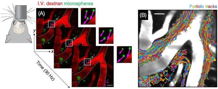
This figure demonstrates cerebrospinal fluid (CSF) bulk flow within the perivascular space (PVS) using in vivo two-photon microscopy in a mouse model. Panel A illustrates the experimental setup with a schematic of a cranial window and cranial microscopy objective. Sequential time-lapse fluorescence images, captured at 30 Hz, show branching cerebral vasculature (labeled in red) and intravenously (I.V.) administered green dextran microspheres. High-magnification insets with purple arrows indicate particle velocity and directionality, demonstrating flow relative to the vessel walls (marked by white dashed lines). Panel B provides a particle tracking velocimetry summary, where multi-colored particle tracks are superimposed on a grayscale anatomical image of the perivascular spaces. The dense, interwoven trajectories visualize the complex, unidirectional transport of solutes through the glymphatic system. This image is used in neurobiology to illustrate the physiological mechanisms of brain waste clearance and CSF-interstitial fluid (ISF) exchange.
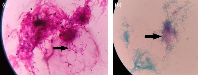
This composite diagnostic image presents two light microscopy views of pleural fluid highlighting the microbiological identification of Nocardia species. Panel (a) shows a Gram stain at high magnification, demonstrating Gram-positive (purple-stained), thin, beaded, and branching filamentous bacteria. Panel (b) illustrates a modified Ziehl-Neelsen (ZN) stain of the same clinical specimen. The bacteria exhibit weak acid-fast properties, characterized by pink-to-red filamentous structures that retain the primary stain against a light blue counterstained background. Arrows in both panels indicate the characteristic filamentous and branching morphology. This combination of Gram-positive branching filaments and partial acid-fastness is a key diagnostic feature used to distinguish Nocardia from other filamentous bacteria like Actinomyces. These findings are clinically significant in diagnosing thoracic nocardiosis in immunocompromised patients or those with pre-existing pulmonary disease.
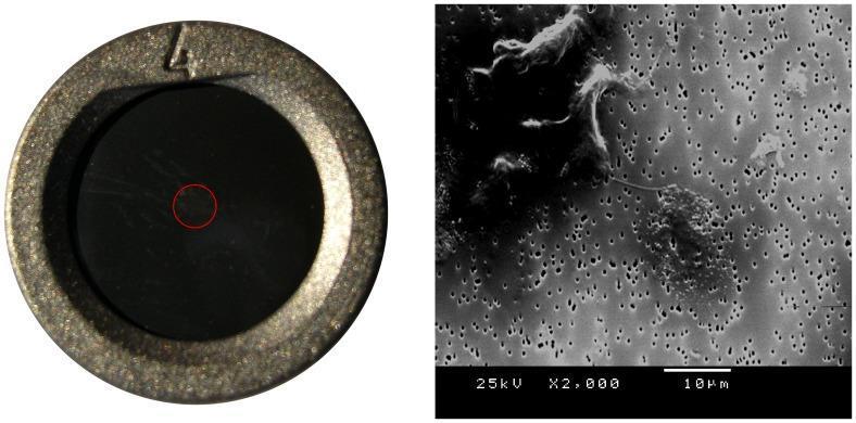
The image consists of two panels showcasing cerebrospinal fluid (CSF) analysis techniques. On the left, a macro photograph displays a 10 mm diameter polycarbonate filter with 0.6 um pores mounted in an airtight device. A red circular annotation in the center highlights the concentrated area of morphological structures obtained via vacuum suction of 200 uL of CSF. On the right, a scanning electron microscopy (SEM) image at 2000x magnification reveals the ultrastructural morphology of a healthy control sample. The SEM view shows a cluster of spherical particles, measuring approximately 0.1–0.2 um in diameter, situated on the porous filter surface. In the upper left quadrant, a phagocytic-type cell is visible, featuring a thin chord-like extension or pseudopod reaching towards the central aggregate of spherical particles. The image illustrates the use of filtration and electron microscopy in clinical research to identify potential biomarkers or atypical morphological structures within the CSF.
Reporting ZN and Gram Staining of CSF in a Clinical Pathology Lab
Pre-Analytical Step: CSF Concentration (Critical)
Always concentrate CSF by centrifugation before making smears.
- Centrifuge at 1500-2000 rpm for 10-15 minutes
- Discard most of the supernatant (keep a small amount to resuspend the pellet)
- Use the resuspended pellet for smear preparation
- Prepare at least 2 smears from the deposit - one for Gram stain, one for ZN/AFB stain
- Allow smears to air-dry, then heat-fix gently
GRAM STAIN OF CSF
Purpose
Sensitivity (know this before reporting)
| Organism | Gram Stain Sensitivity |
|---|---|
| Streptococcus pneumoniae | ~90% |
| Neisseria meningitidis | ~75-85% |
| Gram-negative bacilli (E. coli, Klebsiella) | 50-80% |
| Listeria monocytogenes | ≤50% |
| Overall bacterial meningitis | 60-90% |
Reading the Gram Stain - What to Look For
| Finding | Appearance | Likely Organism |
|---|---|---|
| Gram-positive diplococci (lancet-shaped) | Purple, paired cocci | S. pneumoniae |
| Gram-negative diplococci (intracellular) | Pink, paired cocci inside PMNs | N. meningitidis |
| Gram-positive cocci in clusters | Purple clusters | Staphylococcus spp. |
| Gram-negative coccobacilli or bacilli | Pink rods | E. coli, H. influenzae, Klebsiella |
| Gram-positive short rods | Purple rods (often intracellular in monocytes) | Listeria monocytogenes |
| Gram-positive rods (large, boxcar-shaped) | Purple, squared-off rods | Bacillus anthracis (rare) |
How to Write the Gram Stain Report
Gram Stain of CSF (concentrated deposit):
- No organisms seen on Gram stain
- WBCs: [present / absent / numerous]
- Background: [clear / slightly turbid deposit]
- Culture and sensitivity awaited
Gram Stain of CSF (concentrated deposit):
- Gram-positive diplococci (lancet-shaped) seen - intracellular and extracellular
- Numerous polymorphonuclear leukocytes present
- Morphology consistent with Streptococcus pneumoniae
- Urgent: Result telephoned to clinician at [time]
- Culture and sensitivity in progress
| Descriptor | Meaning |
|---|---|
| Rare / occasional | 1-5 organisms per 100 fields |
| Few | 1-5 per 10 fields |
| Moderate | 5-10 per field |
| Many / numerous | >10 per field |
- Always describe organism morphology (cocci/bacilli/diplococci), Gram reaction (positive/negative), and arrangement (pairs, clusters, chains)
- Note if organisms are intracellular (inside PMNs) - strongly supports pathogenicity
- Note the background cellularity (WBCs: PMNs vs lymphocytes)
- Any positive result must be phoned to the clinician immediately - this is a critical/panic value
ZN (ZIEHL-NEELSEN) STAIN OF CSF
Purpose
Sensitivity - Important Limitation
- The AFB load in CSF is typically very low
- The organism is difficult to concentrate even with centrifugation
- Results depend on the volume of CSF submitted and persistence of searching
- Examining large volumes (≥5 mL, ideally 10 mL) of CSF
- Multiple serial LPs (repeated 2-3 times)
- Examining the fibrin web/pellicle that forms in TBM CSF if the specimen is allowed to stand
Reading the ZN Stain
- Scan at 10x first, then confirm at 40x, and identify at 100x (oil)
- AFB appear as bright red/pink slender rods against a blue counterstain background
- They are often beaded and may be curved
- Search at least 300 fields in a concentrated CSF smear before calling negative (IUATLD recommendation)
- For CSF (a paucibacillary sample), a minimum of 100-200 fields should be examined
WHO/IUATLD Grading Scale for AFB Smear Reporting
| Grade | Number of AFB seen | Report as |
|---|---|---|
| Not seen | 0 AFB in 100 fields (min) | No AFB seen |
| Scanty | 1-9 AFB per 100 fields | Scanty - specify exact number (e.g., "Scanty: 3 AFB seen in 100 fields") |
| 1+ | 10-99 AFB per 100 fields | 1+ |
| 2+ | 1-10 AFB per field (in 50 fields) | 2+ |
| 3+ | >10 AFB per field (in 20 fields) | 3+ |
Note for CSF: Given the very low bacillary load, most positive CSF ZN results will be graded "Scanty" - do not discard a scanty result. Report it with the exact count and it is clinically significant.
How to Write the ZN Stain Report
Ziehl-Neelsen Stain for AFB of CSF (concentrated deposit):
- No acid-fast bacilli seen in [number] fields examined
- Minimum 100 fields examined at oil immersion
- Note: Sensitivity of ZN stain on CSF is low (10-40%).
  A negative result does not exclude tuberculous meningitis.
  Culture for AFB and nucleic acid amplification testing (NAAT/PCR) recommended.
Ziehl-Neelsen Stain for AFB of CSF (concentrated deposit):
- Acid-fast bacilli seen: SCANTY (3 AFB in 100 fields)
  [or 1+ / 2+ / 3+ as applicable]
- Morphology: Slender, beaded, pink-red rods consistent with Mycobacterium spp.
- URGENT: Result telephoned to clinician at [time]
- AFB culture and sensitivity in progress
- Molecular test (GeneXpert MTB/RIF / PCR) recommended for confirmation
Key Points for the Lab Report - Summary
| Feature | Gram Stain | ZN Stain |
|---|---|---|
| Specimen prep | Centrifuge, use deposit | Centrifuge, use deposit |
| Stain used | Crystal violet, Gram's iodine, decolourise, safranin | Carbol fuchsin (heat or cold), acid-alcohol decolourise, methylene blue |
| Reading magnification | 100x oil immersion | 100x oil immersion |
| Minimum fields to scan | 20-30 per smear | 100-200 per smear (CSF) |
| Positive appearance | Gram+ = purple; Gram- = pink | AFB = bright red on blue background |
| Grading system | Rare/Few/Moderate/Many + morphology + arrangement | WHO/IUATLD 0/Scanty/1+/2+/3+ |
| Sensitivity in CSF | 60-90% (bacterial meningitis) | 10-40% (TBM) |
| Panic value action | Phone clinician immediately | Phone clinician immediately |
| Add-on comment | Recommend culture and sensitivity | Recommend AFB culture + GeneXpert/PCR |
General Reporting Reminders
- Always state specimen type and preparation method (concentrated deposit)
- Note the quality of the smear (adequate cellularity, thin smear, well-stained)
- A negative stain never excludes infection - always recommend culture
- CSF results are critical values - positive findings must be communicated verbally to the clinician and documented with time/person notified
- If you see organisms that don't fit a clear category (e.g., branching filaments), consider modified ZN for Nocardia (partially acid-fast = decolourise with 1% H₂SO₄ instead of acid-alcohol)