
Fibroadenoma
fibroadenoma breast histology gross pathology
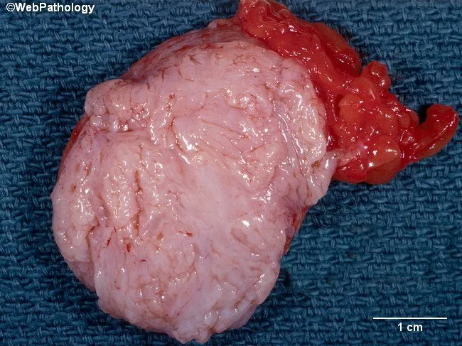
Gross pathology photograph of a breast fibroadenoma. The specimen is a well-circumscribed, firm, pale-tan mass with a smooth surface; surrounding adipose tissue is visible. The cut surface shows lobulated architecture and slit-like spaces consistent with fibroepithelial tumor morphology. Necrosis is not present on gross examination. The lesion measures several centimeters in greatest dimension; a 1 cm scale bar is provided for reference. Histologically, this entity demonstrates biphasic fibroepithelial tissue with a dense fibrous stroma interlacing with benign ductal epithelium, often arranged in compressed or intracanalicular patterns; there is no appreciable cytologic atypia or mitotic activity. Clinically, fibroadenomas are common benign breast tumors in young women, with median age around 25 years and mean age near 30; they typically present as firm, mobile, non-tender masses. Diagnostic significance rests on distinguishing benign fibroadenoma from phyllodes tumor and fibrocystic change; absence of necrosis and orderly architecture support benignity. Differential considerations include phyllodes tumor (benign or malignant), fibrocystic change, adenosis, and ductal hyperplasia. This image is valuable for pathology education, breast disease databases, and radiopathologic correlation studies. Potential clinical applications: teaching gross-pathology correlations, reinforcing macroscopic-histologic relationships, and aiding differential diagnosis for palpable breast masses in premenopausal patients. Useful for examiners, clinicians, researchers, and students.
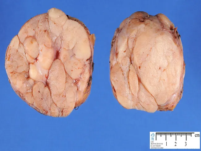
Gross pathology photograph of a bilateral breast fibroadenoma specimen. The two lobulated masses are ex vivo and oriented for frontal viewing, with a clean, pale tan to light pink cut surface. The surface exhibits a multi‑lobulated architecture, with a firm, rubbery consistency and some fibrous septations separating nodular lobules. Each mass measures approximately 2–3 cm in greatest dimension, as estimated against the centimeter scale visible at the lower right. The margins are well circumscribed without overt invasion or necrosis. The lesions are benign fibroepithelial tumors consisting of stromal and epithelial components, and the cut surface is relatively homogeneous, lacking significant hemorrhage. This gross appearance is classic for fibroadenoma, particularly in young or middle‑aged patients, and may be bilateral. Clinically, fibroadenomas present as non tender, mobile breast lumps and often prompt surgical excision for definitive diagnosis. Radiologic correlation can show well circumscribed, oval lesions with smooth margins on ultrasound or mammography. Histologic confirmation is recommended to distinguish fibroadenoma from phyllodes tumor, especially in larger or rapidly growing masses. The image supports diagnostic consideration of benign breast disease and can be used for education, comparison with gross findings, and teaching gross pathology technique. This specimen demonstrates lobulated morphology and firm, well‑circumscribed margins.
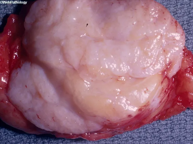
This is a macroscopic photograph of a surgically excised breast mass consistent with a fibroadenoma. Modality and Technique: gross pathology photography of a breast lesion; specimen is opened to expose the cut surface. Location and Anatomical Context: breast parenchyma derived from terminal duct-lobular units; the mass resides within the fibrous breast stroma. Visual features: small, well-demarcated, firm, grayish-pink nodule with a bulging, whorled cut surface; smooth, glistening capsule; homogenous pale color; margins appear noninfiltrative. Notable pathology indicators: the gross appearance reflects biphasic epithelial and stromal proliferation typical of fibroadenoma; microscopic confirmation would demonstrate compressed ducts embedded in fibrous stroma with pericanalicular or intracanalicular patterns (not visible on this image). Diagnostic significance: a classic benign breast tumor with well-defined margins; should be distinguished from phyllodes tumor, fibrocystic change, or malignancy through histology and correlation with imaging. Clinical relevance and use cases: common in reproductive-age women; management usually conservative observation or local excision if symptomatic; imaging correlation with ultrasound or mammography enhances diagnostic confidence; this image serves educational demonstration of gross morphology and surgical pathology. The specimen’s color and texture align with benign stromal-epithelial interactions; radiologic-pathologic concordance supports routine follow-up; prompt recognition reduces unnecessary alarm and guides patient counseling and clinical decision-making.
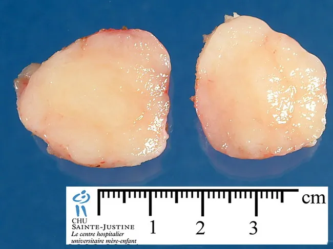
Gross pathology photograph of an excised breast lesion. Modality: gross pathology imaging; technique: external macroscopic photography with a metric ruler for size reference. The specimen is a breast hamartoma composed of mixed mature adipose tissue and fibroglandular elements. Anatomy: mammary gland tissue from the breast, with lobular-adipose admixtures; overall size approximately 2–3 cm based on the ruler. Visual features: well-circumscribed, lobulated mass with a pale pink-tan, homogeneous cut surface; smooth margins; no visible hemorrhage, necrosis, or calcification on this view. Pathology: benign hamartomatous lesion with admixture of adipose tissue and fibroglandular components; preserved architecture; no cytologic atypia is assessable on gross image. Diagnostic significance: supports benign diagnosis; radiologic-pathologic concordance important for differentiation from lipoma, fibroadenoma, or malignant breast neoplasms. Clinical relevance: low recurrence risk after complete excision; findings assist in surgical planning, patient counseling, and educational teaching; image suitable for radiology-pathology correlation, breast surgery planning, and benign breast lesion inventories. Imaging correlation: radiographs often show a well-defined mass with mixed fat and soft tissue density; histology sampling should include both fat and fibroglandular components to confirm admixture. In reports, this lesion is described as benign breast hamartoma or adenolipomatous lesion with no atypia. The image is a valuable teaching aid for radiology-pathology correlation, breast surgery planning, and benign breast lesion inventories.
fibroadenoma breast
Fibroadenoma
Definition
Molecular Pathogenesis
- Two-thirds of fibroadenomas harbor driver mutations in MED12 - a gene encoding a subunit of the mediator complex that regulates RNA polymerase II-dependent transcription. (Notably, uterine leiomyoma - also a hormone-responsive stromal tumor - shares MED12 mutations.)
- Around one-third harbor mutations in RARA (retinoic acid receptor alpha), an estrogen target gene that cooperates with estrogen receptor in transcriptional regulation.
- These mutations likely derange the expression of sex hormone-regulated genes controlling the proliferation and survival of stromal cells. - Robbins, Cotran & Kumar
Morphology
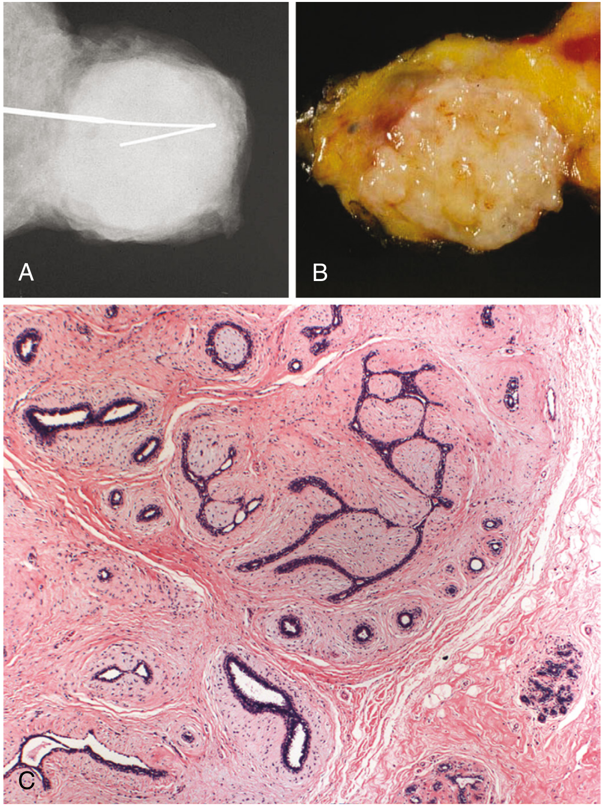
Gross
- Well-circumscribed, rubbery, gray-white nodules that bulge above surrounding tissue
- Slit-like spaces lined by epithelium on cut surface
- Size ranges from <1 cm to large tumors replacing most of the breast
Histology (two patterns)
| Pattern | Description |
|---|---|
| Pericanalicular | Delicate, often myxoid stroma surrounds patent, open ductal structures |
| Intracanalicular | Stroma compresses and distorts ductal elements into cleft-like (slit-like) spaces |
- Stroma resembles normal intralobular stroma; often myxoid
- In older women: stroma becomes densely hyalinized, epithelium becomes atrophic, calcifications may develop
Clinical Features
| Feature | Detail |
|---|---|
| Age | Most commonly 20-35 years; also occurs in teenagers |
| Presentation | Firm, smooth, rubbery, freely mobile, non-tender lump; no skin dimpling or nipple retraction |
| Nickname | "Breast mouse" - due to its high mobility |
| Size | Usually 1-3 cm when detected; may be bilobed (a groove is palpable) |
| Number | Frequently multiple and bilateral |
| Hormonal response | May grow during pregnancy (even undergo infarction, mimicking carcinoma); regresses after menopause |
| Immunosuppression | ~50% of women receiving cyclosporin A after renal transplantation develop multiple bilateral fibroadenomas |
- Berek & Novak's Gynecology; Bailey and Love's Surgery 28th Ed.
Investigations
- Ultrasound (preferred in young women): solid, uniformly hypoechoic, smoothly marginated, oval or round, wider than tall
- Mammography: well-circumscribed density; may show coarse "popcorn" calcifications in older lesions
- Triple assessment: clinical examination + imaging + core needle biopsy (if indicated)
- Biopsy indications: age >25 years, atypical imaging features, or clinical uncertainty
Subtypes
| Subtype | Key Features | Risk |
|---|---|---|
| Simple fibroadenoma | Standard morphology | RR for cancer: 1.5-1.7 |
| Complex fibroadenoma | Contains cysts >0.3 cm, sclerosing adenosis, epithelial calcifications, or papillary apocrine change | RR for cancer: 3.4-3.7 (higher if epithelial hyperplasia present) |
| Giant fibroadenoma | >5 cm; occurs during puberty; rapidly growing | Excision via submammary incision |
| Myxoid fibroadenoma | Myxoid stroma; minority associated with Carney complex (autosomal dominant, PRKAR1A mutations) | Variable |
Natural History
- ~15% regress spontaneously
- Most are static or cease growth at approximately 2-3 cm
- Only 5-10% progress in size
- Rapid growth during pregnancy may cause infarction
- Malignant transformation is rare - Berek & Novak's Gynecology
Management
Conservative (observation)
- Classic appearance on imaging in women <25 years - no biopsy needed
- Follow with serial short-interval imaging to document stability
- Most fibroadenomas do not require surgery
Indications for Surgical Excision
- Age >30 years
- Size >3-5 cm (some guidelines use >3 cm, Bailey & Love uses >5 cm)
- Rapid growth (>20% increase in 6 months)
- Suspicious/atypical features on imaging
- Atypia on core needle biopsy
- Family history of breast cancer
- Patient preference or significant anxiety
- Elderly patients (excision should include a rim of normal tissue as the lesion may contain malignancy or phyllodes tumor)
Minimally Invasive Options
- Ultrasound-guided percutaneous vacuum-assisted biopsy
- Percutaneous cryoablation
Medical (limited role)
- Regression observed with anti-estrogen drugs: tamoxifen and ormeloxifene - Bailey and Love's 28th Ed.
Differential Diagnosis
| Condition | Key Distinguishing Feature |
|---|---|
| Phyllodes tumor | Larger, more rapid growth, leaf-like projections on histology, more cellular stroma with mitoses; requires excision with clear margins |
| Fibrocystic change | Cyclical pain, multiple bilateral cysts, no discrete solid mass |
| Breast cyst | Fluctuant, transilluminates, anechoic on ultrasound |
| Breast carcinoma | Hard, irregular, non-mobile, skin changes, may have lymphadenopathy |
| Adenosis | No discrete mass, part of fibrocystic spectrum |
- Current Surgical Therapy 14e; Robbins
Recent Evidence (PubMed 2025-2026)
- Firm, mobile, non-tender lump
- Intralobular stromal origin
- Biphasic (epithelial + stromal components)
- Regresses post-menopause
- Occurs in women 20-35 years
- Associated with MED12 mutations
- Does not usually need excision if classic
- Excise if >3 cm, rapid growth, atypia, or age >30
- No malignant potential in simple type (low RR)
- Often multiple and bilateral
- Mobile - "breast mouse"
- Atrophic changes + calcification in older women