
Patient complains of selling behind ear and sudden pain in ear which is intense what would be the diagnosis pain radites
mastoiditis swelling behind ear postauricular
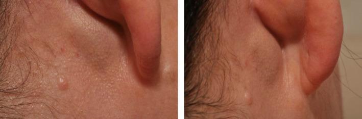
This clinical photograph displays two views of a postauricular swelling behind the right ear of a patient. The left image provides a posterolateral perspective, while the right image offers a direct posterior view. A distinct, rounded mass is visible in the postauricular region, positioned just behind the pinna and below the level of the mastoid. In the posterior view, the swelling is more prominent, causing a noticeable alteration in the natural contour of the neck and ear junction. The overlying skin appears taut and smooth over the peak of the mass, with a slight sheen suggestive of underlying pressure. No significant erythema, ulceration, or acute inflammatory changes are visible on the skin surface. Clinically, such a presentation in the context of pulsatile tinnitus suggests a vascular etiology, such as an arteriovenous malformation or fistula involving the external carotid artery branches and the jugular system.
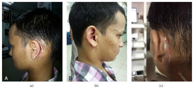
A series of three clinical photographs (a, b, c) demonstrating postoperative complications following ear surgery in a young adult. Panels (a) and (c) show the left postauricular region with clear evidence of wound dehiscence along a previous mastoidectomy incision site. There is active purulent discharge (pus) visible emanating from a focal opening in the wound, accompanied by perilesional erythema and inflammation of the surrounding skin. Panel (b) provides a lateral view of the right neck, highlighting significant soft tissue fullness and swelling in the upper cervical region, which disrupts the normal anatomical contour. These visual findings represent clinical manifestations of postoperative infection and parapharyngeal involvement, specifically highlighting mastoiditis-related complications and secondary abscess formation. This content is highly relevant for medical education concerning Otolaryngology (ENT), surgical site infections, and the diagnostic evaluation of cervical swellings following middle ear procedures.
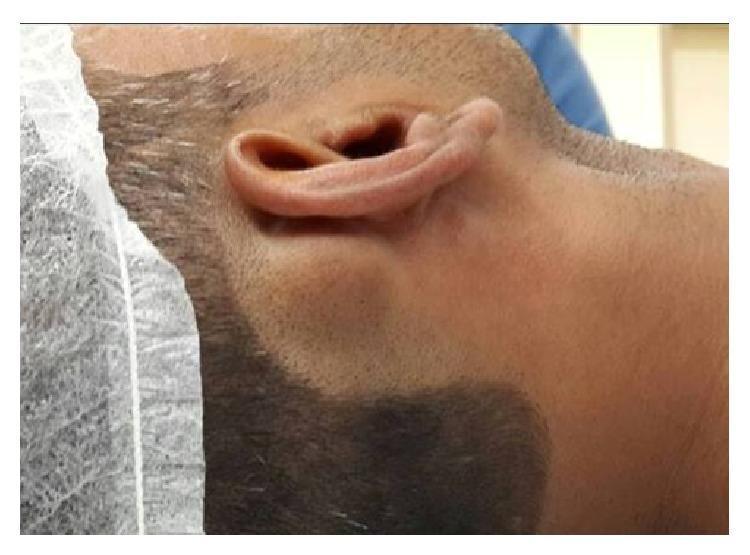
This clinical photograph shows a right-sided profile of a patient’s head, highlighting a significant swelling in the postauricular region. The mass is located directly behind the pinna, over the mastoid area. The swelling appears as a rounded, well-circumscribed, sessile protrusion that alters the natural contour of the head. The overlying skin is intact, smooth, and displays a normal color and texture without signs of erythema, inflammation, ulceration, or scarring. The ear itself appears normally positioned without significant anterior displacement, despite the proximity of the mass. This visual presentation is characteristic of a benign bony growth, such as a mastoid osteoma, and serves as an educational example for Otolaryngology and Dermatology residents in differentiating between soft tissue cysts (like sebaceous cysts) and hard, bony tumors in the postauricular space.
Diagnosis: Acute Mastoiditis (most likely)
Key Clinical Features
| Feature | Detail |
|---|---|
| Postauricular swelling | Erythema and tenderness over mastoid process |
| Ear pain | Intense, sudden; radiates to the temple, jaw, or neck |
| Auricular protrusion | Ear pushed forward/outward by subperiosteal collection |
| Abnormal tympanic membrane | Bulging, erythematous, or perforated TM |
| Fever and toxicity | Elevated temperature, unwell-looking patient |
| Sagging of EAC | Posterosuperior canal wall sagging on otoscopy |
Pathophysiology
- Mucoperiosteal inflammation blocks the aditus
- Purulent debris accumulates under pressure
- Osteoclastic activity decalcifies bony septa (coalescent mastoiditis)
- Pressure expands through the lateral cortex -> subperiosteal abscess behind the ear
Pain Radiation Pattern
- Temporal region - via the auriculotemporal nerve (V3)
- Occipital/neck region - via the greater auricular nerve (C2/C3)
- Jaw/TMJ area - referred via shared trigeminal pathways
- Bezold abscess - if infection tracks down the mastoid tip into the neck (sternocleidomastoid region)
Differential Diagnoses to Consider
| Condition | Differentiating feature |
|---|---|
| Acute Mastoiditis | Postauricular swelling + otalgia + preceding AOM |
| Ramsay Hunt Syndrome | Vesicular rash in ear canal + facial palsy (herpes zoster) |
| Furunculosis of EAC | Localized boil in external canal, no mastoid tenderness |
| Referred otalgia | Dental, TMJ, or pharyngeal pain without mastoid signs |
| Parotid abscess | Swelling in front of ear (preauricular), not behind |
Investigations
- CT temporal bone (gold standard) - shows opacification of mastoid, bony septae destruction, subperiosteal abscess, and any intracranial extension
- Blood culture, CBC, CRP/ESR
- Myringotomy with culture if TM intact
Treatment
- IV antibiotics: Vancomycin 15 mg/kg IV (covers MRSA); add an anti-Pseudomonas agent (e.g., piperacillin-tazobactam) if prior antibiotic use - (Rosen's Emergency Medicine)
- Common pathogens: S. pneumoniae, S. pyogenes, S. aureus, H. influenzae
- Myringotomy with or without tympanostomy tube insertion
- Cortical mastoidectomy (surgical drainage) if: subperiosteal abscess present, no response to IV antibiotics within 24-48h, or intracranial complication
Complications (if untreated)
- Bezold abscess (neck abscess)
- Subperiosteal abscess
- Sigmoid sinus thrombosis
- Meningitis / epidural abscess
- Facial nerve palsy
- Labyrinthitis
It might be swelling of lymp node
posterior auricular lymph node swelling behind ear
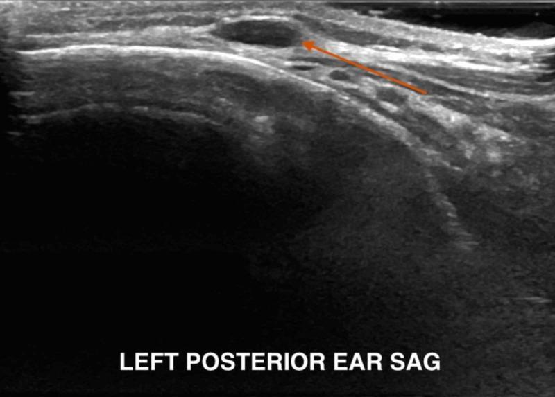
A diagnostic ultrasound image in the sagittal plane capturing the left posterior auricular region. The image demonstrates a well-defined, ovoid, hypoechoic structure (highlighted by an orange arrow) located in the subcutaneous tissue layer, consistent with posterior auricular lymphadenopathy. The structure exhibits distinct borders from the surrounding hyper-echogenic connective and fatty tissues. Notable acoustic features include posterior acoustic enhancement, indicated by increased brightness distal to the lesion, which suggests the presence of fluid or low-density contents within the node. The imaging serves to evaluate localized scalp or ear swelling and differentiates between reactive lymph nodes, cysts, or potential abscesses in a clinical setting involving regional inflammation or infestation.
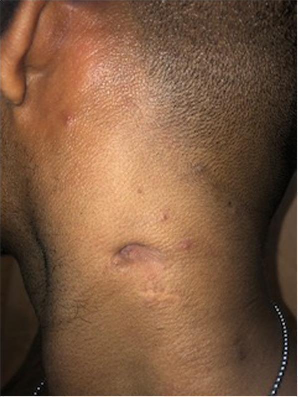
This clinical photograph shows a lateral-posterior view of a patient's neck and auricular region, demonstrating the resolution of cervical lymphadenopathy. A prominent, healed scar is visible on the mid-cervical region, likely representing the site of a formerly discharging sinus tract. The scar is elongated and slightly depressed with a hypopigmented, atrophic appearance compared to the surrounding skin. Several small, scattered, erythematous papules and minor irregularities are also visible on the upper neck and near the ear. The underlying lymphadenopathy has clinically resolved, as evidenced by the lack of visible swelling or active drainage. This image illustrates the skin-level manifestation of healed mycobacterial infection and the resulting cicatricial changes following successful treatment of lymph node involvement.
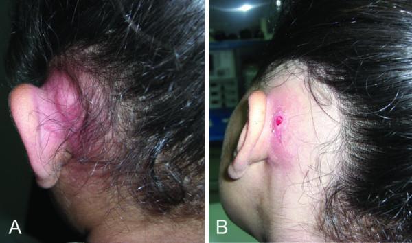
Two side-by-side clinical photographs (labeled A and B) of the posterior auricular and mastoid regions, demonstrating a post-auricular abscess in different stages. Image A shows a large, fluctuant, erythematous, and tense swelling behind the pinna, indicative of a localized abscess resulting from acute mastoiditis. The skin appears intact but highly inflamed. Image B depicts a similar post-auricular inflammatory swelling but with the development of a discharging fistula. The fistula appears as a small, open skin defect with central granulation or active drainage, representing the spontaneous rupture of the underlying subperiosteal abscess. Both images illustrate common extracranial complications of middle ear infections, characterized by auricular protrusion and significant soft tissue inflammation in the mastoid area. This material is suitable for medical education focusing on otolaryngology and infectious diseases.
posterior auricular lymph node swelling causes otalgia differential diagnosis
Swollen Posterior Auricular Lymph Node vs. Mastoiditis
Causes of Posterior Auricular Lymph Node Swelling
1. Infections (most common)
| Cause | Key Feature |
|---|---|
| Scalp infection / folliculitis | Tenderness on scalp, hair follicle involvement |
| Otitis externa | Ear canal inflammation, tragal pain on pressure |
| Acute otitis media | Middle ear infection, bulging TM |
| Rubella (German measles) | Classic: posterior auricular + occipital nodes, maculopapular rash |
| Roseola infantum | Children: fever then rash, postauricular nodes |
| Infectious mononucleosis (EBV) | Sore throat, generalized lymphadenopathy, fatigue |
| Tinea capitis | Scalp ringworm, especially in children |
2. Skin conditions
- Seborrheic dermatitis of the scalp/ear
- Psoriasis of the scalp
- Contact dermatitis behind the ear (e.g., earring reaction)
3. Serious (less common)
- Lymphoma - firm, non-tender, rubbery node, does not resolve
- Metastatic cancer - from scalp/ear primary
How to Distinguish Lymph Node Swelling from Mastoiditis
| Feature | Posterior Auricular Lymph Node | Mastoiditis |
|---|---|---|
| Swelling character | Discrete, round, mobile nodule | Diffuse, tender, fixed swelling over mastoid |
| Ear canal | Normal | Sagging of posterosuperior EAC wall |
| Tympanic membrane | Usually normal | Bulging/erythematous/perforated |
| Preceding AOM | May or may not be present | Almost always present |
| Fever/toxicity | Mild or absent | High fever, unwell patient |
| Auricular protrusion | Absent | Present (ear pushed forward) |
| Tenderness | Node itself is tender | Entire mastoid area tender on percussion |
| CT scan | Lymph node with fatty hilum on imaging | Opacified mastoid air cells, bone destruction |
Most Important Clue
Discrete, palpable round nodule that can be felt separately from the mastoid bone = lymph nodeDiffuse boggy swelling obliterating the postauricular sulcus + forward displacement of the ear = mastoiditis
What Causes the Pain to Radiate in a Swollen Lymph Node?
- The ear canal (via the greater auricular nerve, C2/C3)
- The temporal region (via auriculotemporal nerve branches)
- The occipital region (adjacent occipital lymph nodes share nerve pathways)
Bottom Line
- A scalp or ear infection
- Rubella / viral illness
- Skin condition on the scalp
Pain is raditing fin ramis of mandible and ear
referred otalgia nerve pathways diagram mandible ear
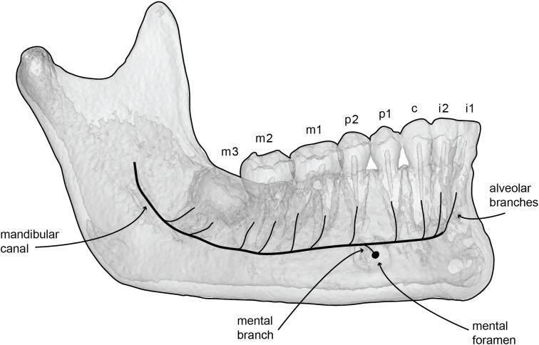
This anatomical diagram features a semi-transparent 3D reconstruction of a human mandible, illustrating the internal neurovascular pathways. The diagram highlights the mandibular canal as a bold black line originating at the ramus and traversing the body of the mandible toward the symphysis. Key neural pathways are depicted through branching patterns: alveolar branches ascend dorsally from the main canal to innervate the roots of the dentition, while a mental branch is shown exiting the bone via the mental foramen. The dental arrangement is labeled from posterior to anterior, identifying the third molar (m3), second molar (m2), first molar (m1), second premolar (p2), first premolar (p1), canine (c), second incisor (i2), and first incisor (i1). This visual aids in understanding the spatial relationship between the inferior alveolar nerve, the mandibular cortical bone, and the alveolar processes, which is critical for dental surgery, anesthesia, and implantology.
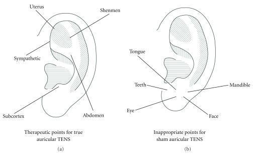
Anatomical diagrams of the human ear illustrating points used for Transcutaneous Electrical Nerve Stimulation (TENS) in an educational or clinical study context. The image is divided into two parts: (a) 'Therapeutic points for true auricular TENS' and (b) 'Inappropriate points for sham auricular TENS.' Diagram (a) highlights five active therapeutic points: Uterus (triangular fossa), Shenmen (superior to the triangular fossa), Sympathetic (internal helix), Abdomen (concha area), and Subcortex (antitragus). Diagram (b) identifies five sham or 'inappropriate' points primarily clustered on the earlobe and lower tragus: Tongue, Teeth, Mandible, Eye, and Face. These diagrams are used to define the placement of electrodes for investigating the analgesic effects of auricular stimulation, distinguishing between specific reflexology-based therapeutic sites and neutral control sites. The illustration utilizes precise anatomical labeling to demonstrate the topographical distribution of acupuncture or TENS points for clinical pain management research.
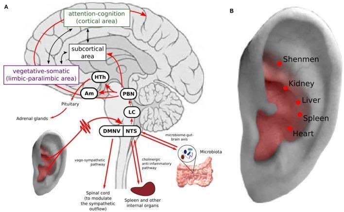
This medical illustration depicts the mechanisms and pathways of transcutaneous auricular vagus nerve stimulation (taVNS) in relation to mental health and systemic inflammation. Part A is a pathophysiology diagram showing a sagittal view of the brain and its connections to the ear and peripheral organs. It illustrates neural pathways starting from the auricular branch of the vagus nerve, projecting to the nucleus tractus solitarius (NTS) and dorsal motor nucleus of the vagus nerve (DMNV). Ascending pathways are shown reaching the locus coeruleus (LC), parabrachial nucleus (PBN), amygdala (Am), hypothalamus (HTh), and cortical areas involved in attention-cognition and vegetative-somatic functions. Efferent pathways highlight the 'cholinergic anti-inflammatory pathway' to the spleen and the 'microbiome-gut-brain axis.' Part B is an anatomical diagram of the external ear (auricle) identifying specific acupuncture points (Shenmen, Kidney, Liver, Spleen, Heart) within the vagus nerve distribution zone. The content integrates concepts from neurobiology, immunology, and integrative medicine, specifically focusing on the treatment of depression through neuromodulation.
Pain Radiating to Ramus of Mandible + Ear - Revised Differential
- The external ear canal and auricle
- The skin over the mandibular ramus and TMJ
- The preauricular region
Top Diagnoses for This Exact Pattern
1. Parotitis / Parotid Abscess (HIGH suspicion)
| Feature | Parotitis |
|---|---|
| Pain location | Constant aching - ear + mandibular ramus region |
| Swelling | Over parotid = preauricular and angle of jaw area |
| Key sign | Earlobe elevated (lifted by parotid swelling) |
| Eating | Pain worse on eating (stimulates salivary secretion against obstruction) |
| Ear | Feeling of pressure in ear |
| Duct | Pus/turbid saliva expressible from Stensen's duct (opposite upper 2nd molar) |
2. Trotter's Syndrome (IMPORTANT - do not miss)
"Aching pain in ear, involving side of face and mandible + deafness + nasal obstruction + cervical lymphadenopathy"
- Firestein & Kelley's Textbook of Rheumatology
| Feature | Detail |
|---|---|
| Pain | Ear + face + mandible/ramus |
| Hearing | Conductive hearing loss (Eustachian tube blocked) |
| Nose | Unilateral nasal obstruction |
| Neck | Cervical lymphadenopathy |
| Cause | Nasopharyngeal carcinoma (NPC) |
3. Eagle's Syndrome
"Mild to sharp stabbing pain in ear, throat, and retrognathic mandible; provoked by swallowing, turning head, or carotid compression; usually post-tonsillectomy"
4. TMJ Disorder (Temporomandibular Dysfunction)
- Pain at the preauricular region (TMJ is just in front of the ear)
- Radiates along the mandibular ramus
- Clicking or locking of jaw
- Pain worse on chewing
- Muscle tenderness on palpation (masseter, temporalis)
5. Trigeminal Neuralgia (V3 branch)
- Sharp, stabbing, electric shock-like pain
- Follows the mandibular nerve territory = mandibular ramus + ear
- Trigger zones (touch, chewing, speaking)
- Short duration bursts
Summary: Most Likely Diagnoses by Probability
| Rank | Diagnosis | Key Distinguishing Feature |
|---|---|---|
| 1 | Parotitis/Parotid abscess | Swelling over jaw angle, earlobe lifted, pain worse eating |
| 2 | TMJ disorder | Clicking jaw, preauricular tenderness, no fever |
| 3 | Trotter's syndrome (NPC) | + Deafness + nasal block + neck node |
| 4 | Eagle's syndrome | Pain on swallowing/head turning, post-tonsillectomy |
| 5 | Trigeminal neuralgia V3 | Electric shock-like, trigger zone |
| 6 | Dental abscess (lower molar) | Tooth tenderness, local swelling |
Recommended Workup
- Examine the parotid - palpate bimanually, check Stensen's duct for pus
- Nasopharyngoscopy - rule out nasopharyngeal carcinoma (Trotter's)
- TMJ palpation - press in front of tragus, open/close jaw
- Dental exam - percuss lower molars
- CT/MRI head and neck - if no obvious cause found
- ESR - if temporal arteritis suspected (older patients)
It started suddenly what would be the etiology and home treatment for it too pass the night
sudden ear pain radiating jaw mandible home treatment relief overnight
Sudden Onset - Most Likely Etiology
Most Probable Causes of Sudden Ear + Mandibular Ramus Pain with Postauricular Swelling
| Rank | Diagnosis | Why Sudden? |
|---|---|---|
| 1. Acute Otitis Media (AOM) | Rapid pressure buildup behind TM from bacterial/viral infection triggers sudden, severe otalgia | Common after URTI; bacteria: S. pneumoniae, H. influenzae, Moraxella catarrhalis |
| 2. TMJ Muscle Spasm | A sudden jaw movement, yawn, hard bite, or overnight grinding (bruxism) can lock/spasm the masseter - immediate intense pain in ear + ramus | Triggered by stress, chewing hard food, wide yawning |
| 3. Acute Parotitis | Salivary duct stone (sialolithiasis) causing sudden obstruction and ductal distension - classically worsens at meal times | Dehydration a common trigger |
| 4. Dental Abscess / Pulpitis | Sudden severe throbbing from a cracked or infected lower molar referred to ear via inferior alveolar nerve (V3) | Often triggered by cold drinks or biting |
| 5. Eustachian Tube Dysfunction | Sudden negative pressure change (flying, diving, cold) causes intense ear pain radiating toward jaw | Air pressure change history |
Pathophysiology of the Pain Pattern
V3 (Mandibular Nerve)
├── Auriculotemporal nerve → EAR + preauricular skin
├── Inferior alveolar nerve → Lower teeth + mandibular ramus
└── Nerve to masseter → TMJ + masseter muscle
Home Treatment to Get Through the Night
Important note: These measures relieve pain temporarily. A doctor must be seen the next morning - especially if fever, jaw locking, or ear discharge develops, as antibiotics or drainage may be needed urgently.
Step 1 - Pain Relief (most important)
| Drug | Dose (adult) | Notes |
|---|---|---|
| Ibuprofen (NSAID) | 400-600 mg every 6-8 hrs with food | Best for inflammatory pain - covers all causes |
| Paracetamol/Acetaminophen | 500-1000 mg every 6 hrs | Safer on empty stomach; combine with ibuprofen if pain is severe |
| Ibuprofen + Paracetamol alternating | Alternate every 3 hrs | Provides near-continuous coverage overnight |
Step 2 - Warm Compress
- Soak a cloth in warm (not hot) water, wring out, and press gently against the ear + jaw/ramus area
- Hold for 10-15 minutes, repeat every 1-2 hours
- Warmth relaxes muscle spasm (TMJ), improves blood flow, reduces parotid duct obstruction, and eases middle ear pressure
- For AOM specifically: warm compress directly on the ear reduces otalgia
Step 3 - Position for Sleep
- Sleep with the painful ear facing UP (not pressed into the pillow)
- Use an extra pillow to elevate the head 30-45 degrees - reduces pressure and venous congestion in the middle ear/mastoid region
- Avoid lying flat - increases Eustachian tube dysfunction and ear pressure
Step 4 - Cause-Specific Measures
| If suspected cause | What to do tonight |
|---|---|
| AOM / Ear infection | Warm compress over ear; keep ear dry; no cotton buds; chew on opposite side |
| TMJ spasm | Eat only soft foods; avoid chewing gum; gentle jaw massage in circular motion at the angle of jaw; apply warmth |
| Parotitis | Sip warm fluids; suck a lemon drop or sour candy (stimulates saliva flow and may dislodge a small stone); stay well hydrated; massage gland toward Stensen's duct |
| Dental pain | Clove oil (eugenol) on a cotton ball pressed against the sore tooth - natural analgesic; rinse with warm salt water |
| Eustachian dysfunction | Try Valsalva maneuver - pinch nose, close mouth, blow gently until ears pop; nasal decongestant spray (xylometazoline) helps open the tube |
Step 5 - What to Avoid Tonight
- Do NOT insert anything into the ear (cotton buds, fingers)
- Do NOT apply ice directly to the ear (worsens muscle spasm)
- Avoid alcohol (vasodilatory, worsens throbbing pain)
- Do not sleep with a hot water bottle directly on the skin (burn risk)
Red Flags - Go to Emergency NOW (do not wait till morning)
- High fever (>38.5°C / 101°F)
- Jaw locking shut (cannot open mouth)
- Swelling spreading rapidly to the neck
- Dizziness, vomiting, or facial weakness
- Pus draining from the ear
- Severe headache or neck stiffness (meningism)
Neck stiffness is seen now
⚠️ MEDICAL EMERGENCY - GO TO HOSPITAL IMMEDIATELY
🚨 THIS IS A MEDICAL EMERGENCY - CALL EMERGENCY SERVICES NOW
What Neck Stiffness Means in This Context
- Postauricular swelling (behind ear)
- Sudden intense ear pain radiating to mandibular ramus
- NEW: Neck stiffness (nuchal rigidity)
"Nuchal rigidity, pain with attempted flexion of the neck, and abnormal abdominal reflexes are OMINOUS CARDINAL SIGNS" of bacterial meningitis as a complication of mastoiditis/otitis.
What Is Happening - The Pathway
Acute Otitis Media / Mastoiditis
         ↓
Infection spreads through mastoid cortex
         ↓
Reaches meninges (lining of brain/spinal cord)
         ↓
OTOGENIC MENINGITIS
         ↓
Nuchal rigidity + headache + fever + photophobia
- Sigmoid sinus thrombosis
- Brain abscess
- Subdural empyema
- Epidural abscess
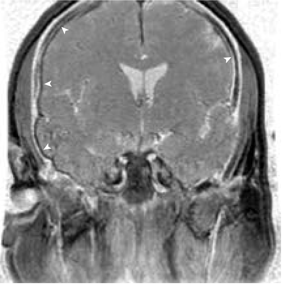
Classic Meningitis Signs to Check RIGHT NOW
| Sign | How to test | Positive if... |
|---|---|---|
| Nuchal rigidity | Try to touch chin to chest | Cannot flex neck - stiff and painful |
| Kernig's sign | Lie flat, flex hip to 90°, try to straighten knee | Pain + resistance = POSITIVE |
| Brudzinski's sign | Lie flat, examiner flexes neck | Both knees involuntarily bend up = POSITIVE |
| Photophobia | Turn on a light | Severe eye pain in light = POSITIVE |
| Fever | Check temperature | High, sustained fever = POSITIVE |
What the Hospital Will Do
- Immediate CT head - to rule out brain abscess before lumbar puncture
- Lumbar puncture (LP) - CSF analysis for cells, glucose, protein, bacteria
- IV antibiotics immediately - Ceftriaxone + Vancomycin (do not delay antibiotics while waiting for CT/LP)
- IV Dexamethasone - reduces meningeal inflammation and risk of hearing loss
- ENT consultation - for mastoidectomy if mastoiditis is the source
"When meningitis develops from COM and mastoiditis, the mastoid must be exenterated... Meningitis is one of the gravest complications of acute or chronic otitis, and it appears to be more lethal when caused by COM and mastoiditis."
⚠️ DO NOT - Under Any Circumstances
- Do NOT give more home pain relief and wait
- Do NOT go back to sleep
- Do NOT attempt a lumbar puncture at home
- Do NOT take antibiotics without hospital assessment (can mask CSF findings)
Summary
| Previous working diagnosis | NOW revised to |
|---|---|
| Mastoiditis / lymphadenopathy | Otogenic Meningitis (intracranial complication of ear infection) |
Minor neck stiffness and no increase in swelling and redness is observed
Reassessment With Updated Information
| Symptom | Status | Significance |
|---|---|---|
| Postauricular swelling | No increase | Reassuring - not rapidly expanding |
| Redness | No increase | Less likely active spreading cellulitis/abscess |
| Neck stiffness | Minor only | Ambiguous - needs careful interpretation |
| Ear pain + jaw radiation | Present | Still needs assessment |
What "Minor Neck Stiffness" Could Mean
1. Reactive Muscle Spasm / Torticollis (Most likely if truly minor)
- Enlarged lymph nodes behind/below the ear irritate the sternocleidomastoid (SCM) muscle
- The SCM attaches to the mastoid process - when a lymph node or mastoid is inflamed there, the muscle goes into protective spasm
- Causes: neck stiffness on the same side only, worse on turning head away from the painful side
- NO fever, NO photophobia, NO headache
- This is the most benign explanation
2. Bezold Abscess (Extracranial - urgent but not brain-threatening)
- Infection tracks from the mastoid tip down into the neck along the SCM
- Causes neck swelling and stiffness from pus in the neck muscles
- Per Scott-Brown's Otolaryngology: "Bezold abscess = neck abscess as a result of direct extension from suppuration in the mastoid tip"
- Still requires urgent surgical drainage - but is extracranial (not inside the skull)
3. Early/Mild Meningismus
- Very early meningeal irritation before frank meningitis
- Minor stiffness can precede full rigidity by hours
- Cannot be safely excluded without CSF examination
How to Distinguish Right Now (Bedside Assessment)
Check 1 - Range of neck movement
| Finding | Suggests |
|---|---|
| Stiff only on one side, can turn/flex somewhat | Muscle spasm / lymph node irritation (less dangerous) |
| Cannot flex chin toward chest at all | Meningeal irritation (go to hospital now) |
| Pain in the neck itself on flexion | Muscle/lymph node origin |
| Pain behind the eyes or in the head on neck flexion | Meningeal origin - go now |
Check 2 - Kernig's sign (do it now)
- Lie flat on back
- Bring one knee up to 90 degrees
- Try to straighten the leg at the knee
- If this causes pain in the back or neck = POSITIVE = go to hospital immediately
- If leg straightens freely with no pain = reassuring
Check 3 - Fever check
- Temperature above 38°C (100.4°F) = go to hospital now regardless of other findings
- No fever = lower risk of bacterial meningitis (but not zero)
Check 4 - Photophobia
- Turn on a bright light
- Severe eye pain or need to look away = meningeal irritation = go now
- No light sensitivity = reassuring
Check 5 - Headache character
- Thunderclap headache (worst headache of life, sudden onset) = emergency
- Dull background headache = less alarming
Current Risk Stratification
| Risk Level | Criteria met? |
|---|---|
| HIGH RISK (go now): Fever + severe headache + photophobia + positive Kernig/Brudzinski | Not confirmed yet |
| MODERATE RISK (go first thing in morning, within hours): Minor stiffness + ear pain + postauricular swelling | THIS PATIENT |
| LOW RISK (routine appointment): Stiffness alone, no ear signs | Not this patient |
What to Do RIGHT NOW (3 AM - moderate risk approach)
If ALL of the following are true - you may monitor carefully until early morning:
- Temperature is below 38°C
- Neck can still move somewhat (not completely rigid)
- No severe headache
- No photophobia
- No vomiting
- No confusion or drowsiness
- Kernig's sign is negative
- No spreading redness or rapidly enlarging swelling
Then for tonight:
- Take ibuprofen 400mg - reduces inflammation in both muscle and lymph node
- Stay in a position of comfort - do not forcibly flex or extend the neck
- Drink water - stay hydrated
- Have someone stay with you to monitor for deterioration
- Set an alarm - reassess every 1-2 hours
Go to hospital IMMEDIATELY if any of these develop:
- Temperature rises above 38°C
- Neck becomes completely rigid (cannot flex at all)
- Severe headache develops or worsens
- Any light sensitivity (photophobia)
- Vomiting
- Drowsiness or confusion
- Rash of any kind (especially purplish/red spots that don't blanch under pressure)
Morning Plan (Non-Negotiable)
- Otoscopy (examine ear drum)
- CT temporal bone (rule out mastoiditis / Bezold abscess)
- Blood tests: CBC, CRP, ESR, blood cultures
- Possible LP if any meningeal signs remain
Parotid gland problem
acute parotitis parotid gland swelling clinical
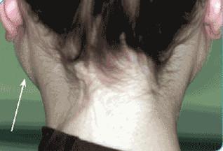
This clinical photograph shows a posterior-oblique view of a patient’s head and neck, highlighting significant bilateral enlargement of the parotid glands. The parotid swelling is visible as pronounced, non-inflammatory masses located anterior and inferior to the ears, extending toward the angle of the mandible. The overlying skin appears normal in texture and color, lacking the erythema or warmth typically associated with acute parotitis. An arrow indicates the area of hypertrophy on the left side. This presentation is characteristic of sialosis (sialadenosis), a non-inflammatory, non-neoplastic, and painless enlargement of the salivary glands. Clinically, this condition is often associated with systemic metabolic disorders, such as diabetes mellitus or chronic alcoholism, leading to acinar cell hypertrophy and adipose infiltration of the gland parenchyma. The image serves as an educational example of how systemic diseases can manifest as specific extraoral anatomical changes in the maxillofacial region.
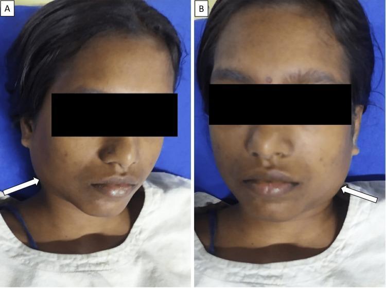
Two clinical photographs (A and B) showing the frontal and slightly oblique facial views of a patient presenting with bilateral parotid swelling. White arrows in each panel highlight large, diffuse, non-erythematous swellings in the preauricular and infraauricular regions, extending down toward the angle of the mandible. The swellings are symmetrical and characteristic of acute parotitis. The patient also exhibits generalized facial puffiness and mild conjunctival congestion. The eyes are obscured by black privacy bars. This clinical presentation is typical of viral parotitis, such as that caused by the mumps virus, which can be associated with systemic complications including myocarditis and pleural effusion in severe cases. The skin tone is consistent with a South Asian phenotype, showing minor post-inflammatory hyperpigmentation spots but no overt primary cutaneous lesions.
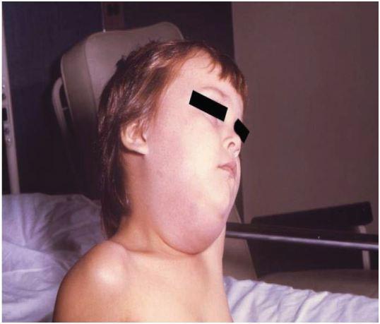
Clinical photograph of a pediatric patient presenting with marked, unilateral swelling of the right parotid region, characteristic of acute mumps parotitis. The image shows a prominent, non-fluctuant bulge extending from the preauricular area, anterior to and below the ear, down to the angle of the mandible and upper cervical region. This localized edema results in the loss of the normal mandibular line and causes the earlobe to be displaced upward and outward. The overlying skin appears taut and mildly erythematous, indicating significant inflammatory glandular enlargement. This finding is the pathognomonic clinical sign of mumps virus infection (Paramyxoviridae family). The visual evidence supports educational recognition of salivary gland pathology and infectious disease manifestations in children. Differential diagnoses for such clinical presentations include bacterial parotitis, salivary calculi, or other viral etiologies like influenza or Epstein-Barr virus.
Confirmed Working Diagnosis: Parotid Gland Pathology
| Symptom | Explained by Parotid |
|---|---|
| Swelling behind/around ear | Parotid tail extends posterior to ear toward mastoid |
| Intense sudden ear pain | Auriculotemporal nerve (V3) runs through parotid |
| Pain radiating to mandibular ramus | Parotid sits directly over the ramus |
| Minor neck stiffness | Reactive spasm of SCM from adjacent parotid inflammation |
| Earlobe displacement | Classic sign - parotid pushes earlobe up and out |
Etiology - What Caused This?
Most Likely Causes (Sudden Onset)
1. Acute Bacterial Sialadenitis (Suppurative Parotitis) - Most likely
"Acute sialoadenitis is a common cause of painful enlargement of the parotid. The organism is usually S. aureus, but can also be S. pneumoniae. Infection is secondary to salivary stasis caused by dehydration or obstruction."
- Dehydration (not drinking enough water)
- Recent illness or fever causing reduced saliva
- Poor oral hygiene
- Prolonged fasting (e.g., overnight, surgery)
- Elderly patients with reduced salivary flow
2. Sialolithiasis (Parotid Duct Stone) - Explains sudden onset perfectly
"Symptoms include recurrent, unilateral, tender salivary enlargement that subsides within 24-48 hours... usually associated with eating."
- Textbook of Family Medicine
- A calcium stone blocks Stensen's duct
- Saliva backs up suddenly behind the blockage
- Causes acute, intense distension pain in ear + jaw + parotid region
- Classic timing: pain comes on sharply at meal times (eating stimulates saliva which cannot drain)
- Parotid stones are radiolucent (less common than submandibular stones)
3. Viral Parotitis (Mumps) - If unvaccinated
- Caused by paramyxovirus
- Incubation 2-3 weeks; illness lasts 7-10 days
- Bilateral or unilateral painful parotid enlargement
- Complications: meningitis, orchitis, pancreatitis, hearing loss
- Diagnosed by viral serology (anti-S and anti-V antibodies)
Anatomy - Why the Pain Radiates Here
Parotid Gland
    │
    ├── Contains: Auriculotemporal nerve (V3 branch)
    │         → Sends pain to EAR + temporal region
    │
    ├── Sits over: Mandibular ramus
    │         → Pressure causes ramus pain
    │
    ├── Adjacent to: SCM muscle attachment at mastoid
    │         → Causes neck stiffness
    │
    └── Stensen's duct opens → inside cheek opposite upper 2nd molar
Treatment
Hospital Treatment (Required - go this morning)
| Treatment | Purpose |
|---|---|
| Anti-staphylococcal antibiotics (flucloxacillin / amoxicillin-clavulanate) | Treat S. aureus infection |
| IV fluids if dehydrated | Restore salivary flow |
| Sialagogues (lemon drops, vitamin C) | Stimulate saliva to flush the duct |
| Ultrasound / CT | Identify stone or abscess |
| Sialoscopy or stone removal | If calculus present |
| Incision & drainage | If abscess has formed |
Home Measures for Right Now (Until You See a Doctor)
1. Stimulate salivary flow (most important)
- Suck a lemon wedge, lemon drop candy, or sour sweet
- Chew sugarless gum
- This stimulates saliva production which helps dislodge a stone and flush bacteria
- Do this every 30-60 minutes
2. Gland massage
- Place warm fingers over the parotid (in front of and below the ear)
- Massage forward toward the cheek in firm circular motions
- This pushes saliva/pus out through Stensen's duct
- Check the inside of your cheek for pus coming out opposite the upper back molar
3. Warm compress
- Apply a warm moist cloth to the swollen parotid area for 10-15 minutes every 1-2 hours
- Reduces pain and promotes drainage
4. Hydration
- Drink at least 2 glasses of water every hour
- Dehydration is a direct cause - reversing it can dramatically improve symptoms
- Avoid alcohol and caffeine (both dehydrate)
5. Pain relief
- Ibuprofen 400mg every 6-8 hours with food (reduces swelling + pain)
- Paracetamol 500-1000mg for breakthrough pain between ibuprofen doses
6. Oral hygiene
- Gently rinse with warm salt water (1/4 tsp salt in a glass of warm water)
- This reduces oral bacterial load feeding into the duct
Red Flags - Go to Emergency Immediately
- Pus visible in the mouth / foul taste suddenly
- Swelling rapidly enlarges or spreads to neck/floor of mouth (Ludwig's angina risk)
- Difficulty swallowing or breathing
- High fever (>38.5°C)
- Jaw locks shut (trismus)
- Neck stiffness worsens
Summary
| Detail | |
|---|---|
| Diagnosis | Acute bacterial sialadenitis OR sialolithiasis of parotid gland |
| Pathogen | S. aureus (most common), S. pneumoniae |
| Root cause | Salivary stasis from dehydration or duct stone |
| Why ear pain | Auriculotemporal nerve (V3) passes through parotid |
| Why jaw pain | Parotid sits over mandibular ramus |
| Why neck stiffness | Reactive SCM spasm - not meningitis |
| Home treatment | Lemon/sour stimulation + warm compress + massage + hydration + ibuprofen |
| Definitive treatment | Anti-staphylococcal antibiotics + hydration + possible stone removal |