
Mitral stenosis
mitral stenosis pathology echocardiography fishmmouth valve
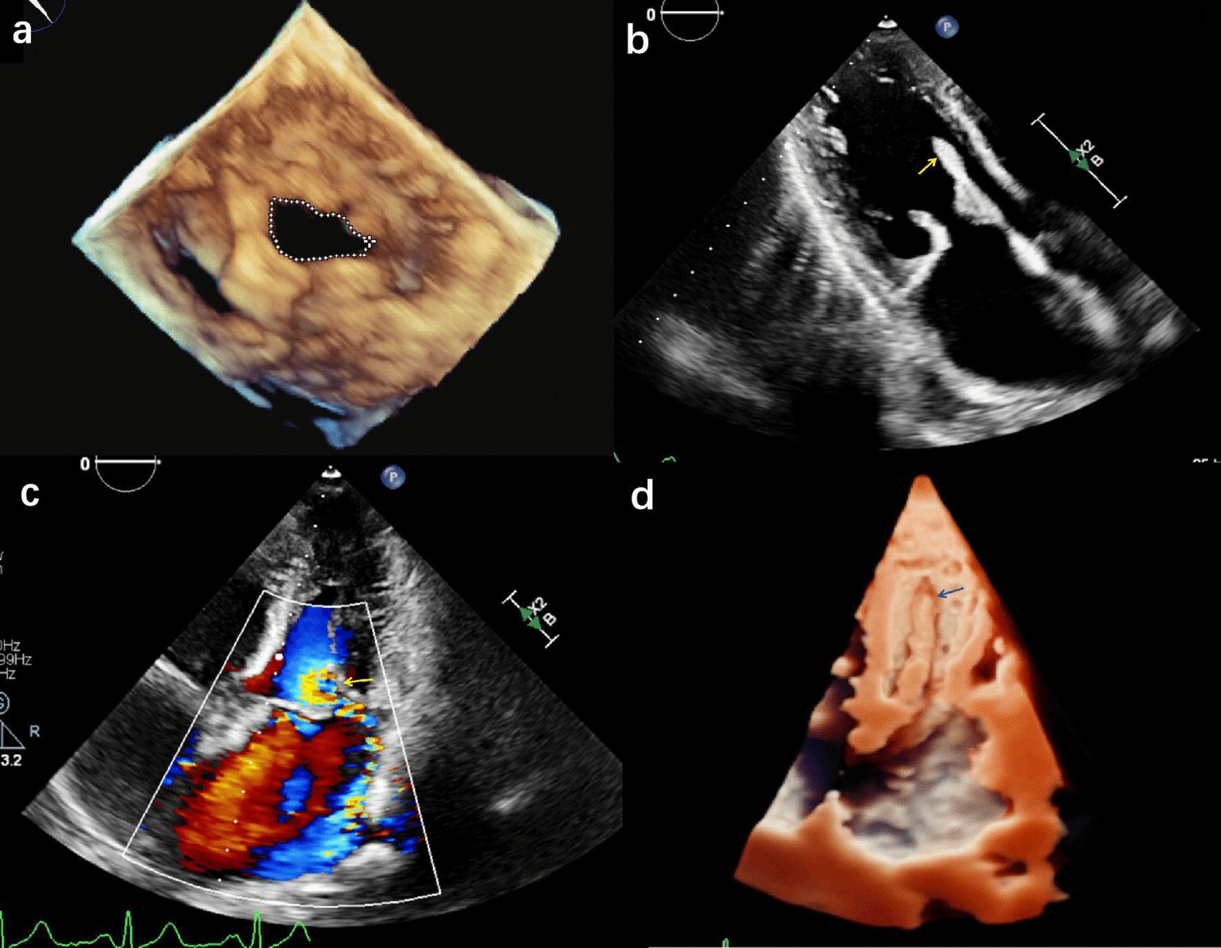
This four-panel image displays diagnostic echocardiographic findings related to mitral valve pathology and its complications. Panel (a) shows a 3D transesophageal echocardiography (TEE) volume rendering of the mitral valve in an atrial view, illustrating mitral stenosis with a thickened, restricted orifice outlined by a dotted line. Panel (b) is a 2D transthoracic echocardiogram (TTE) in an apical four-chamber view, where a yellow arrow identifies a mobile, echogenic mass in the left atrium, suggesting papillary muscle avulsion. Panel (c) utilizes Color Doppler Flow Imaging to demonstrate a large, turbulent regurgitant jet (indicated by a yellow arrow) extending from the mitral valve into the left atrium, diagnostic of severe mitral regurgitation. Panel (d) provides a 3D TTE reconstruction of the left ventricle, where blue arrows highlight the avulsed papillary muscle detached from the ventricular wall. This series illustrates the progression from rheumatic mitral stenosis to acute valvular failure due to subvalvular apparatus injury, emphasizing the role of multi-modal ultrasound in assessing valvular structural integrity and hemodynamic severity.
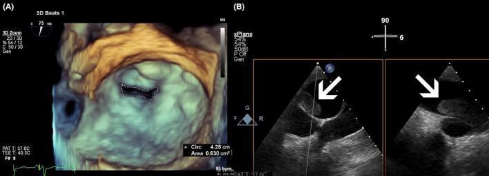
Educational diagnostic imaging compilation showcasing transesophageal echocardiography (TEE) findings in a patient with multi-valvular and vascular pathology. Panel (A) presents a 3D-TEE surgical view of the mitral valve. The image illustrates severe mitral stenosis, characterized by a significantly narrowed, irregular, fish-mouth-shaped orifice. A dotted planimetry line outlines the valve area, with an on-screen measurement of 0.630 cm², confirming a critical reduction in opening. The 3D reconstruction uses a color map of beige, brown, and cyan to provide depth and topographical detail of the valve apparatus. Panel (B) shows a side-by-side X-plane TEE view of the pulmonary artery. White arrows highlight large, echogenic, mobile masses within the proximal right pulmonary artery, diagnostic of pulmonary thromboembolism. The thrombi appear as distinct, heterogeneous structures contrasting against the anechoic blood flow. This composite image is intended for advanced cardiology training, focusing on the integration of 3D and multi-plane TEE for assessing structural heart disease and associated vascular complications like pulmonary hypertension and embolism.
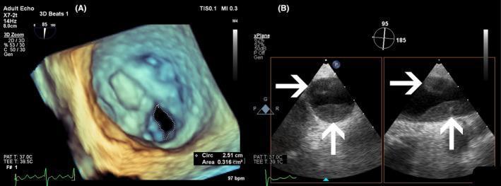
This composite diagnostic image showcases transesophageal echocardiography (TEE) findings in a patient with multi-valvular and vascular pathology. Image (A) presents a three-dimensional (3D) TEE volume rendering of the mitral valve viewed from the left atrium ('surgical view'). It demonstrates severe mitral stenosis (MS), characterized by thickened valve leaflets and a significantly restricted orifice. A dotted white line highlights the planimetered mitral valve area (MVA), measured at 0.316 cm², indicating critical stenosis. Image (B) displays a dual-panel X-plane TEE view of the pulmonary artery. White arrows indicate large, hyperechoic, obstructive masses within the proximal part of the right pulmonary artery, diagnostic of pulmonary thromboembolism (PTE). The educational focus of this visual is the use of advanced echocardiographic modalities, such as 3D rendering and X-plane imaging, to evaluate structural heart disease and associated vascular complications like pulmonary hypertension and thrombosis. The content is highly relevant for cardiology and radiology residents studying valvular hemodynamics and embolic phenomena.
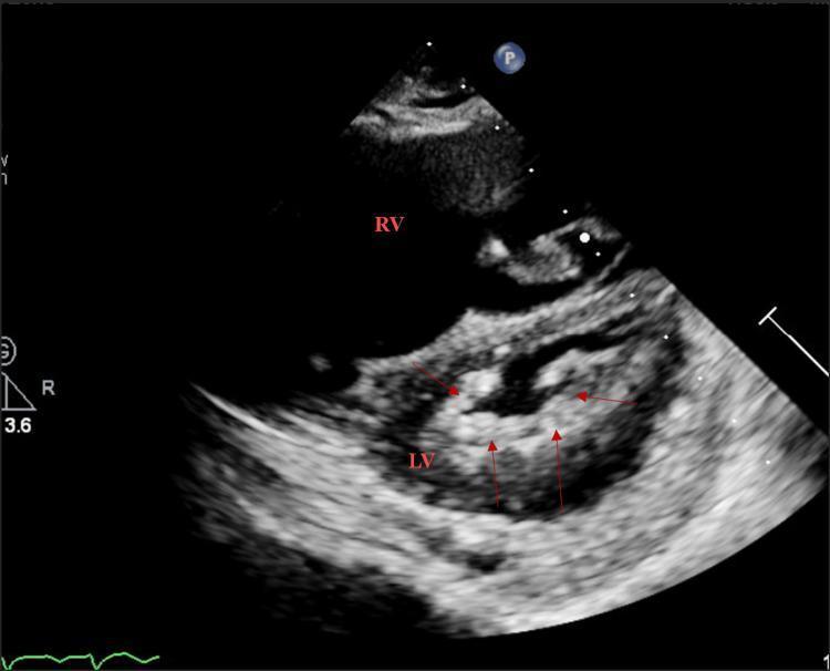
This diagnostic image is a transthoracic echocardiogram in the parasternal short-axis (PSAX) view at the mitral valve level. The image depicts the characteristic cross-sectional anatomy of the heart, showing the right ventricle (RV) positioned anteriorly and the circular left ventricle (LV) containing the mitral valve apparatus. Key pathology is highlighted by red arrows, which indicate diffuse, echogenic thickening of the mitral valve leaflets and surrounding tissue. The mitral orifice is notably restricted and narrowed, a classic visual feature of mitral stenosis, likely of rheumatic etiology given the extent of commissural fusion and leaflet thickening. The LV myocardium displays a typical grainy texture on point-of-care ultrasound (POCUS). This visual is highly relevant for medical education regarding valvular heart disease, specifically demonstrating the morphological changes observed in chronic rheumatic mitral stenosis and the use of echocardiography for assessing cardiac valve orifice patency.
"mitral stenosis"[MeSH Terms] AND management
Mitral Stenosis
Definition and Overview
- Braunwald's Heart Disease, Chapter 75
Etiology
| Cause | Notes |
|---|---|
| Rheumatic (most common worldwide) | Autoimmune response to Group A Streptococcal pharyngitis; valve mimicry by GAS antigens drives humoral + cellular damage |
| Degenerative/Calcific (MAC-related) | Mitral annular calcification; accounts for ~41% of severe MS in Western echocardiography series |
| Congenital | Rare; parachute mitral valve, cor triatriatum |
| Post-procedural | Increasingly seen after ring annuloplasty or prior mitral valve surgery |
| Rare | Carcinoid syndrome, lupus, mucopolysaccharidoses, radiation-induced |
Pathology
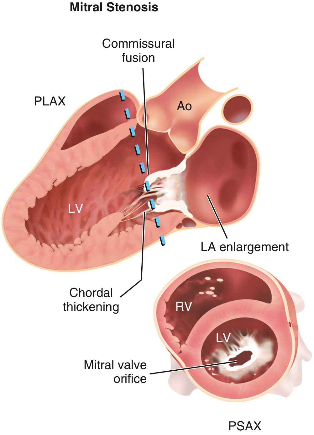
- Diastolic doming/bowing of leaflets (the anterior leaflet bows like a hockey stick in PLAX view)
- Leaflet tip thickening; variable calcification
- Subvalvular involvement: chordal fusion, shortening, fibrosis, and calcification
- The classic "fish-mouth" or "buttonhole" orifice in PSAX view
Pathophysiology
- LA pressure elevation → LA dilation → atrial fibrillation (AF) → stasis in LA appendage → thrombus → systemic thromboembolism/stroke
- Pulmonary venous hypertension → pulmonary congestion → dyspnea, orthopnea, PND, hemoptysis
- Reactive pulmonary arterial hypertension (PAH) → RV pressure overload → RV hypertrophy → tricuspid regurgitation → right heart failure
- LV response: LV is typically underfilled and small; LV systolic function is usually preserved unless there is coexisting disease
Severity Classification (AHA/ACC Stages)
| Stage | Definition | MVA | Gradient | Symptoms |
|---|---|---|---|---|
| A | At risk | Normal | Normal | None |
| B | Progressive | >1.5 cm² | Mean <5-10 mmHg | None or mild |
| C | Severe, asymptomatic | ≤1.5 cm² | Mean ≥5-10 mmHg | None |
| D | Severe, symptomatic | ≤1.5 cm² | Mean ≥5-10 mmHg | NYHA II-IV |
- Mild: MVA 1.5-2.0 cm²
- Moderate: MVA 1.0-1.5 cm²
- Severe: MVA ≤1.5 cm² (guideline intervention threshold); gradients >10 mmHg with resting symptoms at ≤1.0 cm²
Clinical Features
Symptoms
- Dyspnea - initial exertional, progressing to rest; most common presenting symptom
- Paroxysmal nocturnal dyspnea, orthopnea (severe disease)
- Palpitations (due to AF)
- Hemoptysis (pulmonary venous hypertension or rupture of bronchial veins)
- Systemic thromboembolism/stroke
- Fatigue and low-output symptoms (with advanced PAH or over-diuresis)
- Precipitants: AF, pregnancy, fever, anemia, exercise, thyrotoxicosis
Signs
| Finding | Mechanism |
|---|---|
| Loud S1 (palpable "tapping" apex) | Mitral leaflets widely open at diastole, snap shut forcefully |
| Opening snap (OS) | Sudden tensing of fused, still-pliable leaflets at onset of diastole |
| Low-pitched mid-diastolic rumble (MDM) | Turbulent flow across stenotic valve; heard at apex with bell |
| Presystolic accentuation | Atrial systole increases flow (disappears in AF) |
| Malar flush (mitral facies) | Chronic low-output, peripheral vasoconstriction |
| Pulmonary component of S2 loud (P2) | Secondary PAH |
| Signs of RHF | JVD, hepatomegaly, peripheral edema, TR |
Investigations
ECG
- P mitrale - broad, notched P wave in Lead II (>0.12 sec); biphasic P in V1 → LA enlargement
- AF (very common)
- RVH pattern in advanced disease
Chest X-ray
- Straightening of left heart border (LA appendage enlargement - "4th mogul")
- Double density shadow at right cardiac border (LA enlargement)
- Upper lobe blood diversion / Kerley B lines
- Pulmonary hemosiderosis (long-standing cases)
- Valve calcification
Echocardiography (gold standard)
- 2D imaging: Leaflet thickness, mobility, calcification, commissural fusion, subvalvular apparatus
- 3D echo: Superior planimetry of MVA, especially in calcific MS
- MVA measurement methods:
- Planimetry (2D/3D): Direct tracing of orifice in PSAX - reference standard
- Pressure half-time (PHT): MVA = 220/PHT; validated primarily in rheumatic MS (less reliable post-commissurotomy or with abnormal LV compliance)
- Continuity equation
- Mean transmitral gradient by Doppler
- Pulmonary artery pressure estimation
- LA thrombus assessment (TEE if TTE inconclusive, especially before intervention)
Wilkins Score (Echocardiographic Scoring for BMV Suitability)
| Feature | Score 1 | Score 2 | Score 3 | Score 4 |
|---|---|---|---|---|
| Leaflet mobility | Highly mobile | Reduced base/tip | Forward diastolic only | No/minimal movement |
| Valvular thickening | Near normal (4-5mm) | Mid-leaflet thickened | Thickened throughout | Marked (>8-10mm) |
| Subvalvular thickening | Minimal | Thickening 1/3 chordae | Distal 1/3 thickened | Extensive + papillary |
| Calcification | Single echobright area | Scattered margins | Bright extending to mid | Extensive throughout |
Management
Medical Treatment
- Rate control in AF: Beta-blockers, rate-limiting calcium channel blockers (avoid digoxin as first-line)
- Diuretics: For pulmonary congestion
- Anticoagulation: Mandatory with AF, prior embolism, or LA thrombus. Use warfarin (INR 2-3); direct oral anticoagulants (DOACs) are NOT validated in rheumatic MS with AF
- Rheumatic fever prophylaxis: Benzathine penicillin G IM monthly (or oral phenoxymethylpenicillin) until age 40 or for 10 years from last attack
- Avoid: High-intensity exercise; vasodilators (may worsen CO by reducing preload)
Interventional/Surgical Indications (AHA/ACC 2021)
- Procedure of choice for suitable anatomy
- Balloon (Inoue technique) is inflated across the mitral valve, splitting the fused commissures
- Indications: Symptomatic severe MS (Stage D), MVA ≤1.5 cm²; favorable morphology (Wilkins ≤8); no significant MR; no LA thrombus
- Special indication: Severe MS in pregnancy (preferred after 24 weeks gestation)
- Success rate: High (>90%) in younger patients with favorable anatomy; <50% in older patients or calcified valves
- Open mitral commissurotomy: For MS with favorable anatomy but not suitable for BMV (e.g., LA thrombus that cannot be resolved)
- Mitral valve replacement (MVR): For unfavorable anatomy (heavy calcification, significant MR, failed BMV); options - mechanical (requires lifelong anticoagulation) vs. bioprosthetic (no anticoagulation if no AF, but limited durability)
- Surgical mortality ≥10% in elderly; percutaneous options preferred where possible
MS in Pregnancy
- Severe MS (WHO Class IV) - pregnancy is contraindicated; intervention before conception recommended
- Conservative management: Activity restriction, beta-blockers (rate control), diuretics
- AF in pregnancy requires urgent rate control and anticoagulation (heparin in 1st/3rd trimester)
- BMV indicated for NYHA III-IV on optimal medical therapy; optimally after 24 weeks
- Caesarean delivery for NYHA III-IV or moderate MS with PAH
Complications
| Complication | Mechanism |
|---|---|
| Atrial fibrillation | LA dilation + electrophysiologic remodeling |
| Systemic thromboembolism/stroke | LA stasis, especially appendage |
| Pulmonary arterial hypertension | Reactive vasoconstriction + structural remodeling |
| Right heart failure | Sustained RV pressure overload |
| Infective endocarditis | Damaged valve; prophylaxis for high-risk procedures |
| Recurrent MS (post-BMV) | Restenosis at ~5-10 years; re-BMV often possible |
Key Differentials
- Left atrial myxoma (mimics MS perfectly; diagnosed on 2D echo as mass)
- Cor triatriatum (membrane dividing LA)
- Diastolic LV dysfunction
- Pulmonary venoocclusive disease