
Tell me all about puppy’s erythematous
Reading File
The query is about Puppy Strangles (juvenile cellulitis/juvenile pyoderma), which may present with erythematous lesions, or possibly pupillary erythema - but "puppy's erythematous" most likely refers to a dermatological condition in young dogs. Let me search the medical library and web in parallel.
Searching sources...
Finding Sources
Searching the Web
puppy strangles juvenile cellulitis erythematous skin lesions
Lupous erythematous
Reading File
Lupus erythematosus (LE) is a chronic autoimmune disease where the immune system attacks the body's own tissues. It ranges from skin-limited disease to severe multi-organ involvement. Let me pull authoritative textbook content right away.
Searching sources...
Finding Sources
Searching the Web
lupus erythematosus pathophysiology clinical features diagnosis treatment 2024
Excellent - I have strong textbook sources. Let me now read the key sections from Fitzpatrick's dermatology and Andrews' Diseases of the Skin in parallel, plus search for recent evidence.
Reading File
Reading File
Searching PubMed
"lupus erythematosus" AND treatment
Good content so far. Now let me read the main comprehensive LE section from Fitzpatrick's (block31) and the block2 from Andrews', plus read more of the Fitzpatrick block10 for the full overview.
Reading File
Reading File
Now let me read the main comprehensive LE chapter from Fitzpatrick's block10 from the very beginning to get the full overview, epidemiology, pathophysiology, and classification.
Finding Sources
Reading File
Let me read more of the core clinical section from Fitzpatrick's block10 (the main LE chapter content) and the Andrews' block that covers SLE in full.
Reading File
Reading File
I now have very rich textbook content. Let me read a bit more for the treatment section and the key images.
Reading File
I have comprehensive content. Let me now fetch a key clinical image to include in my answer.
Loading Image
I now have all the information needed for a comprehensive answer. Let me compile everything.
Lupus Erythematosus (LE) - Complete Overview
What Is It?
Lupus erythematosus is a chronic autoimmune disease in which the immune system loses tolerance to self-antigens and produces autoantibodies that attack the body's own tissues. It spans a spectrum from purely skin-limited disease to severe multi-organ (systemic) involvement.
Classification
LE is broadly categorized as:
| Type | Abbreviation | Nature |
|---|---|---|
| Cutaneous LE | CLE | Skin-limited or skin-dominant |
| Systemic LE | SLE | Multi-organ involvement |
| Drug-Induced LE | DIL | Triggered by medications |
Cutaneous LE (CLE) is further divided by pace and severity:
- Acute CLE (ACLE) - occurs during active, flaring SLE
- Subacute CLE (SCLE) - intermediate severity and SLE association
- Chronic CLE (CCLE) - often occurs without SLE or with mild, smoldering disease
1. Acute Cutaneous Lupus Erythematosus (ACLE)
The hallmark is the butterfly (malar) rash - confluent symmetric erythema and edema centered over the malar eminences, bridging across the nose.
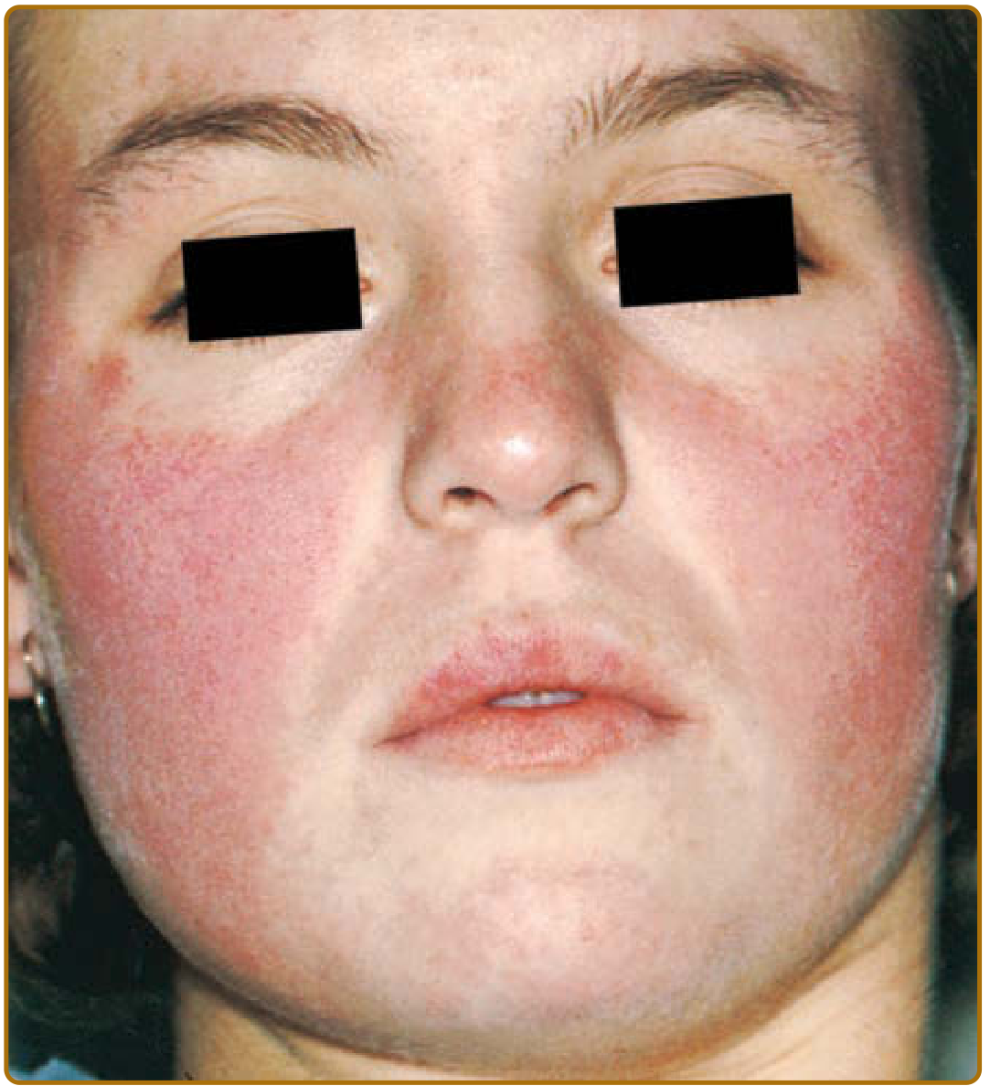
Key features:
- Nasolabial folds are characteristically spared (distinguishes from rosacea and seborrheic dermatitis)
- Forehead, chin, and V-area of neck can also be involved
- Generalized ACLE presents as a widespread morbilliform eruption over extensor surfaces of arms and hands, characteristically sparing the knuckles
- An extreme form can simulate toxic epidermal necrolysis (TEN), but differs by involving sun-exposed skin with more insidious onset
- Precipitated/exacerbated by UV light
- Lesions are ephemeral - lasting hours to weeks
- No atrophic scarring unless complicated by secondary infection
ACLE almost always occurs in the setting of acutely flaring SLE.
2. Subacute Cutaneous Lupus Erythematosus (SCLE)
First characterized as a distinct LE subset in 1979. Begins as erythematous macules/papules that evolve into:
-
Hyperkeratotic papulosquamous plaques, or
-
Annular/polycyclic plaques
-
Strongly associated with anti-Ro/SS-A antibodies
-
Photodistributed but not strictly photodistributed - annular or papulosquamous thin plaques
-
Intermediate risk of underlying SLE
-
SCLE can also be drug-induced (common triggers: hydrochlorothiazide, diltiazem, terbinafine, proton pump inhibitors, TNF inhibitors, ACE inhibitors, statins, paclitaxel, doxycycline)
3. Chronic Cutaneous Lupus Erythematosus (CCLE)
Discoid LE (DLE) - the most common CCLE subtype
Classic DLE lesions progress through three stages:
- Active lesion: erythematous, indurated papule or plaque with adherent scale and follicular plugging
- Progression: central atrophy, hypopigmentation; hyperpigmented peripheral rim
- Burnt-out lesion: atrophic, pale scar
Key anatomical sites:
- Face (cheeks, nose, ears - especially conchal bowl and outer ear canal)
- Scalp: involved in 60% of DLE patients; irreversible scarring alopecia in one-third
- Localized DLE = lesions only on head/neck
- Generalized DLE = lesions above AND below the neck (higher risk of SLE, more treatment-resistant)
Only ~5% of patients with classic localized DLE go on to develop unequivocal SLE. Roughly one-quarter of SLE patients develop DLE lesions at some point.
Other CCLE Variants
Hypertrophic/Verrucous DLE: thick, hyperkeratotic plaques that can mimic squamous cell carcinoma (and rarely transform into SCC).
LE Panniculitis (LE Profundus / Kaposi-Irrgang disease):
- Inflammatory lesions of the lower dermis and subcutis
- Firm nodules 1-3 cm on head, proximal arms, chest, breasts, buttocks, thighs
- Overlying skin becomes attached, creating deep saucerized depressions
- Breast involvement can mimic carcinoma (lupus mastitis)
- ~50% of LE panniculitis patients have evidence of SLE
Chilblain LE:
- Purple-red patches, papules, plaques on toes, fingers, and face precipitated by cold/damp conditions
- Associated with anti-Ro/SS-A antibodies and Raynaud phenomenon
- ~20% later develop SLE
4. Systemic Lupus Erythematosus (SLE)
SLE is a multi-system autoimmune disease that predominantly affects young to middle-aged women. Skin involvement occurs in 80% of cases.
ACR Diagnostic Criteria (11 criteria; 4+ required)
Four of the 11 criteria are mucocutaneous:
| # | Criterion |
|---|---|
| 1 | Malar rash |
| 2 | Discoid rash |
| 3 | Photosensitivity |
| 4 | Oral ulcers |
| 5 | Arthritis |
| 6 | Proteinuria >0.5 g/day or casts |
| 7 | Neurologic disorders (seizures or psychosis) |
| 8 | Pleuritis/pericarditis |
| 9 | Blood abnormalities (hemolytic anemia, leukopenia, thrombocytopenia) |
| 10 | Immunologic: anti-dsDNA, anti-Sm, antiphospholipid antibodies |
| 11 | Positive ANA |
The updated SLICC criteria require at least 4 criteria (including ≥1 clinical + ≥1 immunologic) OR biopsy-proven lupus nephritis with positive ANA or anti-dsDNA.
Systemic Manifestations
- Musculoskeletal: non-erosive arthritis/arthralgias (most common)
- Renal: lupus nephritis (major cause of morbidity); WHO/ISN classes I-VI
- Neuropsychiatric: seizures, psychosis, cognitive dysfunction, stroke
- Hematologic: hemolytic anemia, leukopenia, lymphopenia, thrombocytopenia
- Cardiovascular: pericarditis, Libman-Sacks endocarditis, accelerated atherosclerosis
- Pulmonary: pleuritis, pneumonitis, pulmonary hypertension
- Serositis: pleuritis/pericarditis
Hair Loss in SLE
- Lupus hairs: short miniaturized hairs on the anterior scalp
- Diffuse non-scarring alopecia (telogen effluvium) during systemic flares - reversible
- Scarring alopecia from DLE involvement - irreversible
5. Drug-Induced Lupus (DIL)
- Occurs in older patients; affects men as frequently as women
- Symptoms generally mild: fever, myalgias/arthralgias, serositis
- Antibody pattern: positive ANA (homogeneous), antihistone antibodies; negative anti-dsDNA, normal complement
- Resolves on drug withdrawal
Common triggering drugs: procainamide, hydralazine, quinidine, captopril, isoniazid, minocycline, carbamazepine, propylthiouracil, sulfasalazine, statins, TNF inhibitors (etanercept - this form can have anti-dsDNA and renal/CNS involvement).
Serology / Antibodies
| Antibody | Association |
|---|---|
| ANA | Positive in >95% SLE (sensitive but not specific) |
| Anti-dsDNA | Specific for SLE; correlates with disease activity (especially nephritis) |
| Anti-Sm | Highly specific for SLE |
| Anti-Ro/SS-A | SCLE, chilblain LE, neonatal LE, photosensitivity |
| Anti-La/SS-B | Often with anti-Ro; neonatal LE |
| Antihistone | Drug-induced LE |
| Antiphospholipid | Thrombosis, recurrent pregnancy loss, thrombocytopenia |
Histopathology
DLE (established lesions) shows:
- Hyperkeratosis, follicular plugging
- Interface (vacuolar) dermatitis at the dermoepidermal junction (DEJ)
- Basement membrane zone thickening
- Pigment incontinence
- Dermal mucin deposition
- Perivascular and periadnexal lymphoid infiltrates
Direct immunofluorescence (DIF): Granular deposition of IgG, IgA, IgM, and C3 at the DEJ - the "full house" pattern (positive lupus band test).
Treatment
Sun Protection (All CLE Subtypes)
- Strict UV avoidance, broad-spectrum SPF ≥50 sunscreen, protective clothing
- Vitamin D supplementation (as patients avoid sun)
Topical/Intralesional
- Potent topical corticosteroids (first line for limited disease)
- Intralesional corticosteroids (for resistant DLE plaques)
- Topical calcineurin inhibitors (limited benefit)
- Topical tazarotene (generally disappointing)
Systemic - First Line
- Hydroxychloroquine (HCQ): cornerstone of SLE and CLE management; antimalarial, reduces flares and organ damage; requires ophthalmologic monitoring
- Chloroquine: alternative antimalarial
Systemic - Second Line (CLE and SLE)
- Corticosteroids: for acute flares; long-term use limited by side effects
- Methotrexate
- Mycophenolate mofetil (especially lupus nephritis)
- Azathioprine
- Dapsone
- Thalidomide (highly effective for refractory CLE; strict teratogenicity precautions)
- Retinoids (acitretin)
- Sulfasalazine
Biologics
- Belimumab (anti-BLyS/BAFF): FDA-approved for SLE; reduces flares
- Anifrolumab (anti-IFN-α receptor): FDA-approved for SLE, particularly effective for skin disease
- Rituximab (anti-CD20): used off-label for refractory SLE and lupus nephritis
- Voclosporin + mycophenolate: approved for lupus nephritis
Emerging (2024-2026)
A recent systematic review shows CAR T-cell therapy targeting CD19 is achieving deep, sustained remissions in refractory SLE, including patients who had failed belimumab and standard immunosuppression. The 2023 EULAR recommendations update (informed by a systematic literature review) emphasizes treating to target (remission or lupus low disease activity state - LLDAS) to reduce long-term organ damage.
Prognosis
- Most patients with localized DLE have a good prognosis; only ~5% develop SLE
- Generalized DLE carries higher risk of SLE progression
- SLE prognosis has improved dramatically; 10-year survival is now >90% in many cohorts
- Main causes of morbidity and mortality: lupus nephritis, cardiovascular disease (accelerated atherosclerosis), infections from immunosuppression, and neuropsychiatric lupus
- Flares are unpredictable; maintaining low disease activity (LLDAS) significantly reduces damage accrual
Sources: Fitzpatrick's Dermatology, 9th ed., pp. 1071-1100; Andrews' Diseases of the Skin, 13th ed., pp. 189-192; Brenner and Rector's The Kidney; EULAR 2023 recommendations (PMID 38777375)
This is a shared conversation. Sign in to Orris to start your own chat.