I now have rich content from multiple authoritative sources. Let me compile the comprehensive answer.
Airway Management: Adult vs Pediatric Anatomy and Rapid Sequence Intubation (RSI)
Sources: Rosen's Emergency Medicine, Tintinalli's Emergency Medicine, Sabiston Textbook of Surgery
PART 1: ADULT AIRWAY - Overview
The adult airway is composed of the upper airway (nose, pharynx, larynx) and the lower airway (trachea, bronchi). Key landmarks for the emergency physician:
- Tongue is the most common cause of airway obstruction in the unconscious adult
- Larynx sits at C4-C6 level
- Narrowest point is the glottis (vocal cords) - relevant for ETT sizing
- Cricothyroid membrane is the site for emergency surgical airway (2-3 cm wide x 0.9 cm tall in adults) - easily palpable between thyroid and cricoid cartilages
- Sniffing position (head extension + neck flexion) aligns the oral-pharyngeal-laryngeal (OPL) axes for laryngoscopy
Key adult ETT size: 7.5-8.0 mm for women, 8.0-8.5 mm for men. Insert to approximately 23 cm (men) and 21 cm (women) at the teeth.
PART 2: PEDIATRIC AIRWAY ANATOMY - Detailed with Exam Focus
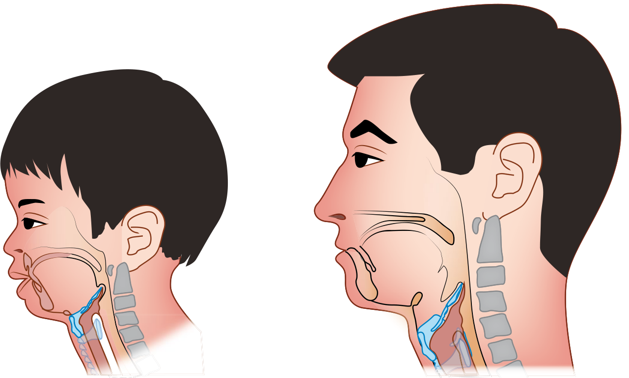
Visual Comparison
Fig. FIGURE 42.1 - Differences in anatomy between the pediatric and adult airways (Sabiston Textbook of Surgery)
The anatomic differences between pediatric and adult airways are most pronounced in the first 2 years of life. Children 2-8 years represent a transitional phase. By age ~8-10, the airway approaches adult proportions.
Key Anatomic Differences (HIGH-YIELD TABLE)
| Anatomic Feature | Pediatric | Clinical Implication |
|---|
| Occiput/Head | Large relative to body | Neck flexes when supine - need shoulder roll in infants <6 months |
| Tongue | Proportionally larger | Obstructs airway easily; jaw thrust + OPA helpful |
| Larynx position | Higher and more anterior (C3-C4 vs C4-C6 in adults) | Cords harder to visualize - "anterior airway" |
| Epiglottis | Omega-shaped (Ω), floppy, angled 45° | Straight blade (Miller) preferred to lift epiglottis directly |
| Narrowest point | Subglottic (cricoid ring) - circular, complete ring | Traditionally uncuffed tubes; cuffed tubes now safe if pressure monitored |
| Tonsils/Adenoids | Large | Bleed easily - blind nasotracheal intubation contraindicated <10 years |
| Trachea | Short, narrow, soft | Risk of mainstem intubation; dynamic collapse possible |
| Cricothyroid membrane | Very small | Surgical cricothyrotomy difficult - needle cricothyrotomy preferred in infants/young children |
| Oxygen consumption | Higher (6-8 mL/kg/min vs 3-4 mL/kg/min in adults) | Desaturate MUCH faster during apnea |
| FRC | Smaller relative to body weight | Less oxygen reserve; closing capacity exceeds FRC |
Key mnemonic: SLOPE - Small mouth, Large tongue/occiput, Obliqueglottis/anterior, Pediatric cricoid = narrowest, Epiglottis floppy
Positioning for Pediatric Laryngoscopy
- Infant (<6 months): Shoulder roll to overcome neck flexion from large occiput
- Child (6 months - 5 years): Typically no support needed; neutral position adequate
- Older child/adolescent: Head elevation (as in adult sniffing position)
- The external auditory canal-to-anterior shoulder line should be horizontal to the bed
ETT Sizing (Pediatric)
- Neonates/infants: 3.0-3.5 mm uncuffed ETT
- Older children (cuffed): (Age/4) + 3.5 mm (cuffed) or (Age/4) + 4.0 mm (uncuffed)
- Depth of insertion: ETT size × 3 cm
- Alternative sizing: Diameter of child's pinky finger or nostril
- Oral airway size: Distance from central incisors to angle of mandible
- Nasal airway size: Distance from nose to tragus
PART 3: RAPID SEQUENCE INTUBATION (RSI) - Emergency Medicine Exam Focus
Definition
RSI is the nearly simultaneous administration of a potent induction (sedative) agent and neuromuscular blocking agent (NMBA) after preoxygenation and cardiopulmonary optimization to achieve tracheal intubation. It is used in 85% of all ED intubations.
Goal: Take a conscious, spontaneously breathing patient to unconscious + completely paralyzed without interposed bag-mask ventilation, then intubate.
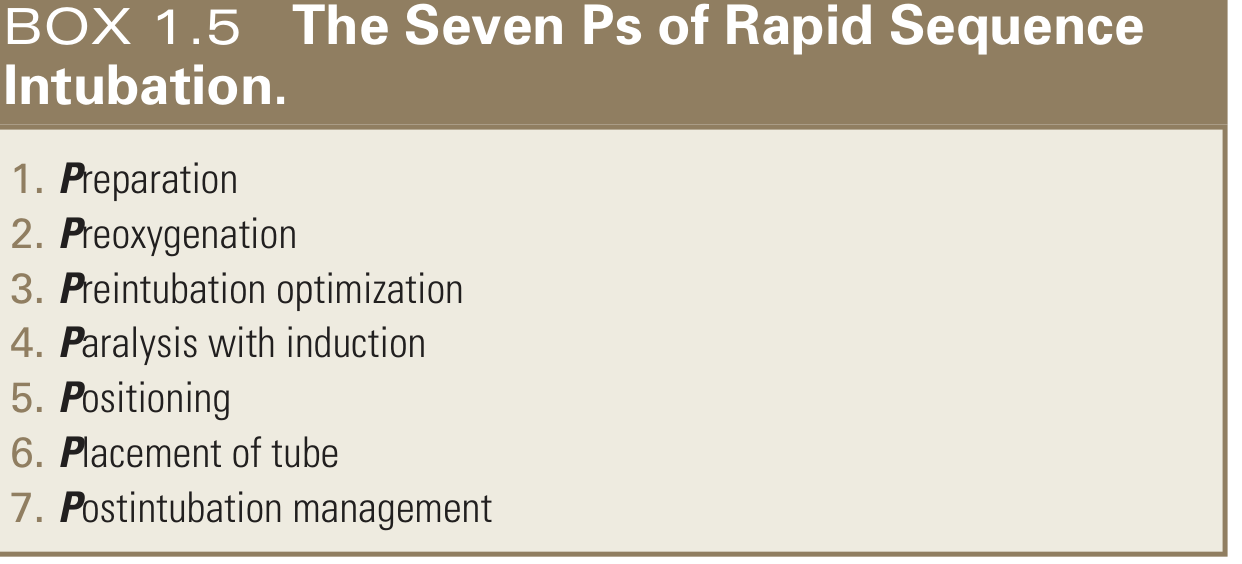
THE SEVEN Ps OF RSI (High Yield)
| Step | "P" | Details |
|---|
| 1 | Preparation | Assess airway difficulty, calculate drug doses, assemble equipment (ETT, stylet, suction, BVM, rescue airway), 2 IV lines, monitors, rescue plan |
| 2 | Preoxygenation | Goal: replace FRC nitrogen with O2. Use flush-rate O2 (40-70 L/min) via non-rebreather. 3 minutes at tidal breathing OR 8 vital capacity breaths. Target SpO2 >95% |
| 3 | Preintubation optimization | Correct hypotension, hypoxia, acidosis before drugs. "Crash airway" = skip this step |
| 4 | Paralysis with induction | Give sedative agent + NMBA simultaneously (or within 30-60 sec). Time = 0 |
| 5 | Positioning | Head elevated 20-30° (sniffing position) - reduces aspiration risk, improves view |
| 6 | Placement of tube | Intubate at 45-60 sec (succinylcholine) or 60-75 sec (rocuronium). Confirm with waveform capnography |
| 7 | Postintubation management | Sedate + analgesia + NMBA as needed, confirm placement, CXR, set ventilator |
RSI DRUGS - INDUCTION AGENTS
| Drug | Dose | Key Points |
|---|
| Etomidate | 0.3 mg/kg IV | Hemodynamically stable, minimal CV effects; single dose adrenal suppression (controversial); drug of choice for most ED patients |
| Ketamine | 1-2 mg/kg IV | Cardiovascular stimulant, bronchodilator, preserves airway reflexes; drug of choice for asthma, hypotension, early sepsis, status asthmaticus, pediatric RSI |
| Propofol | 1.5-2 mg/kg IV | Rapid push; can cause hypotension; excellent for hemodynamically stable patients |
| Midazolam | 0.1-0.3 mg/kg IV | Slower onset, not ideal for RSI induction alone |
| Thiopental | 3-5 mg/kg IV | Historically used; reduces ICP; causes hypotension; largely replaced |
RSI DRUGS - NEUROMUSCULAR BLOCKING AGENTS
Succinylcholine (Depolarizing)
- Dose: 1.5 mg/kg IV (children <10 kg: 2 mg/kg; >10 kg: 1-1.5 mg/kg)
- Onset: 45-60 seconds; Duration: 8-10 minutes
- Advantages: Fastest onset, shortest duration, allows rapid return of spontaneous ventilation
- Contraindications (Hyperkalemia Risk):
| Condition | Period of Risk |
|---|
| Burns >10% BSA | >5 days after injury until healed |
| Crush injury | >5 days after injury until healed |
| Denervation (stroke, SCI) | >5 days until 6 months post-injury |
| Neuromuscular disease (ALS, MS, MD) | Indefinitely |
| Intraabdominal sepsis | >5 days until resolved |
- Other side effects: Fasciculations, myalgia, bradycardia (especially in children), masseter spasm, malignant hyperthermia trigger, increased IOP, increased intragastric pressure
Exam pearl: Succinylcholine is NOT contraindicated in acute (<5 days) burn, trauma, stroke, or SCI. Risk begins at 5 days post-injury.
Rocuronium (Non-depolarizing)
- Dose for RSI: 1.2 mg/kg IV (high-dose)
- Onset: ~60-75 seconds at 1.2 mg/kg
- Duration: 60-90 minutes (much longer than succinylcholine)
- Reversal: Sugammadex 16 mg/kg reverses even high-dose rocuronium within 3 minutes - making it a safe alternative to succinylcholine
- Preferred in pediatrics due to risk of succinylcholine-induced hyperkalemic cardiac arrest in children with undiagnosed neuromuscular disease
- No contraindications for hyperkalemia
PRETREATMENT AGENTS (3 minutes before RSI)
Controversial - not routinely recommended; used in specific situations
| Agent | Dose | Indication |
|---|
| Lidocaine | 1.5 mg/kg IV | Blunt ICP rise with laryngoscopy (head injury, status asthmaticus) - limited evidence |
| Fentanyl | 3 mcg/kg IV | Blunt sympathetic response in hypertensive emergencies, ICP elevation |
| Atropine | 0.02 mg/kg IV (min 0.1 mg) | Pediatric bradycardia prevention - NOT routinely recommended; bradycardia = sign of hypoxia, address that first |
PEDIATRIC RSI - SPECIFIC POINTS
- RSI is the preferred method in children in the ED - highest success and lowest complication rates
- Rocuronium is now the paralytic of choice in most pediatric centers (risk of fatal hyperkalemia with succinylcholine in undiagnosed myopathy)
- Ketamine is the preferred induction agent in most pediatric emergencies
- No evidence to support pretreatment agents in children
- Atropine does NOT prevent succinylcholine-associated bradycardia and should NOT be given prophylactically
FAILED AIRWAY MANAGEMENT
When RSI fails (can't intubate after 3 attempts):
- Call for help immediately
- BVM ventilation to maintain oxygenation
- Supraglottic airway (LMA/King LT) as rescue
- If can't ventilate + can't intubate: Surgical airway (cricothyrotomy)
- Adults: Surgical cricothyrotomy
- Children <8-10 years/infants: Needle cricothyrotomy (surgical CTM too small)
POSTINTUBATION CONFIRMATION
- Waveform capnography is the gold standard (continuous EtCO2)
- Children <15 kg: small colorimetric detector; >15 kg: adult-sized detector
- Chest X-ray to confirm ETT tip position (should be 3-5 cm above carina)
COMPLICATIONS OF RSI (Exam Table)
| Complication | Prevention | Management |
|---|
| Oxygen desaturation | Preoxygenation | Verify ETT position, hyperventilate |
| Hypotension | Fluid resuscitation before RSI | Trendelenburg, IV fluids, pressors |
| Esophageal intubation | View ETT pass through cords | Remove ETT, reoxygenate |
| Mainstem intubation | Know correct depth | Withdraw ETT |
| Aspiration | Avoid aggressive BVM ventilation | Suction ETT and oropharynx |
| Cardiac arrest | Ensure adequate HR/BP before intubation | CPR |
HIGH-YIELD EXAM SUMMARY
| Point | Key Fact |
|---|
| Most used technique in ED | RSI (85% of intubations) |
| Narrowest point adult airway | Glottis (vocal cords) |
| Narrowest point pediatric airway | Subglottis (cricoid ring) |
| Fastest NMBA | Succinylcholine (45-60 sec) |
| Reversal agent for rocuronium | Sugammadex 16 mg/kg |
| Succinylcholine hyperkalemia risk starts | >5 days post injury |
| Pediatric preferred NMBA | Rocuronium (avoids hyperkalemia risk) |
| Pediatric preferred induction | Ketamine |
| ETT size formula (cuffed, child) | (Age/4) + 3.5 |
| Surgical airway in infant | Needle cricothyrotomy (not surgical) |
| Preoxygenation gold standard | Flush-rate O2 (40-70 L/min) via NRM |
| RSI contraindicated when | Can't intubate AND can't ventilate predicted |
Sources: Rosen's Emergency Medicine (9e), Tintinalli's Emergency Medicine (9e), Sabiston Textbook of Surgery (21e)