
Chemical properties of monosaccharides
Reading File
Now let me search the medical library for monosaccharide chemical properties.
Finding Sources
Finding Sources
Finding Sources
Finding Sources
Reading File
Reading File
Reading File
Reading File
Reading File
Reading File
Reading File
Finding Sources
Reading File
Finding Sources
I now have sufficient information from both Lippincott's Biochemistry and Harper's Illustrated Biochemistry to give a thorough answer. Let me also fetch a couple of key diagrams.
Loading Image
Loading Image
Loading Image
I now have comprehensive information from both Lippincott's and Harper's Biochemistry to give a detailed, well-structured answer.
Chemical Properties of Monosaccharides
Monosaccharides are the simplest, non-hydrolyzable carbohydrates. Their empirical formula is (CH₂O)n, where n ≥ 3. Below is a systematic coverage of all their key chemical properties.
1. Classification by Carbon Number
Monosaccharides are named according to how many carbons they contain:
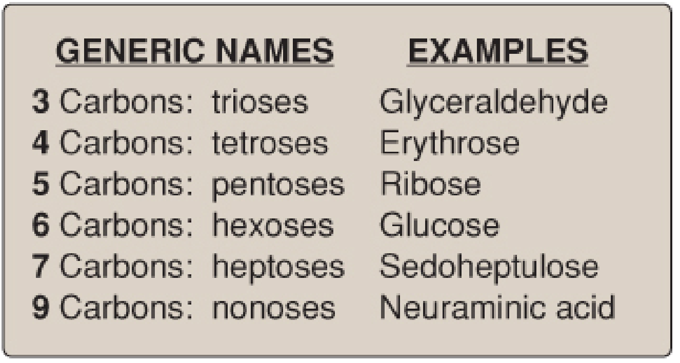
| Carbons | Name | Example |
|---|---|---|
| 3 | Triose | Glyceraldehyde |
| 4 | Tetrose | Erythrose |
| 5 | Pentose | Ribose |
| 6 | Hexose | Glucose |
| 7 | Heptose | Sedoheptulose |
- Biochemistry, 8th ed. Lippincott Illustrated Reviews, p. 256
2. Carbonyl Group: Aldoses vs. Ketoses
The nature of the carbonyl group defines two broad classes:
- Aldoses - contain an aldehyde group (e.g., glyceraldehyde, glucose). The carbonyl is at C-1.
- Ketoses - contain a keto group (e.g., dihydroxyacetone, fructose). The carbonyl is at C-2.
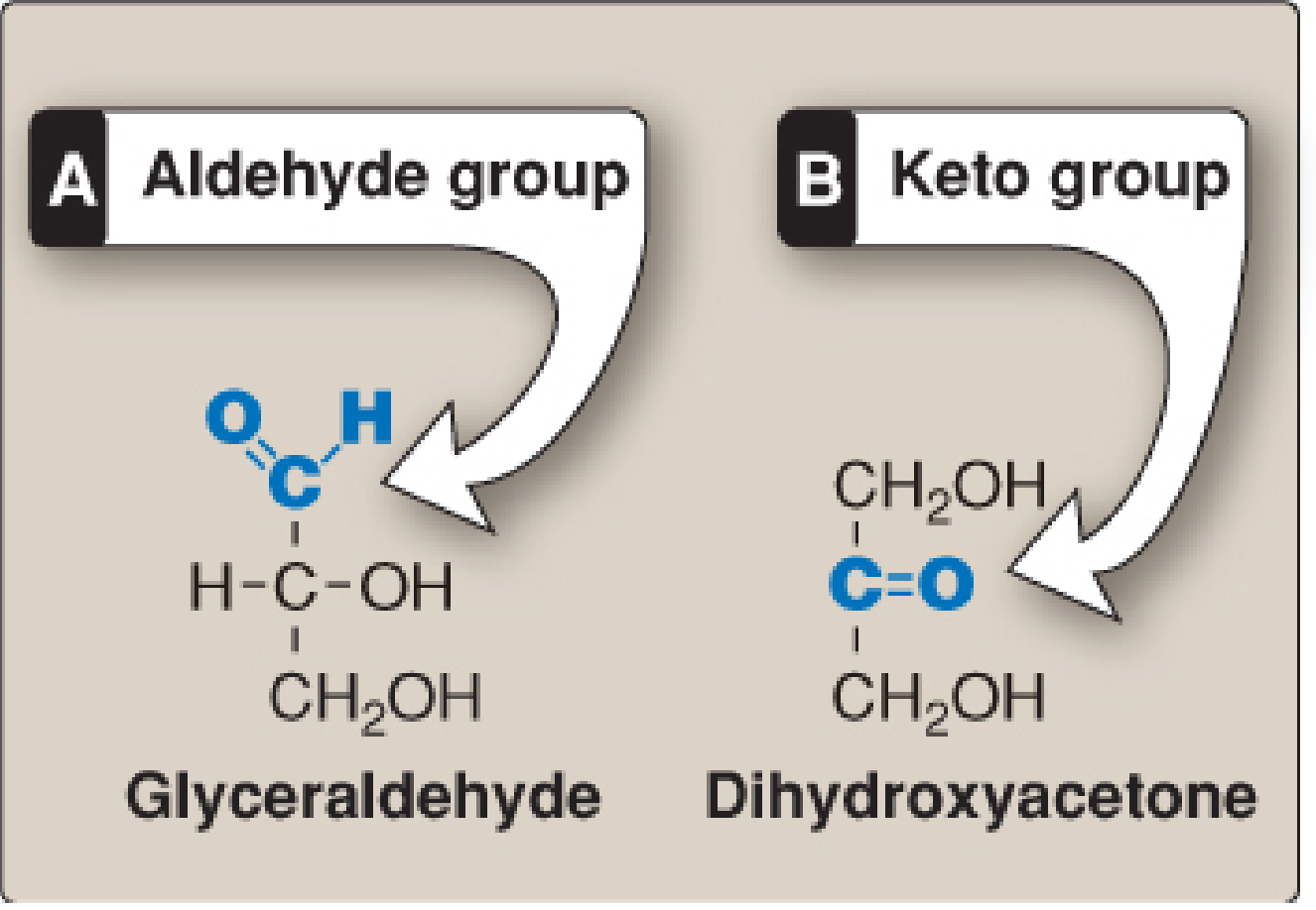
Aldoses are reducing compounds ("reducing sugars") because their free aldehyde group can reduce alkaline copper solutions, forming a brick-red precipitate. This was classically used to detect glucose in urine in poorly controlled diabetes. Fructose, though a ketose, is also a reducing sugar because it can be isomerized to an aldose. - Harper's Illustrated Biochemistry, 32nd Ed., p. 161
3. Stereoisomerism
D- and L-Isomers (Enantiomers)
The configuration of the -OH group on the asymmetric carbon farthest from the carbonyl carbon determines whether a sugar is D or L:
- D-form: -OH is on the right in Fischer projection
- L-form: -OH is on the left
The vast majority of naturally occurring monosaccharides in humans are D-isomers, and metabolic enzymes are generally specific for this configuration. - Lippincott, p. 258; Harper's, p. 160
Optical Activity
Monosaccharides rotate plane-polarized light:
- Dextrorotatory (+): rotates light to the right (e.g., glucose is D(+), also called dextrose)
- Levorotatory (-): rotates light to the left (e.g., fructose is D(-))
The direction of optical rotation is independent of D/L designation - a sugar can be D(-) or L(+). - Harper's, p. 160
Epimers
Epimers are isomers that differ in the configuration of -OH and -H at only one specific carbon (other than the anomeric carbon). Examples:
- Glucose and galactose are C-4 epimers (differ only at carbon 4)
- Glucose and mannose are C-2 epimers (differ only at carbon 2)
- Galactose and mannose differ at two carbons (C-2 and C-4), so they are simply isomers, not epimers
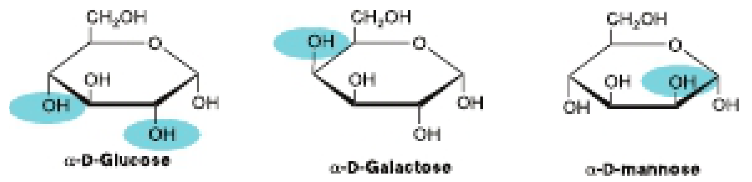
- Lippincott, p. 257; Harper's, p. 161
4. Cyclization (Ring Formation)
In solution, less than 1% of monosaccharides with five or more carbons exist in open-chain form. The aldehyde (or keto) group reacts intramolecularly with a hydroxyl group on the same sugar to form a cyclic hemiacetal (aldose) or hemiketal (ketose):
- Pyranose rings: 6-membered (similar to pyran) - e.g., glucopyranose. For glucose in solution, >99% is in the pyranose form.
- Furanose rings: 5-membered (similar to furan) - common for fructose in some glycosides.
The ring is typically drawn as a Haworth projection (planar) or a chair conformation (more accurate 3D representation). - Harper's, p. 160; Lippincott, p. 258
5. Anomers (α and β Forms)
When the ring closes, the former carbonyl carbon becomes a new chiral center called the anomeric carbon. This creates two new configurations:
- α-anomer: -OH on the anomeric carbon projects to the same side as the ring in Fischer projection; it is trans to the -CH₂OH group in Haworth projection.
- β-anomer: -OH projects to the opposite side.
These two forms are not mirror images - they are called anomers of each other. Crystalline glucose is α-D-glucopyranose. - Lippincott, p. 258; Harper's, p. 161
Mutarotation
In solution, the α and β anomers spontaneously interconvert through the open-chain form - this equilibrium process is called mutarotation. At equilibrium, glucose in solution contains approximately 36% α and 64% β forms. The specific rotation of glucose changes from its initial value until equilibrium is reached. - Harper's, p. 161
6. Reducing Sugar Property
All monosaccharides are reducing sugars because they all possess a free anomeric (hemiacetal or hemiketal) -OH group that can open to regenerate the carbonyl group, which then acts as a reducing agent. This property is the basis for:
- Benedict's test (alkaline copper reduction - turns brick red/orange)
- Fehling's test
- Old clinical urine glucose testing
Note: When two monosaccharides are joined via their anomeric carbons (as in sucrose), neither has a free anomeric -OH, so the disaccharide is non-reducing. - Lippincott, p. 261; Harper's, p. 161
7. Glycoside Formation
The -OH group on the anomeric carbon can react with a hydroxyl group of another compound (saccharide or non-saccharide) to form a glycosidic bond (an acetal linkage), releasing water:
- If the aglycone (second group) is another sugar: O-glycosidic bond forms - this is how disaccharides and polysaccharides are built.
- If the second group is an amine (e.g., adenine): an N-glycosidic bond forms (as in nucleotides like ATP).
Once a glycosidic bond forms, the anomeric carbon is locked and mutarotation no longer occurs at that position. - Harper's, p. 162
8. Important Monosaccharide Derivatives
Monosaccharides can be chemically modified to yield biologically important derivatives:
| Derivative | Modification | Example | Significance |
|---|---|---|---|
| Sugar phosphates | Phosphate esterification | Glucose-6-phosphate | Key metabolic intermediates (glycolysis) |
| Deoxy sugars | -OH replaced by -H | 2-Deoxyribose | DNA backbone |
| Amino sugars | -OH replaced by -NH₂ | Glucosamine, Galactosamine | Glycoproteins, glycosaminoglycans |
| Uronic acids | Terminal -CH₂OH oxidized to -COOH | D-Glucuronate | Glucuronide formation, glycosaminoglycans |
| Sugar alcohols | Carbonyl reduced to -OH | Sorbitol, Ribitol | Metabolic byproducts |
Amino sugars are also found in several antibiotics (e.g., erythromycin), where they are important for antibiotic activity. - Harper's, p. 162
Summary of Key Chemical Properties
| Property | Description |
|---|---|
| Molecular formula | (CH₂O)n, n ≥ 3 |
| Carbonyl type | Aldehyde (aldoses) or ketone (ketoses) |
| Chirality | D- and L-forms; most biological forms are D |
| Optical activity | Dextrorotatory (+) or levorotatory (-) |
| Predominant form in solution | Cyclic (ring) form |
| Ring size | Pyranose (6-membered) or furanose (5-membered) |
| Anomers | α and β forms at anomeric carbon |
| Mutarotation | Interconversion of anomers in solution |
| Reducing property | All are reducing sugars (free anomeric -OH) |
| Glycoside bonds | React via anomeric -OH to form O- or N-glycosidic bonds |
Sources: Biochemistry, 8th ed. Lippincott Illustrated Reviews, pp. 255-265; Harper's Illustrated Biochemistry, 32nd Ed., pp. 159-163
Explore glycosaminoglycans and their amino sugar components
Reading File
This is a broad biochemistry topic. Let me run parallel searches across the library.
Finding Sources
Finding Sources
Reading File
Finding Sources
Reading File
Finding Sources
Reading File
Reading File
Reading File
Reading File
Reading File
Reading File
Reading File
Reading File
Now let me fetch the key diagrams for the answer.
Loading Image
Loading Image
Loading Image
Excellent - I now have comprehensive, well-illustrated content from both Lippincott's and Basic Medical Biochemistry. Here is a thorough answer:
Glycosaminoglycans (GAGs) and Their Amino Sugar Components
1. Overview
Glycosaminoglycans (GAGs) are large, negatively charged heteropolysaccharide chains. They are generally associated with a small amount of protein to form proteoglycans (up to 95% carbohydrate by weight). GAGs were originally called mucopolysaccharides because of their viscous, lubricating properties in mucous secretions.
Their key biological roles include:
- Forming the gel-like ground substance of the extracellular matrix (ECM)
- Binding large amounts of water, acting as a flexible support
- Acting as a molecular sieve, influencing movement of materials through the ECM
- Providing viscous lubrication in synovial fluid and mucous secretions
- Lippincott Biochemistry, 8th ed., p. 457; Basic Medical Biochemistry, 6th ed., p. 1671
2. The Repeating Disaccharide Unit - Core Structure
The defining structural feature of all GAGs is a repeating disaccharide unit composed of two alternating monosaccharide residues:
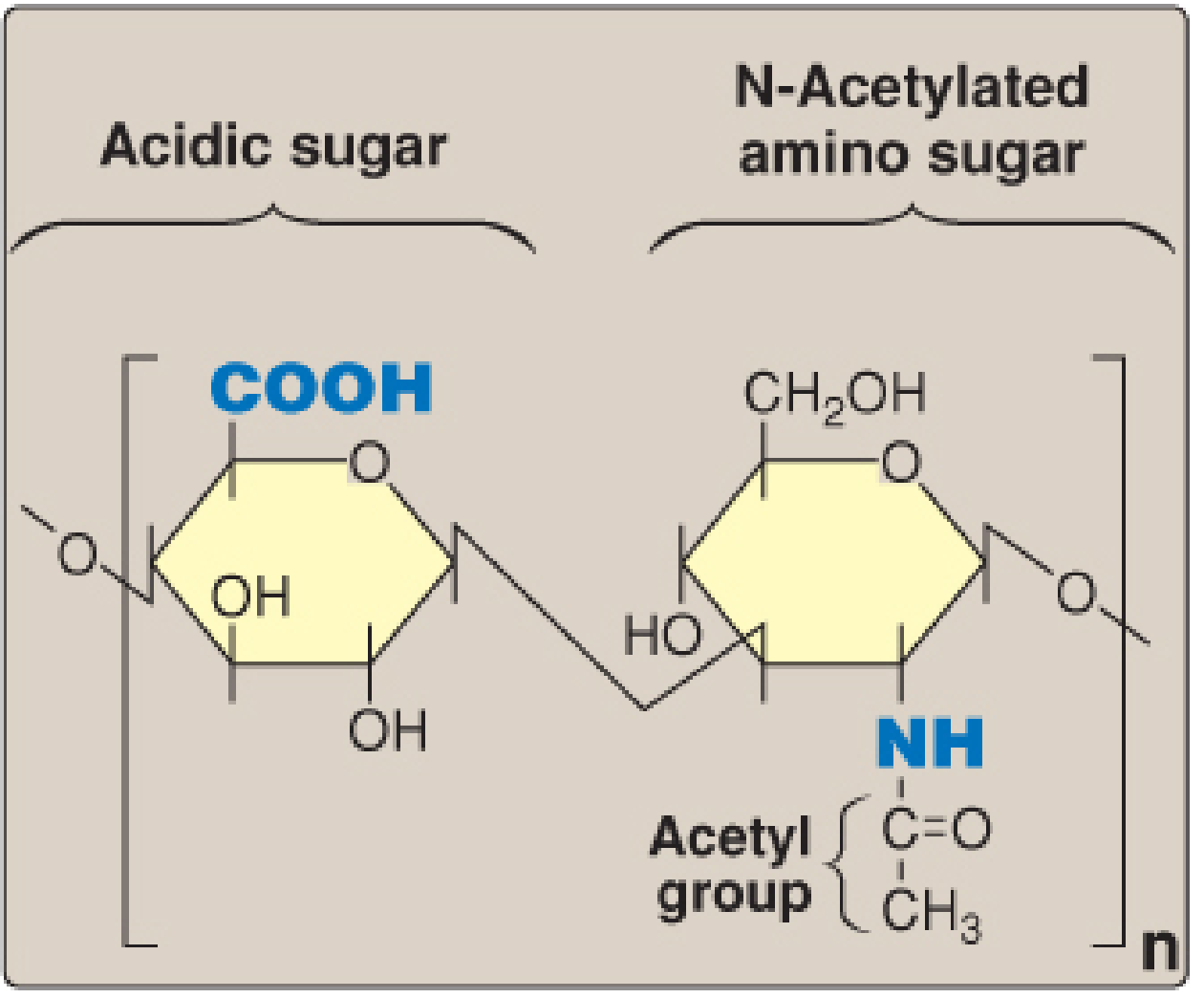
- One sugar = an N-acetylated amino sugar (either GlcNAc or GalNAc)
- Other sugar = an acidic sugar (D-glucuronic acid or L-iduronic acid)
- Exception: Keratan sulfate uses galactose instead of an acidic sugar
- Lippincott, p. 458
3. Amino Sugar Components
What are amino sugars?
Amino sugars (hexosamines) are monosaccharides in which a hydroxyl (-OH) group is replaced by an amino (-NH₂) group. In GAGs, the amino group is almost always N-acetylated, eliminating its positive charge.
The two principal amino sugars in GAGs are:
| Amino Sugar | Also Found In | Notes |
|---|---|---|
| D-Glucosamine (GlcN) | Hyaluronic acid (skeleton), heparin, heparan sulfate | Can be N-acetylated (GlcNAc) or N-sulfonated in heparin |
| D-Galactosamine (GalNAc) | Chondroitin sulfate, dermatan sulfate | Always N-acetylated in these GAGs |
Additional modification - sulfation
The amino sugar may also be sulfated at:
- Carbon 4 (C-4)
- Carbon 6 (C-6)
- On a non-acetylated nitrogen (as in heparin - N-sulfamate linkage)
These sulfate groups, together with the carboxyl groups on the acidic sugars, give GAGs their strongly anionic (negative) character at physiologic pH.
A third amino sugar - N-acetylneuraminic acid (NANA/sialic acid) - is a 9-carbon amino sugar also produced from fructose 6-phosphate. It appears as a terminal residue in glycoproteins and glycolipids, and occasionally in GAGs.
4. Acidic Sugar Components
The acidic sugars that pair with the amino sugars are:
- D-Glucuronic acid (GlcUA) - glucose with C-6 oxidized (-CH₂OH → -COOH)
- L-Iduronic acid (IdUA) - the C-5 epimer of glucuronic acid (produced post-incorporation by uronosyl 5-epimerase)
Glucuronic acid also serves in detoxification (glucuronidation of bilirubin, steroids, drugs) and in the uronic acid pathway.
5. The Six Major Classes of GAGs
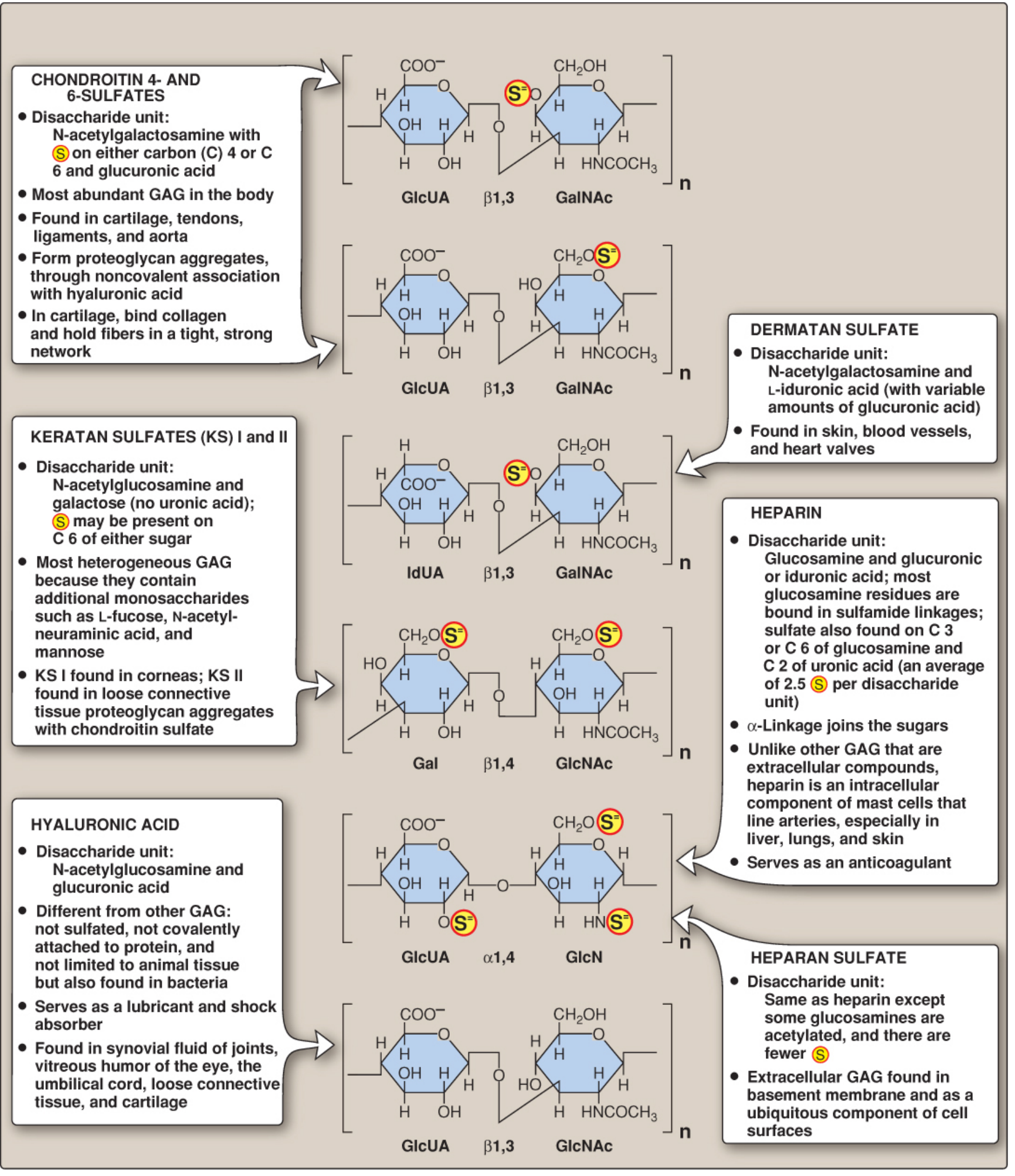
| GAG | Amino Sugar | Acidic/Other Sugar | Sulfated? | Protein-Linked? | Location |
|---|---|---|---|---|---|
| Hyaluronic acid | GlcNAc | GlcUA | No | No (unique) | Synovial fluid, vitreous humor, umbilical cord, loose CT |
| Chondroitin 4- and 6-sulfate | GalNAc | GlcUA | Yes (C-4 or C-6) | Yes | Cartilage, tendon, ligaments, aorta - most abundant GAG |
| Dermatan sulfate | GalNAc | L-IdUA (+ some GlcUA) | Yes | Yes | Skin, blood vessels, heart valves |
| Heparin | GlcN (N-sulfonated) | GlcUA or IdUA | Yes (avg 2.5 per unit) | Yes (intracellular) | Mast cells in liver, lung, skin |
| Heparan sulfate | GlcNAc (some N-sulfonated) | GlcUA | Yes (fewer than heparin) | Yes | Basement membranes, cell surfaces |
| Keratan sulfate I & II | GlcNAc | Galactose (no uronic acid) | Yes (C-6) | Yes (I: Asn; II: Ser/Thr) | KS-I: cornea; KS-II: loose CT with chondroitin sulfate |
- Lippincott, pp. 459-460; Basic Medical Biochemistry, p. 1671
Key features of each:
Hyaluronic acid is the only GAG that is:
- Not sulfated
- Not covalently attached to a protein core
- Found in bacteria as well as animal tissues
- Serves as the backbone of proteoglycan aggregates (many monomers non-covalently bind to one hyaluronate chain via link proteins)
Heparin is unique in being:
- An intracellular compound (stored in mast cell granules)
- The most highly sulfated GAG (avg 2.5 sulfate groups per disaccharide)
- A clinical anticoagulant - binds and activates antithrombin III; also releases lipoprotein lipase from capillary walls
6. Proteoglycans - GAG-Protein Complexes
When GAGs (except hyaluronic acid) are covalently attached to a core protein, the resulting structure is a proteoglycan.
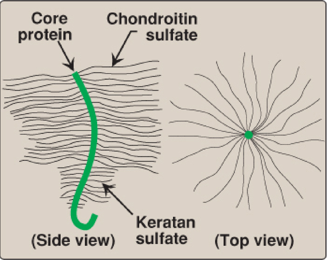
Monomer structure (Bottle-brush model)
A proteoglycan monomer from cartilage has:
- A core protein
- Up to 100 linear GAG chains radiating outward
- Each GAG chain: up to 200 disaccharide units long
- Mutual charge repulsion keeps chains separated
GAG-protein linkage
The most common linkage is an O-glycosidic bond between:
- Xylose (attached to GAG chain) → hydroxyl of serine in core protein
- The full linker: Xylose-Galactose-Galactose (trihexoside) bridging GAG to serine
- Exception: Keratan sulfate I attaches to asparagine (N-linkage)
Aggregate formation
Many proteoglycan monomers associate non-covalently with one long hyaluronic acid backbone to form massive proteoglycan aggregates, stabilized by small link proteins. The aggrecan family (aggrecan, versican, neurocan, brevican) is a major example.
7. Synthesis of GAGs
Synthesis occurs primarily in the Golgi apparatus and follows these steps:
- Amino sugar synthesis: Fructose 6-phosphate → (glutamine amidotransferase, acetylation, isomerization, activation) → UDP-GlcNAc. UDP-GalNAc is formed by epimerization of UDP-GlcNAc.
- Acidic sugar synthesis: UDP-Glucose → (2 NAD+-dependent oxidations) → UDP-Glucuronic acid. L-Iduronic acid is formed by epimerization of glucuronic acid after it has been incorporated into the chain.
- Core protein synthesis: Ribosome → RER → Golgi, where glycosylation occurs.
- Chain elongation: Sequential addition of alternating acidic + amino sugars via specific glycosyltransferases, using their UDP-activated nucleotide sugar donors. The chain is initiated by a xylose-serine O-glycosidic linkage.
- Sulfation: After incorporation, sulfate groups are added from PAPS (3'-phosphoadenosyl-5'-phosphosulfate, "active sulfate") by sulfotransferases. This is the same sulfate donor used in glycosphingolipid synthesis.
8. Degradation of GAGs
GAGs are degraded in lysosomes by acid hydrolases (optimal pH ~5, a protective mechanism against cytoplasmic damage). Half-lives range from minutes to months.
Steps:
- Phagocytosis and delivery to lysosomes
- Endoglycosidases cleave long chains into shorter oligosaccharides
- Exoglycosidases and sulfatases sequentially remove residues from the non-reducing end (last group added is first removed)
9. Clinical Relevance - Mucopolysaccharidoses (MPS)
Deficiencies of individual lysosomal enzymes that degrade GAGs lead to the mucopolysaccharidoses - lysosomal storage diseases in which partially degraded GAGs accumulate, causing multiorgan damage (skeletal deformity, cognitive impairment, hepatosplenomegaly):
| Disease | Enzyme Deficiency | Accumulated GAG |
|---|---|---|
| Hurler / Scheie (MPS I) | α-L-Iduronidase | Heparan sulfate, dermatan sulfate |
| Hunter (MPS II) | Iduronate sulfatase | Heparan sulfate, dermatan sulfate |
| Sanfilippo A (MPS IIIA) | Heparan sulfamidase | Heparan sulfate |
| Sanfilippo B (MPS IIIB) | N-acetylglucosaminidase | Heparan sulfate |
| Morquio A (MPS IVA) | Galactosamine 6-sulfatase | Keratan sulfate |
| Morquio B (MPS IVB) | β-Galactosidase | Keratan sulfate |
| Maroteaux-Lamy (MPS VI) | Arylsulfatase B (N-Ac-GalNAc sulfatase) | Dermatan sulfate |
| Sly (MPS VII / Mucolipidosis VII) | β-Glucuronidase | Heparan/dermatan sulfate |
| Austin disease | All sulfatases (formylglycine defect) | Multiple GAGs |
- Lippincott, pp. 472-473; Basic Medical Biochemistry, p. 1675-1676
Summary Diagram of Key Relationships
GAG Core Structure:
[Acidic sugar — Amino sugar]n
   ↓                ↓
GlcUA or IdUA    GlcNAc or GalNAc
(COOH at C6)     (NH-acetyl or NH-sulfonate)
+ Sulfate groups on C4/C6 of amino sugar, C2 of uronic acid
= Strongly polyanionic chain

↓ Covalently linked to core protein (via Xyl-Gal-Gal-Ser)
= PROTEOGLYCAN MONOMER (bottle brush)

↓ Non-covalently associated with hyaluronic acid via link proteins
= PROTEOGLYCAN AGGREGATE
Sources: Biochemistry, 8th ed. Lippincott Illustrated Reviews, pp. 457-474; Basic Medical Biochemistry - A Clinical Approach, 6th ed., pp. 1671-1676
This is a shared conversation. Sign in to Orris to start your own chat.