I now have all the images and text needed. Let me compile a complete, well-structured answer.
Posterior Compartment of the Arm
1. Muscle of the Posterior Compartment of the Arm
The posterior compartment of the arm contains one primary muscle - the Triceps Brachii - plus the small Anconeus muscle at the elbow.
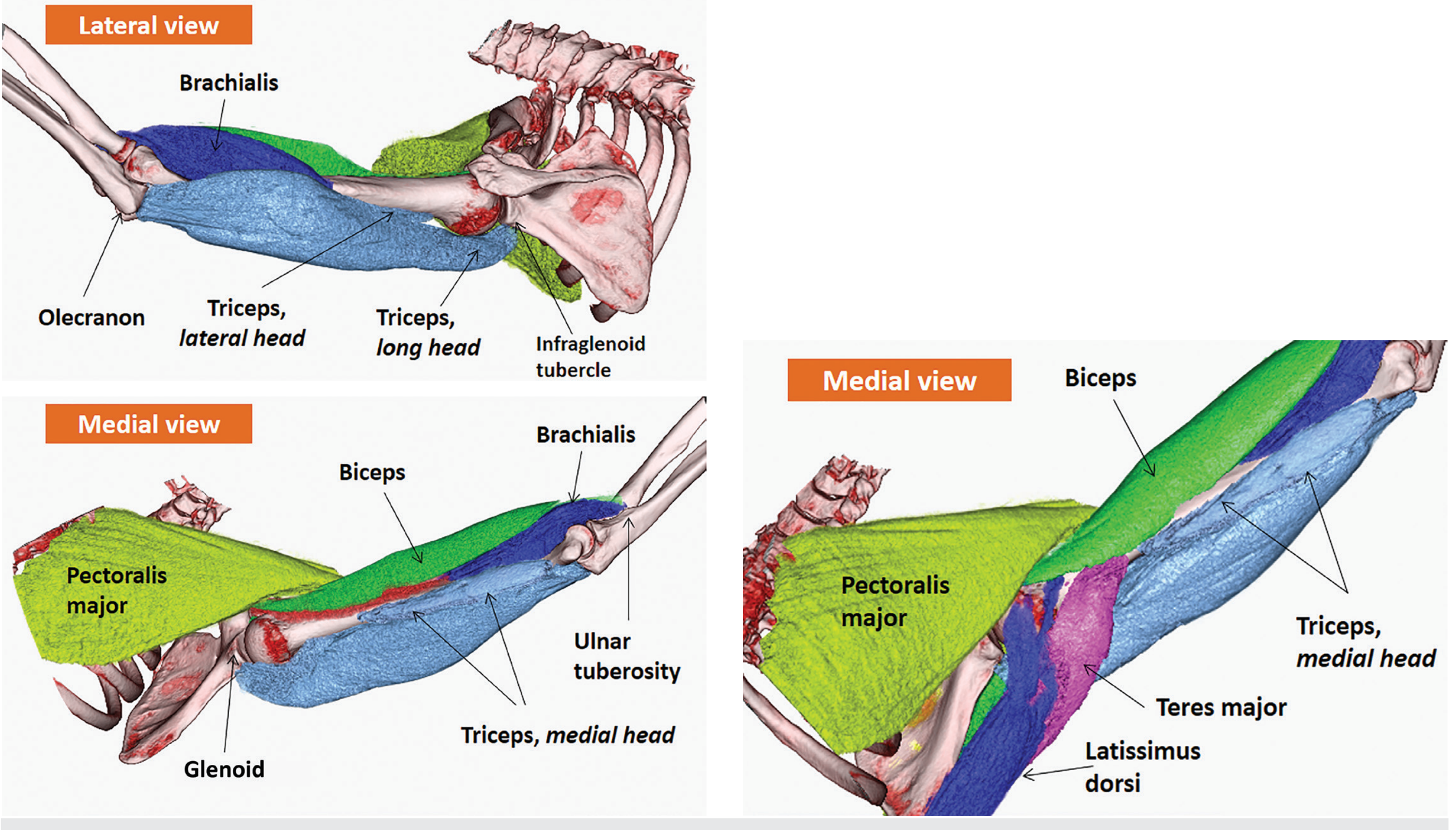
3D Rendered Cadaveric Image - All three heads of Triceps (lateral, medial, and long views):
Fig. 3.16 - Triceps brachii muscle. Top-left (lateral view): Long head arising from infraglenoid tubercle, lateral head from posterior humerus superior to the radial groove, both converging to the olecranon. Bottom-left and right (medial views): Medial head visible deep to long head, with surrounding muscles (biceps, brachialis, pectoralis major, teres major, latissimus dorsi) for orientation.
(Imaging Anatomy Text and Atlas, Vol. 3)
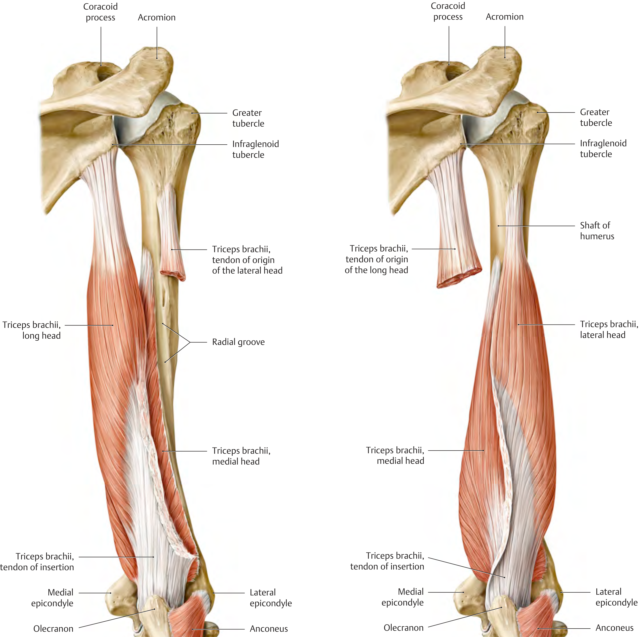
Posterior anatomical diagram showing all three heads and the radial groove:
Fig. B/C - Right arm, posterior view. Left panel: Intact triceps with radial groove clearly labeled between long and medial heads. Right panel: Lateral head partially removed to expose the medial head underneath. Note the tendon of insertion converging to olecranon and the small anconeus muscle laterally.
(THIEME Atlas of Anatomy - General Anatomy and Musculoskeletal System)
2. Origin and Insertion of Triceps Brachii
The triceps brachii is the only muscle of the posterior compartment of the arm. It has three heads, each with a distinct origin.
Origins
| Head | Origin |
|---|
| Long Head | Infraglenoid tubercle of the scapula (below the glenoid) - the only head that crosses the shoulder joint |
| Lateral Head | Posterior surface of the humerus superior (proximal) to the radial groove + lateral intermuscular septum |
| Medial Head | Posterior surface of the humerus inferior (distal) to the radial groove + medial intermuscular septum |
Key relationship: The radial groove is the anatomical landmark that separates the lateral head origin (above) from the medial head origin (below). The long head is the only head that originates from the scapula, making it the only head that acts at the shoulder joint.
Insertion
All three heads converge into a single common tendon that inserts onto the upper (posterior) surface of the olecranon process of the ulna.
- The tendon has a bipartite appearance at insertion:
- The common tendon of the long + lateral heads = superficial and tendinous
- The medial head = deeper and mainly muscular at insertion
3. Nerve Supply of the Three Heads of Triceps
The triceps brachii receives all its motor innervation from the radial nerve (C6, C7, C8), but there are distinct branches to each head:
| Head | Branch from Radial Nerve | Level of Origin |
|---|
| Long head | Branch arising before the radial nerve enters the posterior compartment | Proximal - in the axilla/at triangular interval |
| Medial head | Branch arising before the radial nerve enters the posterior compartment (passes with ulnar nerve) | Proximal - also arises early, passes down medially |
| Lateral head | Branch arising as radial nerve passes through the radial groove | In the posterior compartment |
| Medial head (lower part) | Branch from radial nerve in the radial groove | Distal part of posterior compartment |
Clinical note: Because the branch to the medial head of triceps arises proximal to the radial groove, the medial head is often spared in midshaft humeral fractures (Saturday night palsy), even when the rest of the radial nerve is damaged. This is why elbow extension is preserved in radial nerve injuries at the radial groove.
Major spinal segments: C7 is the dominant root (C6 and C8 contribute as well). The branching pattern may vary between individuals.
4. Actions of Triceps Brachii
| Joint | Action | Head Responsible |
|---|
| Elbow joint | Extension of the forearm (primary action) | All three heads |
| Shoulder joint | Backward movement (extension) of the arm | Long head only |
| Shoulder joint | Adduction of the arm | Long head only |
- The long head also acts as a dynamic stabilizer of the glenohumeral joint, particularly in resisting inferior subluxation
- Triceps is the primary antagonist of the biceps brachii and brachialis
- The medial head is considered the "workhorse" of extension during low-load activities; the long and lateral heads are recruited during high-load extension
5. Radial Groove and Its Contents
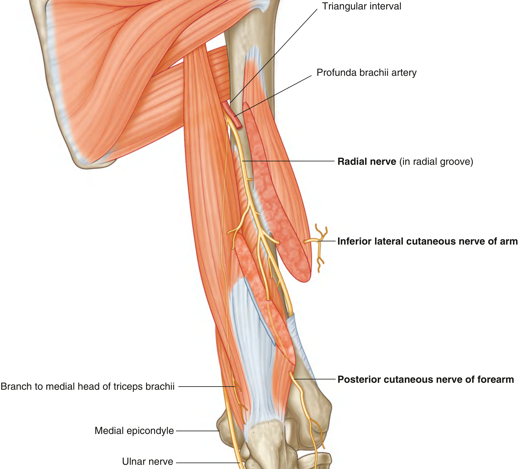
Diagram - Radial nerve in the radial groove with profunda brachii artery:
Fig. 7.69 - Radial Nerve in the Arm, posterior view. (Gray's Anatomy for Students)
The Radial Groove (Spiral Groove / Musculospiral Groove)
- A shallow groove on the posterior surface of the humerus, running obliquely from medial-proximal to lateral-distal
- Located between the origins of the lateral head (above) and medial head (below) of triceps
- The groove represents the path where structures pass diagonally around the back of the humerus
Contents of the Radial Groove
- Radial nerve - lying directly on bone in the groove
- Profunda brachii artery (deep brachial artery) - accompanies the radial nerve throughout
Clinical importance: Both the radial nerve and profunda brachii artery are tightly bound together between the medial and lateral heads of triceps in the radial groove. A midshaft humeral fracture can stretch or transect the radial nerve here, causing wrist drop and loss of sensation over the dorsum of the hand.
6. Branches of the Radial Nerve at the Radial Groove
As the radial nerve passes diagonally through the posterior compartment in the radial groove, it gives off both muscular and cutaneous branches:
Muscular Branches (in the groove)
- Branches to lateral head of triceps brachii
- Branches to medial head of triceps brachii (distal part - some branches arise more proximally before entering the groove)
- Branch to brachioradialis (arises as radial nerve exits the groove anterolaterally)
- Branch to extensor carpi radialis longus (arises as nerve exits the groove)
- Small contribution to brachialis (lateral part - C7 fibers)
Cutaneous Branches (arising in the posterior compartment)
- Inferior lateral cutaneous nerve of the arm - supplies skin on the lateral arm distally
- Posterior cutaneous nerve of the forearm - supplies skin on the posterior forearm
Both cutaneous branches pierce through the lateral head of triceps and the overlying deep fascia to become subcutaneous.
Note: Before entering the groove, the radial nerve gives off branches to the long head and medial head (proximal) of triceps, plus the posterior cutaneous nerve of the arm - all from the axilla/triangular interval level.
7. Profunda Brachii Artery
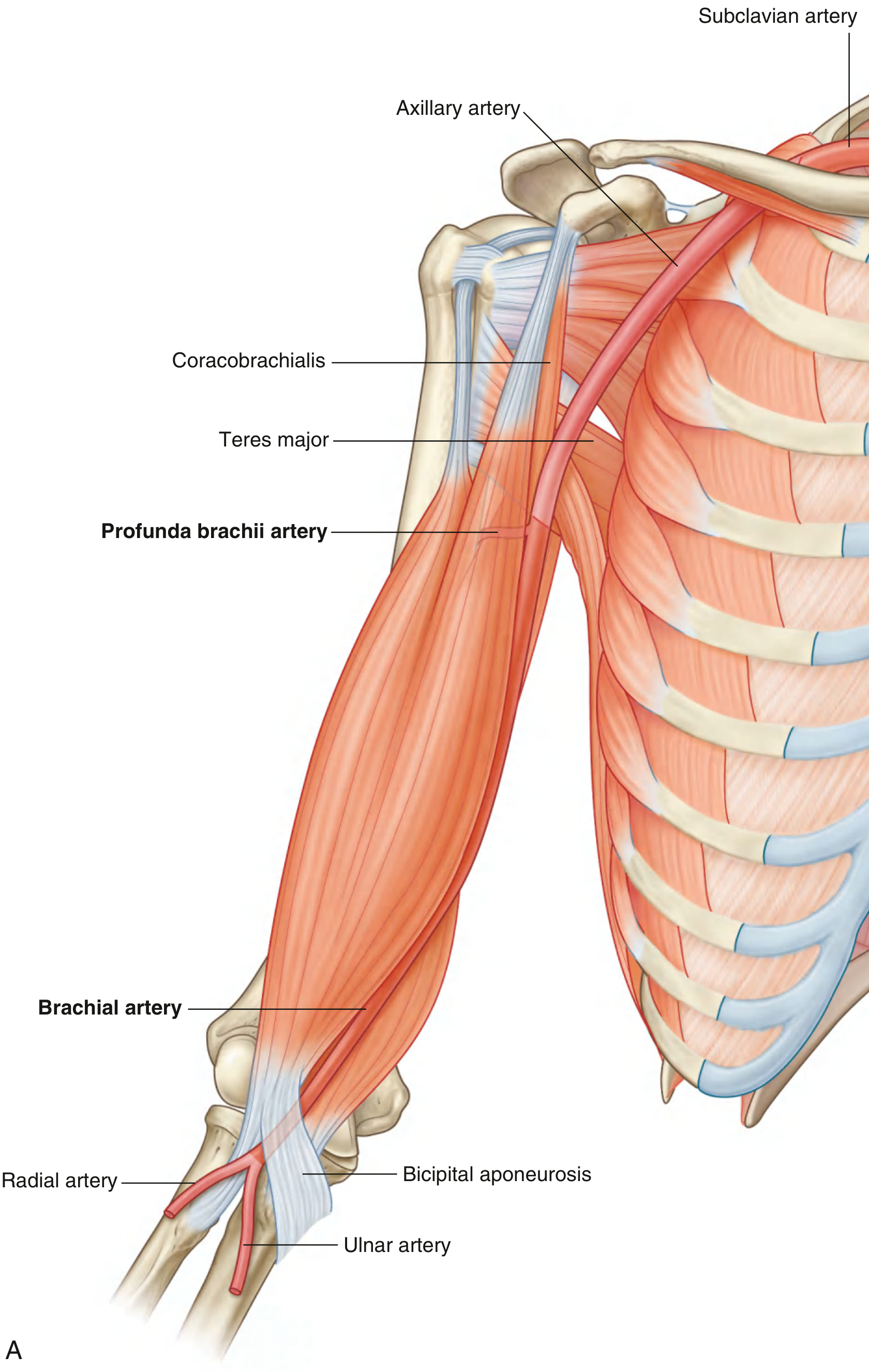
Brachial artery and profunda brachii - anterior contextual view:
Fig. 7.66A - Brachial Artery in context, showing profunda brachii artery as the first major branch. (Gray's Anatomy for Students)
Key Facts about the Profunda Brachii Artery
| Feature | Detail |
|---|
| Origin | Largest branch of the brachial artery, arising at the level of the tendon of teres major (in 55-70% as a single trunk) |
| Entry to posterior compartment | Passes with the radial nerve through the triangular interval (bounded by: shaft of humerus, inferior margin of teres major, lateral margin of long head of triceps) |
| Course | Travels along the radial groove on the posterior surface of the humerus, deep to the lateral head of triceps |
| Branches | Muscular branches to triceps + anastomoses with posterior circumflex humeral artery |
| Terminal branches | Divides into: middle collateral artery and radial collateral artery |
| Anastomosis | Radial collateral artery anastomoses with the radial recurrent artery (branch of radial artery) around the elbow - contributes to the periarticular anastomosis |
The profunda brachii artery is the main blood supply to the posterior compartment of the arm and is the vascular companion of the radial nerve throughout the radial groove. Both structures are vulnerable together in midshaft humeral fractures.
Sources:
- Gray's Anatomy for Students, Figs. 7.66, 7.69, Table 7.9
- Imaging Anatomy Text and Atlas, Vol. 3 (Figs. 3.16, 3.17)
- THIEME Atlas of Anatomy - General Anatomy and Musculoskeletal System