
comprehensively explain about tremors and their types
tremor types classification pathophysiology clinical features 2024
tremor classification types management
essential tremor pathophysiology treatment
Tremors: A Comprehensive Overview
Definition
- Adams and Victor's Principles of Neurology, 12th Edition
Classification Framework
- Activation conditions - when the tremor appears (rest vs. action)
- Etiology/Syndrome - the underlying cause
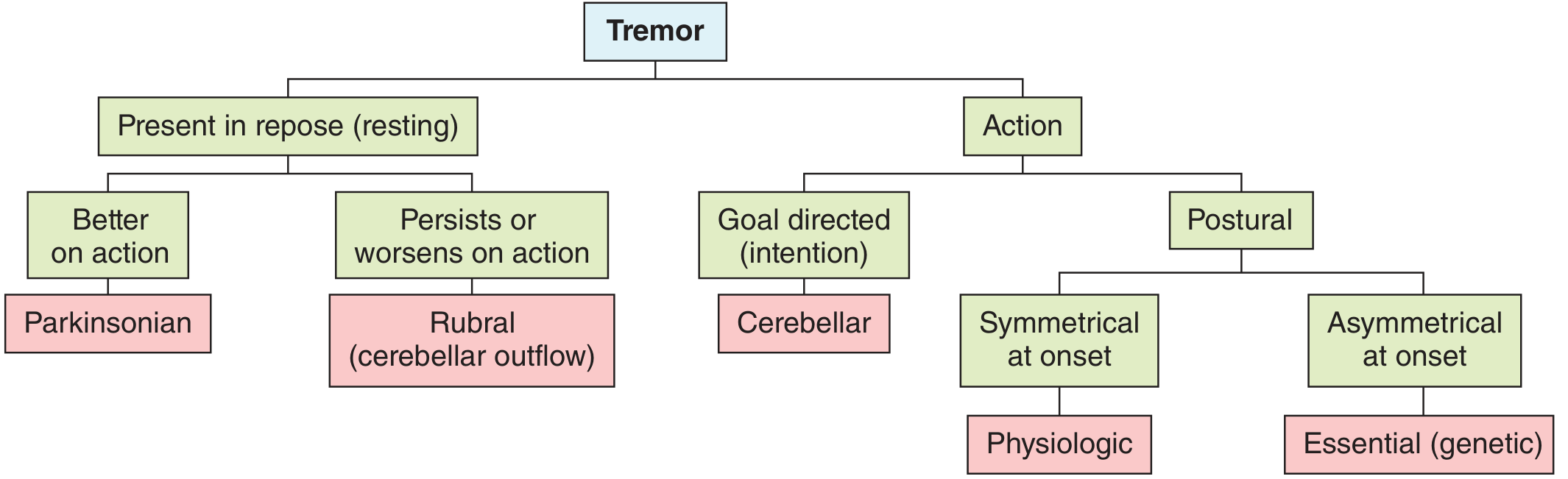
Major Types of Tremor
Summary Table (from Adams and Victor's)
| Type | Frequency (Hz) | Location | Enhanced by | Attenuated by |
|---|---|---|---|---|
| Physiologic (enhanced) | 8-13 | Hands | Epinephrine, beta-adrenergic | Alcohol, beta-blockers |
| Parkinsonian (rest) | 3-5 | Hands, forearms, fingers, feet, lips, tongue | Emotional stress | L-dopa, anticholinergics |
| Cerebellar (intention/rubral) | 2-4 | Limbs, trunk, head | Emotional stress | - |
| Postural/action | 5-8 | Hands | Anxiety, fright, alcohol withdrawal, xanthines, exercise | Beta-blockers (some cases) |
| Essential (familial/senile) | 4-8 | Hands, head, vocal cords | Anxiety, exercise, fatigue | Alcohol, propranolol, primidone |
| Orthostatic | 14-16 | Legs | Quiet standing | Repose, walking, clonazepam |
| Neuropathic | 4-7 | Hands | - | - |
| Palatal | 1-2 (60-100/min) | Palate, sometimes face, pharynx | - | Clonazepam, valproate |
| Dystonic | Irregular | Concordant with focal dystonia | - | Botulinum toxin |
1. Physiologic Tremor
- Frequency: 8-13 Hz in the hands; as slow as 6.5 Hz elsewhere
- Mechanism: Driven peripherally by mechanical-reflex oscillations and centrally by synchronized motor neuron discharges
- Not pathologic in itself
Enhanced (Exaggerated) Physiologic Tremor
- Physiologic states: Anxiety, fear, fright, exercise, fatigue
- Metabolic: Hyperthyroidism, hypoglycemia, hypercortisolism, pheochromocytoma, hypothermia
- Drugs and toxins: Lithium, nicotinic acid, xanthines (caffeine, aminophylline), cocaine, methamphetamine, corticosteroids, valproate, SSRIs, bronchodilators, cyclosporine, tacrolimus
- Withdrawal: Alcohol, benzodiazepines, barbiturates
- Mechanism: Enhancement in metabolic/toxic states is primarily due to stimulation of muscular beta-adrenergic receptors by elevated circulating catecholamines - NOT a CNS-mediated effect
- Treatment: Remove the cause; beta-adrenergic antagonists (propranolol)
2. Resting Tremor (Parkinsonian Tremor)
- Frequency: 3-5 Hz (some sources state 5-7 Hz)
- Location: Hands and forearms (most common), fingers, feet, lips, tongue, chin - notably not the head (head tremor favors essential tremor)
- Classic appearance: "Pill-rolling" tremor - appears as if the patient is rolling something between the thumb and other fingers
- Pattern: Asymmetrical at onset
- Enhanced by: Emotional stress, mental distraction (e.g., asking the patient to perform mental arithmetic while hands rest)
- Decreased by: Voluntary movement
3. Action Tremors
a) Postural Tremor
- Occurs when a body part is maintained against gravity (e.g., arms held outstretched)
- Absent at rest; appears with muscle activation
- Best examples: Essential tremor, enhanced physiologic tremor
b) Kinetic Tremor
- Occurs during voluntary movement (not specifically goal-directed)
- Simple kinetic tremor: during any movement
- Task-specific tremor: only during particular tasks (e.g., writing, speaking)
- Primary writing tremor - a task-specific tremor that only occurs when writing or adopting a writing posture; no tremor in other conditions; frequency ~5-7 Hz
c) Intention Tremor (Goal-directed kinetic tremor)
- Tremor that increases in amplitude as the limb approaches a target
- Classic sign of cerebellar disease (appendicular cerebellar ataxia)
- Demonstrated by: Finger-nose-finger test, heel-shin test
- Also accompanied by: Dysmetria, dysdiadochokinesia, dysarthria, wide-based gait
- Frequency: 2-4 Hz (lower frequency, coarser)
- Pathophysiology: Loss of cerebellar modulation of motor output; the lateral cerebellar hemispheres and dentato-rubro-thalamo-cortical pathway are disrupted
d) Isometric Tremor
- Occurs during sustained muscle contraction without movement (e.g., squeezing a rigid object)
4. Essential Tremor
- Frequency: 4-8 Hz (usually lower end of range), lower than physiologic tremor
- Location: Hands/arms (most common), head (titubation), vocal cords ("shaky voice"), tongue, lips; legs and trunk less often
- Pattern: Bilateral but may be asymmetrical
- Classic trigger: Posture and movement (postural + kinetic); absent at rest early on
- Worsened by: Anxiety, emotion, exercise, fatigue, caffeine
- Characteristic: Improves transiently with alcohol (a key differentiator from Parkinsonian tremor)
- Genetics: Autosomal dominant with high penetrance; previously called familial, benign, or senile tremor
- Functional impact: Can progress to illegible handwriting, inability to bring utensils or a glass to the mouth without spillage
- First-line: Propranolol (beta-blocker) or primidone (antiepileptic)
- Second-line: Topiramate, gabapentin, clonazepam
- Alcohol (temporary, not a clinical option due to risk)
- Surgical: Thalamotomy (lesion of Vim) or DBS of the Vim nucleus - highly effective
- Focused ultrasound thalamotomy (newer option)
5. Cerebellar Tremor
- Intention tremor is the hallmark (see above), but cerebellar lesions also cause:
- Postural tremor at rest with limbs held in space (trunk and head titubation - associated with vermis lesions)
- Frequency: 2-4 Hz, coarse, low-frequency
- Associated signs: Dysmetria, dysarthria, nystagmus, ataxic gait, hypotonia
- Causes: Multiple sclerosis, stroke, tumors, alcohol-related cerebellar degeneration, spinocerebellar ataxias
6. Holmes Tremor (Rubral Tremor / Midbrain Tremor)
- A combination of rest + postural + intention tremor - the only tremor type with all three components
- Frequency: 2-4 Hz, low-frequency, often violent
- Usually unilateral
- Low amplitude at rest, but becomes coarse and violent as the limb is raised slightly or when movement is attempted
- Mechanism: Lesion involving the superior cerebellar peduncle (dentato-rubro-thalamic tract) near the red nucleus (midbrain)
- Causes: Multiple sclerosis, brainstem infarcts (midbrain stroke), trauma
- Can resemble the wing-beating tremor of Wilson's disease
- Neuroanatomy through Clinical Cases, 3rd Edition
7. Orthostatic Tremor
- A distinctive high-frequency (14-16 Hz) tremor, the fastest of all tremor types
- Occurs exclusively in the legs shortly after standing still; disappears when sitting, lying down, or walking
- Patients feel unsteady and have a tendency to fall if stationary
- Difficult to see visually; best diagnosed by palpating the thigh muscles while the patient stands or by EMG showing characteristic ~16 Hz bursts
- Treatment: Clonazepam, valproate, gabapentin
- Adams and Victor's Principles of Neurology
8. Palatal Tremor (formerly Palatal Myoclonus)
- Rhythmic movement of the soft palate at 0.5-5 Hz (60-100/min)
- Can extend to the face, pharynx, and proximal upper extremities
- Famous for persisting during sleep - a distinguishing feature from most other tremors
- May cause ear clicking due to Eustachian tube movement
- Two forms:
- Symptomatic: Due to lesions of the central tegmental tract or dentate-red nucleus-inferior olive circuit (infarcts, MS, trauma); associated with olivary hypertrophy on MRI
- Essential palatal tremor: No known cause; accompanied by ear clicking
- Treatment: Botulinum toxin injections into the tensor or levator veli palatini
- Neuroanatomy through Clinical Cases, 3rd Edition
9. Neuropathic Tremor
- Action tremor (relatively symmetric) associated with peripheral neuropathies
- Especially linked to chronic inflammatory demyelinating polyneuropathy (CIDP) and IgM paraproteinemic neuropathy
- Frequency: 4-7 Hz, involves hands
- Associated with positive Romberg sign and other neuropathy features
- Mechanism: Disruption of peripheral sensory feedback loops that normally damp tremor
10. Dystonic Tremor
- Tremor occurring in a body part affected by dystonia
- Irregular, jerky rhythm (not smooth sinusoidal oscillation)
- Location is concordant with the focal dystonia (e.g., cervical dystonic tremor with torticollis)
- May appear as head tremor, hand tremor, or voice tremor in the context of dystonia
- Treatment: Botulinum toxin injections; geste antagoniste (sensory trick) may temporarily suppress it
11. Drug-Induced Tremors
- Neuroleptics/antipsychotics - parkinsonian rest tremor
- Lithium - postural/action tremor, toxic doses → coarse tremor
- Valproate - action tremor (dose-dependent)
- Beta-adrenergic agonists (salbutamol, terbutaline) - enhanced physiologic
- SSRIs, tricyclics - enhanced physiologic
- Corticosteroids - enhanced physiologic
- Calcium channel blockers, amiodarone
- Immunosuppressants: cyclosporine, tacrolimus - can cause severe tremor
- Alcohol withdrawal - coarse postural/action tremor
- Management: Dose reduction or withdrawal of offending agent; beta-blockers if needed
12. Functional (Psychogenic) Tremor
- Abrupt onset, static course, spontaneous remissions
- Variable frequency (more specific than variable amplitude alone)
- Entrainment sign: When asked to copy a rhythmic tapping with the unaffected hand at a different frequency - the affected limb entrains to the same rhythm, stops, or the patient cannot copy the movement
- Ballistic movement test: Brief stopping of tremor during fast ballistic movements with the other hand
- Distractibility: Tremor diminishes with mental distraction (mental arithmetic) - opposite to organic tremor
- Loading sign: Adding wrist weights worsens functional tremor (organic tremor improves with loading)
- Coactivation sign: Simultaneous agonist-antagonist activation, resembling voluntary shivering
- Coherence: If bilateral, both limbs tremor at the exact same frequency (organic tremors have slightly different frequencies in different limbs)
- Unresponsive to antitremor drugs; responds to psychotherapy/physiotherapy
- Normal dopamine transporter (DaT) scan
- Bradley and Daroff's Neurology in Clinical Practice
Pathophysiology: Common Neural Circuits
- Are arranged somatotopically
- Respond to kinesthetic inputs from involved muscles/joints
- Project to premotor cortex via the ventrolateral thalamo-cortical pathway
- Ultimately drive tremor via the lateral corticospinal tract
Differential Diagnosis: Key Distinguishing Features
| Feature | Parkinsonian | Essential | Cerebellar (Intention) | Functional |
|---|---|---|---|---|
| Present at rest | Yes | No (mild later) | No | Variable |
| Worsens with movement | No (improves) | Yes (postural) | Yes (goal-directed) | Variable |
| Frequency | 3-5 Hz | 4-8 Hz | 2-4 Hz | Variable |
| Symmetry | Asymmetric | Bilateral | Unilateral | Variable |
| Head tremor | No | Yes | Yes (titubation) | Yes |
| Alcohol response | No effect | Improves | No effect | Variable |
| Associated signs | Bradykinesia, rigidity | Usually none | Dysmetria, ataxia | Distractible |
Treatment Overview
| Tremor Type | First-Line Treatment | Second-Line / Surgical |
|---|---|---|
| Essential | Propranolol, primidone | Topiramate, DBS (Vim), focused ultrasound |
| Parkinsonian | Levodopa, dopamine agonists | DBS (STN or GPi), anticholinergics |
| Cerebellar/intention | Clonazepam (limited) | Weights on limbs; DBS (less effective) |
| Orthostatic | Clonazepam, gabapentin | Valproate |
| Palatal | Botulinum toxin | Clonazepam, valproate |
| Dystonic | Botulinum toxin | Anticholinergics |
| Enhanced physiologic | Remove cause; propranolol | - |
| Functional | Physiotherapy, CBT | No antitremor drugs |
Recent Evidence Note
- Adams and Victor's Principles of Neurology, 12th Edition - Chapter 4: Tremor, Myoclonus, Focal Dystonias
- Neuroanatomy through Clinical Cases, 3rd Edition - Chapter 16: Movement Disorders
- Bradley and Daroff's Neurology in Clinical Practice - Functional/Psychogenic Movement Disorders
- MDS Task Force Classification of Tremor (Frontiers in Neurology, 2021)
- RACGP Tremor Review 2024
talk comprehensively about brain hemorrhagges
brain hemorrhage types classification management guidelines 2024 2025
intracerebral hemorrhage management outcome
Overview and Classification
| Type | Location of Bleeding |
|---|---|
| Epidural hematoma (EDH) | Between skull and outer dura mater |
| Subdural hematoma (SDH) | Between dura and arachnoid membrane |
| Subarachnoid hemorrhage (SAH) | Within the subarachnoid space (CSF space) |
| Intracerebral hemorrhage (ICH) | Within brain parenchyma itself |
| Intraventricular hemorrhage (IVH) | Within the brain ventricles |
- Primary (hypertensive) intracerebral hemorrhage
- Ruptured saccular aneurysm
- Ruptured arteriovenous malformation (AVM)
- Cavernous angioma
- Trauma
- Hemorrhagic disorders: leukemia, thrombocytopenia, anticoagulants, thrombolytics, hemophilia
- Hemorrhage into brain tumors (primary or metastatic)
- Septic embolism, mycotic aneurysm
- Hemorrhagic infarction
- Cerebrovascular amyloidosis (CAA)
- Pituitary apoplexy
- Reversible cerebral vasoconstriction syndrome (RCVS)
- Cocaine, vasopressor drugs, PRES, moyamoya
1. Epidural Hematoma (EDH)
Anatomy and Pathophysiology
- Because the bleeding is arterial, it does not self-tamponade and blood rapidly accumulates
- Can progress from headache → herniation within hours
- Less common cause: venous epidural hematoma (from torn dural venous sinuses) - slower course
- Rare causes: skull metastases (especially hepatocellular carcinoma), eosinophilic granuloma, craniofacial infections
Classic Clinical Presentation
- Initial impact → brief loss of consciousness (due to concussion)
- Lucid interval → patient regains consciousness, appears relatively normal, complains only of headache
- Deterioration → progressive decline in consciousness, ipsilateral pupil dilation (CN III compression from uncal herniation), contralateral hemiplegia
- Herniation → coma, bilateral pupil changes, decerebrate posturing, death if untreated
Signs of associated basal skull fracture:
- Battle's sign - ecchymosis behind the ear (mastoid)
- Raccoon eyes - periorbital ecchymosis
- Blood behind tympanic membrane (hemotympanum)
Imaging
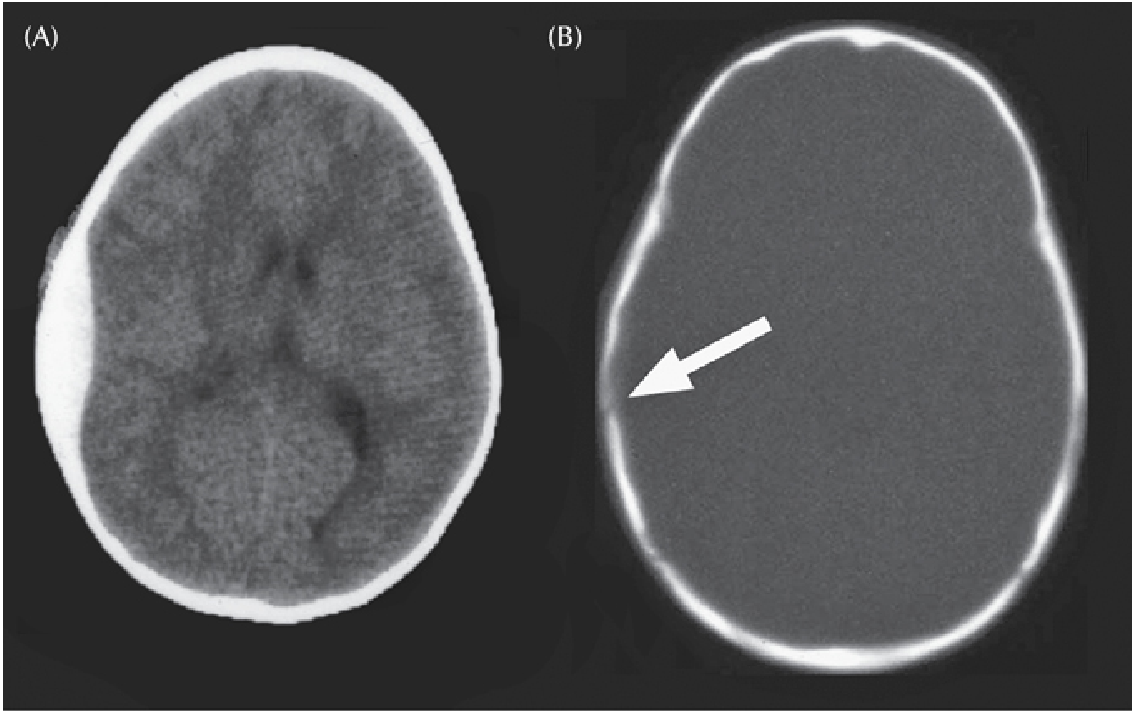
- CT: Biconvex (lens-shaped) hyperdense collection between skull and brain; does NOT cross suture lines (dura attached at sutures limits spread)
- Does not cross the midline falx
Treatment
- Surgical evacuation (craniotomy and hematoma drainage) - indicated for significant EDH with neurological deficit or large volume
- Small EDH in neurologically intact patients can be managed conservatively with close monitoring
- Outcome is excellent if treated before herniation develops
2. Subdural Hematoma (SDH)
Acute Subdural Hematoma
- Blood accumulates faster than the brain can accommodate → severe mass effect and herniation
- Frequently associated with underlying brain injury (contusion, diffuse axonal injury)
- Patients often present in coma
- Anticoagulant use dramatically increases risk - must be reversed promptly
- Rarely: aneurysm rupture into subdural space (no subarachnoid blood)
Chronic Subdural Hematoma
- Headache, often dull and fluctuating
- Cognitive decline, memory impairment
- Fluctuating level of consciousness (characteristically fluctuates)
- Hemiparesis, dysphasia
- Can mimic dementia or TIA in elderly patients
- Bilateral in ~20% of cases
- Acute SDH: Hyperdense (white) crescent-shaped collection on CT, following the brain surface
- Chronic SDH: Hypodense (dark) or isodense collection on CT (isodense to brain makes it tricky to spot)
- Follows a crescent shape along the inner surface; unlike EDH, can cross suture lines and may be bilateral
- Symptomatic SDH: Burr hole drainage or craniotomy
- Small asymptomatic chronic SDH may resorb spontaneously
- Correct anticoagulation before any procedure
3. Subarachnoid Hemorrhage (SAH)
Etiology
- Aneurysmal SAH (80%): Rupture of a saccular (berry) aneurysm - the most dangerous cause
- Perimesencephalic (non-aneurysmal) SAH (~10%): Venous or unknown origin; blood limited to cisterns around the midbrain; more benign course
- Other (~10%): AVM, trauma, vasculitis, cocaine, tumors, RCVS
Saccular Aneurysms
- Size: typically average 7.5 mm; those that rupture are usually ≥10 mm
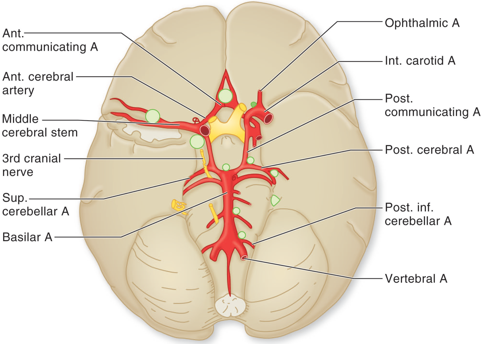
- Location: Typically at vessel bifurcations (see diagram below)
- Rupture site: the dome of the aneurysm
- Multiple aneurysms in 20% of patients
- Incidence of unruptured aneurysms at autopsy: ~2%
- Autosomal dominant polycystic kidney disease (ADPKD)
- Ehlers-Danlos syndrome
- Fibromuscular dysplasia
- Coarctation of the aorta
- Moyamoya disease
Clinical Presentation
- Loss of consciousness at onset in ~40% (from acute massive rise in ICP and transient cessation of cerebral blood flow)
- Nausea and vomiting
- Meningismus - neck stiffness (from blood irritating meninges; develops over hours)
- Photophobia
- Sentinel headache: A severe headache 1-4 weeks before the major bleed (warning leak) in ~30-40% of patients - critical to recognize
- Subhyaloid/preretinal hemorrhages on fundoscopy (Terson syndrome)
- Third nerve palsy (posterior communicating artery aneurysm compressing CN III) - presents as ipsilateral ptosis, mydriasis, and "down and out" eye
Hunt and Hess Grading Scale (SAH severity)
| Grade | Clinical Features |
|---|---|
| I | Asymptomatic or minimal headache, slight nuchal rigidity |
| II | Moderate to severe headache, nuchal rigidity, no neuro deficit except cranial nerve palsy |
| III | Drowsiness, confusion, mild focal deficit |
| IV | Stupor, moderate to severe hemiparesis, possible decerebrate rigidity |
| V | Deep coma, decerebrate rigidity, moribund |
Complications of SAH
- Rebleeding: Greatest risk in the first 24-48 hours (20-30% within the first day if untreated); peak 7-10 days. The major reason why aneurysms must be secured urgently.
- Vasospasm / Delayed Cerebral Ischemia (DCI): Begins 3-4 days after SAH, peaks at day 7-10, lasts ~3 weeks. Caused by breakdown products of subarachnoid blood inducing smooth muscle contraction in adjacent arteries. Leads to ischemic stroke and worsening neurological deficits.
- Hydrocephalus:
- Acute: Within hours-days; blood obstructs arachnoid granulations and ventricular outflow
- Chronic: Months after SAH; communicating hydrocephalus from impaired CSF absorption
- Hyponatremia: From SIADH or cerebral salt wasting
- Cardiac complications: ECG changes (ST elevation, T-wave inversion, prolonged QT), neurogenic stunned myocardium, Takotsubo cardiomyopathy
- Seizures: Early (~5-10%) and late (long-term epilepsy risk)
Diagnosis
- Non-contrast CT head: Detects blood in >90% if done within 24 hours; blood appears as high-density (white) in the basal cisterns, Sylvian fissures, and sulci
- CT angiography (CTA): First-line for identifying aneurysm location and morphology
- Lumbar puncture: If CT negative but SAH clinically suspected (done ≥12 hours after symptom onset to allow xanthochromia to develop); xanthochromia (yellow discoloration of CSF from oxyhemoglobin/bilirubin) is diagnostic
- Digital subtraction angiography (DSA): Gold standard for aneurysm characterization before treatment
- Fisher Scale: Grades thickness/distribution of subarachnoid blood on CT; predicts risk of vasospasm
Treatment
- Endovascular coiling (most common): Catheter-based insertion of platinum microcoils into the aneurysm sac to induce thrombosis; preferred for most aneurysms
- Surgical clipping: Open craniotomy with clip placed at the aneurysm neck; preferred for some complex aneurysms or those with an associated intracerebral hematoma requiring evacuation
- Nimodipine (oral, 60 mg every 4 h × 21 days): Calcium channel blocker - standard of care for preventing DCI; reduces neurological deficits and improves outcomes
- Blood pressure control: Before aneurysm is secured, target SBP <160 mmHg to prevent rebleeding while maintaining cerebral perfusion
- Euvolemia: Avoid dehydration; triple-H therapy (hypertension, hypervolemia, hemodilution) for established vasospasm
- Angioplasty/intra-arterial vasodilators for refractory vasospasm
- Ventriculostomy (EVD) for acute hydrocephalus
4. Intracerebral Hemorrhage (ICH) / Primary Brain Parenchymal Hemorrhage
Causes: Primary vs. Secondary
- Hypertension (~50%): Chronic hypertension causes Charcot-Bouchard microaneurysms on small penetrating arteries (particularly lenticulostriate, thalamoperforating, pontine perforators, and cerebellar perforators). These arteries rupture, especially during acute blood pressure elevations
- Cerebral amyloid angiopathy (CAA): Deposition of beta-amyloid in walls of cortical and leptomeningeal arteries - increasingly important as the population ages; causes lobar hemorrhages (cortical/subcortical), often multiple and recurrent; associated with ApoE2 and E4 alleles
- AVMs, cavernous malformations
- Anticoagulant therapy (warfarin, DOACs) - causes hemorrhages in atypical locations
- Thrombolytics (tPA)
- Hemorrhage into tumor
- Venous sinus thrombosis → hemorrhagic infarction
- Mycotic aneurysm
- Cocaine and amphetamines
Location of Hemorrhage
- Putamen and adjacent internal capsule (~50%) - the most common site; hypertensive
- Lobar (temporal, parietal, frontal white matter) - not strictly hypertensive; often CAA, AVM, tumor
- Thalamus
- Cerebellum
- Pons (brainstem)
Pathological Evolution of the Hematoma
| Phase | Time | Hemoglobin Form | T1 | T2 |
|---|---|---|---|---|
| Hyperacute | <24h | Oxyhemoglobin (intracellular) | Iso/hypointense | Hyperintense |
| Acute | 1-3 days | Deoxyhemoglobin (intracellular) | Iso/hypointense | Hypointense |
| Early subacute | >3 days | Methemoglobin (intracellular) | Hyperintense | Hypointense |
| Late subacute | >7 days | Methemoglobin (extracellular) | Hyperintense | Hyperintense |
| Chronic | >14 days | Hemosiderin (extracellular) | Iso/hypointense | Hypointense ("blooming") |
Clinical Syndromes by Location
- Contralateral hemiplegia (corticospinal tract involvement)
- Contralateral hemisensory loss
- Homonymous hemianopia
- Eyes deviate toward the side of the lesion
- If large: progressive obtundation, coma from herniation
- Contralateral sensory loss (all modalities)
- Contralateral hemiparesis (if internal capsule involved)
- Characteristic ocular signs: Eyes deviated downward and inward, convergence paresis, retraction nystagmus, unequal pupils, ipsilateral Horner syndrome
- Extension into third ventricle → hydrocephalus
- Deep coma within minutes (almost invariably)
- Pinpoint (1 mm) pupils that still react to light (bilaterally)
- Bilateral Babinski signs, decerebrate rigidity, total paralysis
- Absent or impaired lateral eye movements (MLF/PPRF involvement)
- Usually fatal within hours
- Onset over minutes, typically without early loss of consciousness
- Repeated vomiting - a prominent and characteristic feature
- Occipital headache
- Inability to sit, stand, or walk (gait ataxia)
- Minimal limb signs early
- Ipsilateral facial weakness, 6th nerve palsy, gaze palsy in larger bleeds
- Risk of sudden deterioration as 4th ventricle compresses → obstructive hydrocephalus → herniation
- Surgical emergency if large - posterior fossa decompression can be life-saving
- Headache characteristic
- Focal deficits depending on lobe: motor (frontal), sensory (parietal), visual (occipital), aphasia (left temporal/parietal)
- Seizures more common than deep hemorrhages (~20%)
- ~50% have decreased consciousness, 20% in coma at admission
Prognostic Score: ICH Score
| Variable | Points |
|---|---|
| GCS 3-4 | 2 |
| GCS 5-12 | 1 |
| GCS 13-15 | 0 |
| ICH volume ≥30 mL | 1 |
| Intraventricular hemorrhage | 1 |
| Infratentorial origin | 1 |
| Age ≥80 years | 1 |
Management of ICH
- Target SBP 130-150 mmHg within the first hour (intensive BP reduction to ≤140 mmHg safe and may limit hematoma expansion)
- If initial SBP >220 mmHg or large hematoma: caution - reduce BP gradually, maintain CPP >60-70 mmHg; SBP reduction should not exceed 70 mmHg from baseline
- IV agents: labetalol, nicardipine, clevidipine
- Warfarin + elevated INR: IV Vitamin K + 4-factor PCC (prothrombin complex concentrate) - preferred over FFP
- Dabigatran (DOAC): Idarucizumab (specific reversal agent)
- Factor Xa inhibitors (rivaroxaban, apixaban): Andexanet alfa or 4-factor PCC
- Head of bed 30°
- Osmotic therapy: hypertonic saline or mannitol
- Ventriculostomy (EVD) for associated hydrocephalus
- Tranexamic acid (TXA): May be considered to reduce hematoma expansion; ESO 2025 acknowledges it as expert consensus option
- Recombinant Factor VIIa: Can reduce hematoma expansion but has NOT improved functional outcomes and increases thrombotic risk - not recommended routinely
- Cerebellar ICH >3 cm or deteriorating: Surgical evacuation - strongest surgical indication in ICH; can be life-saving
- Supratentorial ICH: Remains controversial
- Minimally invasive surgery (MIS): Stereotactic aspiration ± tPA (MISTIE trial, ENRICH trial 2024) - shows promise for lobar hematomas, especially within 24 hours and volume 30-80 mL
- Traditional craniotomy: Evidence mixed; may worsen outcome in some patients from additional brain trauma
- Intraventricular hemorrhage (IVH): EVD + intraventricular tPA (CLEAR trial) to dissolve clot
- 2025 Cochrane review (PMID 40673401) confirms surgery for supratentorial ICH remains under investigation
5. Intraventricular Hemorrhage (IVH)
- Bleeding into the ventricular system (lateral, third, fourth ventricles)
- May be primary (rare; from AVM or cavernoma directly in ventricle) or secondary (extension from ICH - especially thalamic or putaminal hemorrhage)
- Causes acute obstructive hydrocephalus
- Clinical: Sudden headache, altered consciousness, meningismus; worse prognosis than isolated ICH
- Treatment: EVD (external ventricular drain); intraventricular thrombolytics (urokinase, tPA) to dissolve the clot and restore CSF flow
6. Cerebral Amyloid Angiopathy (CAA) - Special Cause
- Beta-amyloid deposits in walls of cortical and leptomeningeal arteries → fragility → rupture
- Causes lobar hemorrhages (cortical/subcortical), typically in the posterior > anterior cerebral hemispheres
- Multiple hemorrhages (simultaneous or sequential) are characteristic
- Microbleeds on susceptibility-weighted MRI (SWI) are a hallmark
- Associated with APOE ε2 and ε4 alleles
- No specific treatment; manage risk factors; anticoagulants are especially dangerous in CAA
Comparison Table: Key Distinguishing Features
| Feature | Epidural | Subdural | SAH | ICH |
|---|---|---|---|---|
| Cause | Skull fracture/artery | Bridging veins/trauma | Aneurysm rupture | HTN, CAA, AVM |
| CT appearance | Biconvex (lens-shaped) hyperdense | Crescent-shaped hyperdense | Hyperdense in basal cisterns | Hyperdense intraparenchymal mass |
| Crosses sutures | No | Yes | N/A | N/A |
| Lucid interval | Classic (50%) | May occur | Rare | Rare |
| Headache | Post-traumatic | Variable | "Worst headache of life" | Sudden, severe |
| Consciousness | Progressive loss | Fluctuating | Lost in 40% | Variable |
| Seizures | Uncommon | Uncommon | ~5-10% early | More common (lobar) |
| Primary treatment | Craniotomy | Burr holes/craniotomy | Coiling/clipping + nimodipine | BP control, reversal of anticoagulation, select surgery |
MRI Signal Evolution of Hemorrhage (Summary)
- <24 hours: T2 bright (oxyhemoglobin)
- 1-3 days: T2 dark / T1 dark (deoxyhemoglobin - "acute")
- >3 days: T1 bright / T2 dark (intracellular methemoglobin - "early subacute")
- >7 days: T1 bright / T2 bright (extracellular methemoglobin - "late subacute")
- >14 days: T1 dark / T2 dark ("blooming" from hemosiderin on GRE/SWI - "chronic")
- Adams and Victor's Principles of Neurology, 12th Edition - Ch. 33: Cerebrovascular Diseases
- Plum and Posner's Diagnosis and Treatment of Stupor and Coma - Ch. 4: Structural Causes of Stupor and Coma
- Neuroanatomy through Clinical Cases, 3rd Edition
- AHA/ASA 2022 ICH Management Guidelines
- ESO/EANS Guidelines 2025 on spontaneous ICH
- Cochrane Review 2025: Surgery for supratentorial ICH (PMID 40673401)
tell me comprehensively about vertigo
vertigo causes types diagnosis treatment comprehensive review 2024 2025
vertigo vestibular disorders diagnosis treatment
Vertigo: A Comprehensive Guide
Definition
- Vertigo - true spinning/rotation illusion
- Presyncope - impending faintness, lightheadedness
- Disequilibrium - unsteadiness without spinning
- Non-specific dizziness - vague floating or giddiness
- Textbook of Family Medicine, 9th Edition
The Vestibular System: Anatomy and Physiology
- Labyrinth (inner ear): Utricle, saccule, and three semicircular canals (posterior, anterior/superior, horizontal/lateral)
- Otolithic organs (utricle, saccule) detect linear acceleration and head tilt via otoconia (calcium carbonate crystals embedded in a gelatinous membrane)
- Semicircular canals detect rotational head movement via cupula deflection
- Vestibulocochlear nerve (CN VIII): Transmits vestibular signals to the brainstem
- Vestibular nuclei in the pons and medulla
- Cerebellum (particularly the flocculonodular lobe - the "vestibular cerebellum")
- Thalamus and vestibular cortex (parieto-insular region)
- Vestibulo-ocular reflex (VOR): The key reflex keeping gaze stable during head movement; dysfunction causes nystagmus and oscillopsia
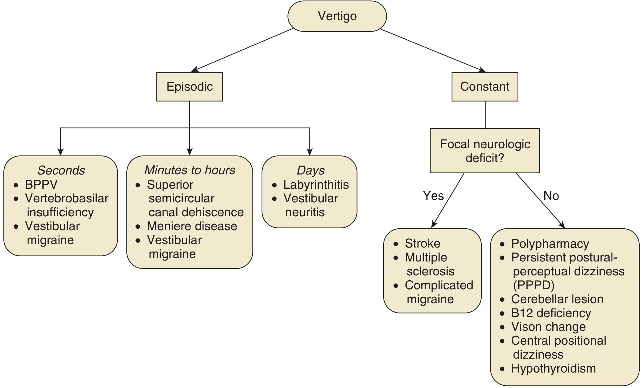
Classification of Vertigo
- Location: Peripheral (inner ear/CN VIII) vs. Central (brainstem/cerebellum)
- Duration of episodes: Seconds, minutes-hours, days, or constant
- Trigger: Positional vs. spontaneous
Peripheral vs. Central Vertigo: Key Distinctions
| Feature | Peripheral Vertigo | Central Vertigo |
|---|---|---|
| Onset | Abrupt, sudden | Gradual or abrupt |
| Severity | Often intense | Mild to moderate |
| Course | Episodic, with remissions | May be constant |
| Nystagmus | Horizontal or rotatory; unidirectional (fast component toward unaffected side); suppressed by fixation | Vertical, bidirectional, or direction-changing; NOT suppressed by fixation |
| Hearing loss / tinnitus | May be present (cochlear involvement) | Usually absent |
| Neurological symptoms | Absent | May be present (dysarthria, diplopia, dysphagia, ataxia, facial numbness) |
| Head impulse test | Abnormal (catch-up saccade) | Normal (concerning sign!) |
| Nausea/vomiting | Often severe | Variable |
| Latency in Dix-Hallpike | Yes (3-20 sec) | No latency |
| Fatigability | Fatigues with repetition | Does not fatigue |
| Examples | BPPV, vestibular neuritis, Menière's | Cerebellar stroke, MS, posterior fossa tumor |
Critical rule: In a patient with acute continuous vertigo and nystagmus, a normal head impulse test is a red flag for a central cause (stroke) - not the reassuring finding one might expect.
Peripheral Vestibular Disorders
1. Benign Paroxysmal Positional Vertigo (BPPV)
- Sudden brief episodes of vertigo (typically <1 minute, often 15-30 seconds)
- Always triggered by changes in head position relative to gravity: lying down, rolling over in bed, getting up, tilting head back to look up
- No hearing loss or tinnitus
- Rolling over in bed is a classic trigger (helps distinguish from orthostatic hypotension)
- Nausea may accompany attacks
- Upbeat-torsional nystagmus on Dix-Hallpike (upper poles of eyes beat toward the affected downward ear)
- Latency: 3-20 seconds before onset
- Duration: <1 minute
- Fatigues with repeated testing
- Reverses on returning patient to sitting
- Epley maneuver (canalith repositioning procedure) - first-line for posterior canal BPPV; highly effective; often resolves in single office visit
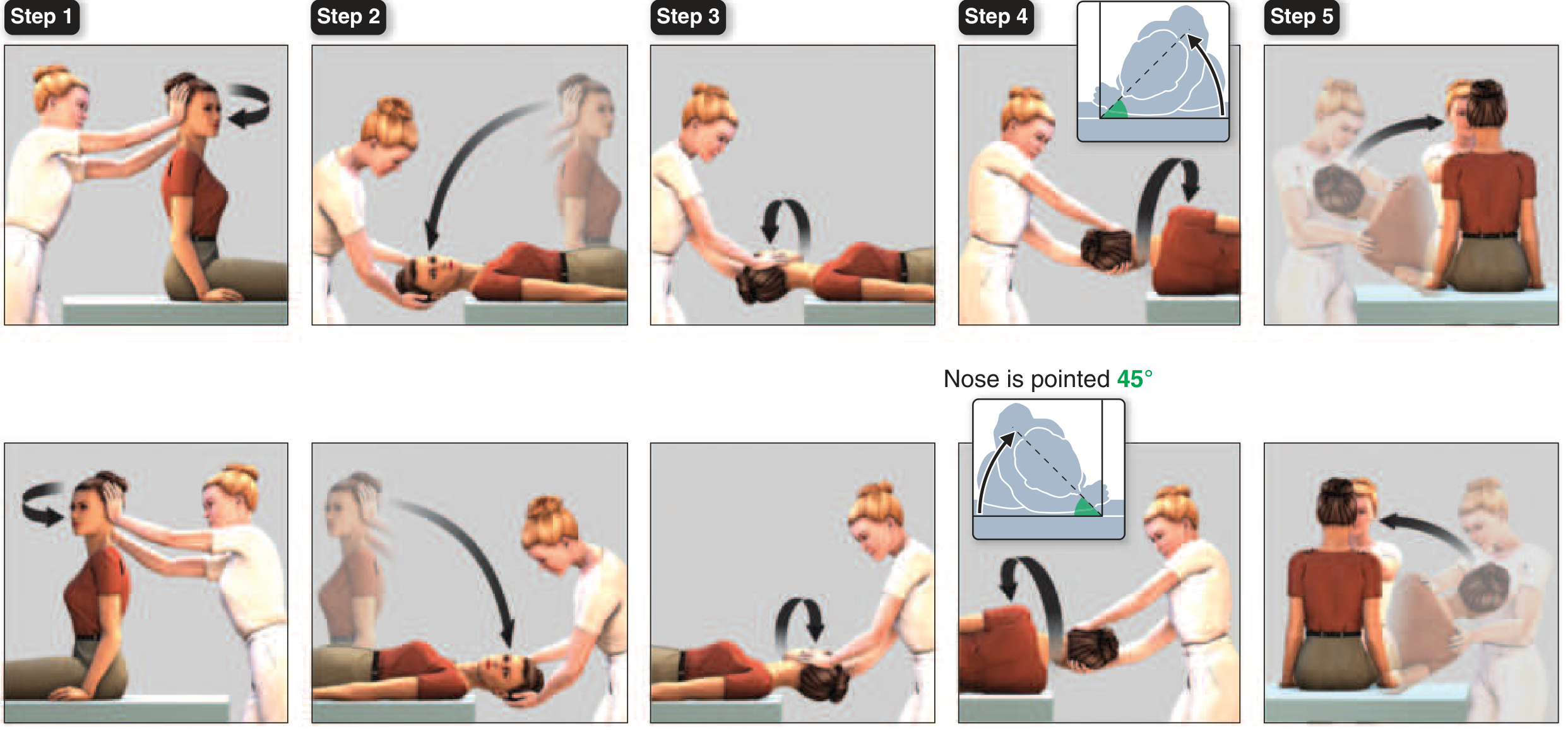
- Semont maneuver: Alternative repositioning procedure
- Home exercises (Brandt-Daroff exercises): Self-repositioning exercises, repeated twice daily until symptoms resolve
- Vestibular suppressants (meclizine, benzodiazepines): NOT recommended routinely for BPPV by AAO-HNS guidelines - they interfere with vestibular habituation and compensation
- Natural course: Often resolves in weeks to months; recurrence rate ~30-50%
2. Vestibular Neuritis (Vestibular Neuronitis)
- Sudden severe constant vertigo, lasting 1-2 days acutely, with gradual resolution over weeks to months
- No hearing loss (distinguishes from labyrinthitis)
- No tinnitus
- Spontaneous nystagmus (horizontal + rotary) with fast phase beating away from the affected ear (toward the healthy side)
- Nausea and vomiting often severe
- Positive head impulse test toward the affected ear (pathological catch-up saccade)
- The affected ear is on the side opposite to the direction of spontaneous nystagmus
- Symptomatic relief (acute phase):
- IV ondansetron 4 mg - recommended first-line parenteral antiemetic
- IM promethazine 25 mg (effective but more side effects)
- IV lorazepam 1-2 mg for severe intractable vertigo
- Meclizine 12.5-50 mg q4-6h orally (onset ~1 hour)
- Steroids: Methylprednisolone may improve recovery of vestibular function (evidence limited)
- Antivirals (valacyclovir): Not shown to improve outcomes - not recommended
- Vestibular rehabilitation exercises: Start early; help the brain compensate for the unilateral loss. Avoid prolonged vestibular suppressants as they impair central compensation
- Long-term: Most patients compensate well within weeks; some develop persistent postural-perceptual dizziness (PPPD)
3. Labyrinthitis
- Sudden severe vertigo PLUS tinnitus and hearing loss (sensorineural, often severe and potentially permanent)
- Inflammation involves both the vestibular and cochlear portions of the labyrinth
- Most commonly viral (upper respiratory tract viruses)
- Bacterial: Extension of bacterial otitis media into the inner ear (a serious complication requiring urgent treatment)
- Serous (toxic) labyrinthitis: From adjacent inflammation without actual infection of the labyrinth (e.g., following acute otitis media)
- Less common: Syphilis (treponema), Lyme disease (rickettsia)
4. Ménière's Disease (Endolymphatic Hydrops)
- Episodic vertigo - attacks lasting 20 minutes to several hours (20 min to 12 hours); severe, often disabling
- Fluctuating low-frequency sensorineural hearing loss - characteristic audiometric finding on affected side; may improve between attacks early in disease, but permanent loss develops over time
- Tinnitus - typically low-pitched roaring in the affected ear
- Aural fullness/pressure - a sense of pressure or pain in the affected ear, often preceding the attack
- Step 1 - Lifestyle and dietary:
- Low-sodium diet (<1500-2000 mg/day) - reduces endolymph pressure
- Avoid caffeine, alcohol, stress
- Step 2 - Medical:
- Diuretics (hydrochlorothiazide, acetazolamide) - reduce endolymph production
- Betahistine (widely used in Europe; evidence mixed in large RCTs)
- Step 3 - Intratympanic injections:
- Intratympanic glucocorticoids (dexamethasone) - non-ablative; reduces attack frequency
- Intratympanic gentamicin - ablative; chemical labyrinthectomy of the affected ear; highly effective for vertigo control but risks ipsilateral hearing loss
- Step 4 - Surgery:
- Endolymphatic sac decompression/shunting - non-ablative
- Vestibular nerve section - ablative; preserves hearing, eliminates vertigo
- Labyrinthectomy - ablative; for patients with no serviceable hearing; complete elimination of attacks
- Harrison's Principles of Internal Medicine, 22nd Edition
5. Vestibular Migraine
- Episodes of vertigo lasting minutes to hours (occasionally days of disequilibrium)
- May precede, accompany, or occur completely without headache (the key diagnostic challenge)
- Motion sensitivity and sensitivity to visual motion (e.g., scrolling screens, movies, crowds) are prominent
- Even without headache, migrainous features may accompany: photophobia, phonophobia, visual aura, history of previous migraines
- Patients often have a past history of classical migraine headaches
- Acute attack: Triptans (if headache present), antiemetics (prochlorperazine, ondansetron)
- Prophylaxis: Beta-blockers (propranolol, metoprolol), tricyclics (amitriptyline), topiramate, valproate, verapamil, SSRIs/SNRIs, lifestyle modifications (sleep, diet, stress)
- A 2025 systematic review (PMID 41166161) on prophylactic management of vestibular migraine confirms the above agents but notes limited high-quality RCT evidence
6. Perilymph Fistula
- Cause: Rupture of the oval or round window membrane → leakage of perilymph fluid from inner ear into middle ear
- Triggers: Barotrauma (scuba diving, flying, heavy lifting), violent nose blowing, sneezing, head trauma, ear surgery
- Features: Sudden pop in ear, then hearing loss, vertigo, tinnitus; can be fluctuating
- Diagnosis: Fistula test - pneumatic otoscopy applies pressure to tympanic membrane, provoking nystagmus and vertigo (Hennebert sign/Tullio phenomenon)
- Treatment: Bed rest, avoid Valsalva; surgical exploration and patching if persistent
7. Superior Semicircular Canal Dehiscence (SSCD)
- Thinning or absence of bone overlying the superior semicircular canal
- Creates a "third window" in the inner ear, causing:
- Vertigo and nystagmus provoked by loud sounds (Tullio phenomenon) or pressure changes (Valsalva)
- Low-frequency conductive hearing loss (despite intact middle ear)
- Autophony - hearing own voice/heartbeat abnormally loudly
- Diagnosis: High-resolution CT temporal bones (0.5 mm slices); confirmed by VEMP (vestibular evoked myogenic potentials) showing abnormally low thresholds
- Treatment: Surgical repair (middle fossa craniotomy or transmastoid approach to plug/resurface the dehiscence)
8. Vestibular Schwannoma (Acoustic Neuroma)
- Benign tumor of the Schwann cells of the vestibular nerve at the cerebellopontine angle (CPA)
- Does NOT typically cause vertigo because the gradual vestibular loss allows central compensation as it develops
- Clinical: Slowly progressive unilateral sensorineural hearing loss + unilateral tinnitus; vertigo is uncommon
- Vestibular examination: Deficient head impulse test toward affected side; minimal nystagmus
- Diagnosis: MRI with gadolinium of the internal auditory canals - test of choice
- Indication for MRI: Any unexplained unilateral sensorineural hearing loss or unilateral vestibular hypofunction
- Treatment: Observation (small tumors), stereotactic radiosurgery (Gamma Knife), or microsurgical resection
Central Vestibular Disorders
9. Posterior Circulation Stroke (Cerebellar/Brainstem Stroke)
- Sudden vertigo, severe nausea, inability to walk (truncal ataxia is prominent)
- Gait ataxia is a key finding
- Ipsilateral limb ataxia, dysarthria
- Danger: Cerebellar edema → posterior fossa herniation (can occur hours to days later); requires urgent surgical decompression
- Vertigo, nystagmus, nausea (vestibular nucleus involvement)
- Ipsilateral facial numbness, contralateral body numbness (crossed sensory findings - classic)
- Ipsilateral Horner syndrome
- Dysphagia, dysarthria, hoarseness
- Ipsilateral cerebellar ataxia
The HINTS Exam: Distinguishing Stroke from Vestibular Neuritis
HINTS Components:
- Hold patient's head and rapidly rotate it 20° left then right, randomly
- Normal HIT (eyes stay on target): Suggests central cause - WORRYING sign
- Abnormal HIT (catch-up saccade toward affected ear): Suggests peripheral vestibular neuritis - reassuring
- Unidirectional nystagmus that does not change direction with gaze = peripheral (reassuring)
- Direction-changing nystagmus (fast component reverses with gaze direction) = central (worrying)
- Vertical or purely torsional spontaneous nystagmus = central
- Cover-uncover test while patient fixates on examiner's nose
- Vertical refixation movement (skew deviation) when eye is uncovered = central (worrying)
- No vertical movement = peripheral (reassuring)
- Impulse Normal → central
- FAst phase Alternating (direction-changing nystagmus) → central
- Refixation on cover test (skew deviation) → central
- Tintinalli's Emergency Medicine
10. Multiple Sclerosis (MS)
- Demyelinating plaques in brainstem/cerebellum cause vertigo
- May be presenting symptom
- Associated INO (internuclear ophthalmoplegia), other cranial nerve signs
- MRI brain with gadolinium shows enhancing plaques in characteristic periventricular and juxtacortical locations
11. Persistent Postural-Perceptual Dizziness (PPPD)
- Chronic dizziness ≥3 months, fluctuating, present at rest but worse while standing
- Heightened sensitivity to self-motion and visual motion (movies, supermarkets, crowds)
- Often preceded by an acute vestibular event (vestibular neuritis, BPPV, panic attack)
- Neurologic exam and vestibular testing are normal or show compensated deficit
- Treatment: SSRIs, cognitive-behavioral therapy (CBT), vestibular rehabilitation. Avoid vestibular suppressants - they worsen the condition
12. Other Central Causes
- Cerebellar tumors: Gradual onset, persistent vertigo, cerebellar signs
- Arnold-Chiari malformation: Downbeat nystagmus, occipital headache
- Vertebrobasilar insufficiency (TIA): Brief episodes of vertigo with other brainstem symptoms (diplopia, dysarthria, drop attacks); rarely isolated
- Cervicogenic vertigo: Controversial; from abnormal proprioceptive input from upper cervical spine; associated with neck pain and head movement
Diagnostic Approach
History Priorities
- Duration of each episode (the most critical question)
- Trigger: Positional vs. spontaneous
- Associated hearing loss, tinnitus, aural fullness
- Neurological symptoms: headache, diplopia, dysarthria, facial weakness, ataxia
- Medications (aminoglycosides, anticonvulsants, antihypertensives, lithium)
- Cardiovascular symptoms (palpitations, syncope)
Three-Step Clinical Algorithm
- Seconds triggered by head movement → Almost always BPPV → Dix-Hallpike → Epley
- Minutes to hours, spontaneous → Ménière's disease, vestibular migraine, TIA
- Days, acute continuous → Vestibular neuritis/labyrinthitis vs. stroke → HINTS exam
Physical Examination
- Orthostatic BP (to exclude presyncope)
- Ocular examination: Nystagmus type, direction, fixation suppression
- Tuning fork tests: Weber and Rinne (to lateralize/characterize hearing loss)
- Pneumatic otoscopy: Fistula test
- Dix-Hallpike maneuver: For BPPV
- Head impulse test: VOR integrity
- Romberg and tandem gait: Cerebellar/proprioceptive integrity
- Cranial nerve examination: Particularly CN V, VI, VII, IX, X, XII
- Cover-uncover test: Test of skew (for central vertigo)
Investigations
- Audiometry: Sensorineural vs. conductive hearing loss; lateralization
- MRI brain with DWI and gadolinium: For central causes; internal auditory canals for schwannoma
- CT temporal bones (high resolution): For semicircular canal dehiscence
- Electronystagmography (ENG)/Videonystagmography (VNG): Objective nystagmus recording; caloric testing for bilateral vestibular hypofunction
- Video head impulse test (vHIT): Quantitative measurement of VOR
- Vestibular evoked myogenic potentials (VEMP): Otolith and saccule/utricle function
- Rotary chair testing: Bilateral vestibular function
Drug Treatment Summary
| Drug Class | Drug | Dose | Indication |
|---|---|---|---|
| Antihistamines | Meclizine | 25-50 mg 3×/day | Acute peripheral vertigo |
| Dimenhydrinate | 50 mg 1-2×/day | Acute peripheral vertigo | |
| Promethazine | 25 mg 2-3×/day (also IM/rectal) | Acute severe vertigo/vomiting | |
| Benzodiazepines | Diazepam | 2.5 mg 1-3×/day | Acute severe vertigo |
| Clonazepam | 0.25 mg 1-3×/day | Acute severe vertigo | |
| Anticholinergic | Scopolamine patch | Transdermal | Motion sickness only |
| Antiemetic | Ondansetron IV | 4 mg | First-line IV for acute vertigo |
| Physical therapy | Epley maneuver | - | BPPV |
| Vestibular rehabilitation | - | Vestibular neuritis, PPPD | |
| Diuretics + low Na diet | Hydrochlorothiazide, acetazolamide | - | Ménière's disease |
| Antimigraine drugs | Propranolol, amitriptyline, topiramate | - | Vestibular migraine prophylaxis |
| SSRIs | Sertraline, fluoxetine | - | PPPD and anxiety comorbidity |
Important: Benzodiazepines should not be prescribed for discharge in vestibular neuritis - they impair central vestibular compensation. Vestibular suppressants are generally contraindicated in BPPV (interfere with habituation) and in PPPD.
Quick Reference: Differential Diagnosis by Episode Duration
| Episode Duration | Top Diagnoses |
|---|---|
| <1 min, triggered by position | BPPV |
| <1 min, spontaneous | Vertebrobasilar TIA, vestibular paroxysmia (CN VIII compression) |
| Minutes | Vestibular migraine, TIA |
| 20 min to 12 hours, spontaneous | Ménière's disease, vestibular migraine |
| Hours to days, acute onset | Vestibular neuritis, labyrinthitis, posterior fossa stroke |
| Constant (weeks-months) | Compensating vestibular neuritis, PPPD, central lesion (tumor, MS) |
- Harrison's Principles of Internal Medicine, 22nd Edition (2025) - Ch. 24: Vertigo
- Textbook of Family Medicine, 9th Edition - Ch. 18: Ear, Nose, and Throat
- Cummings Otolaryngology Head and Neck Surgery - Ch. 137: Vertigo
- Tintinalli's Emergency Medicine, 9th Edition - Ch. 170: Vertigo and Dizziness
- Rosen's Emergency Medicine - Ch. 15: Dizziness and Vertigo
- Shambaugh Surgery of the Ear - Ch. on BPPV
- Symptom to Diagnosis: An Evidence-Based Guide, 4th Edition - Ch. 14
- Systematic review: Vestibular migraine prophylaxis (PMID 41166161); Vestibular paroxysmia (PMID 39932594)
give me comprehensive info on paresis and paralysis
paresis paralysis UMN LMN classification causes clinical features comprehensive neurology 2024
spinal cord injury rehabilitation motor recovery
Paresis and Paralysis: A Comprehensive Guide
Definitions
- Paresis - partial or incomplete loss of voluntary motor function (weakness); some motor control remains
- Paralysis (plegia) - complete loss of voluntary motor function; no movement possible
- Palsy - used interchangeably with paralysis/paresis in clinical practice (e.g., Bell's palsy, cerebral palsy)
- Plegia - suffix denoting complete paralysis (e.g., hemiplegia, paraplegia)
- Paresis - suffix/standalone term denoting incomplete motor loss
The Motor System: Anatomical Basis
The Motor Cortex (Upper Motor Neuron Origin)
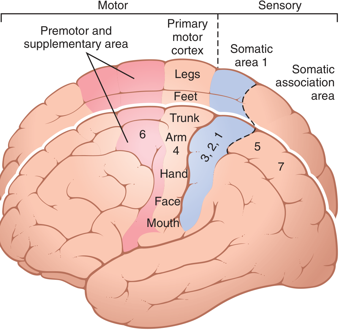
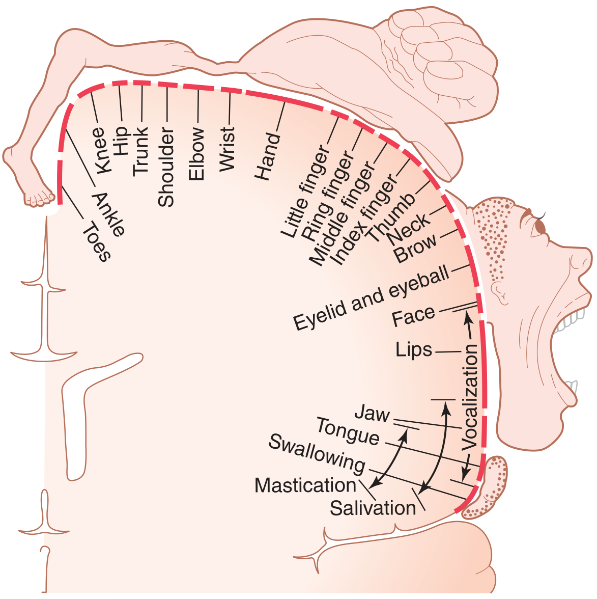
Descending Motor Pathways
- Arises from primary motor cortex (area 4), premotor cortex (area 6), and somatosensory cortex
- Descends through the internal capsule (posterior limb) → cerebral peduncles → pons → medullary pyramids
- At the pyramidal decussation in the medulla: ~85% cross to form the lateral corticospinal tract (contralateral spinal cord, controls distal limb muscles)
- ~15% remain ipsilateral as the anterior corticospinal tract (cross at spinal level, control axial/trunk muscles)
- The lateral corticospinal tract synapses on alpha motor neurons in the anterior horn
- Controls muscles of face, jaw, tongue, pharynx, larynx
- Most cranial motor nuclei receive bilateral corticobulbar input (so unilateral cortical lesions spare most cranial muscles)
- Exception: The lower face (CN VII) and tongue (CN XII) - these receive predominantly contralateral cortical input, so a unilateral UMN lesion causes contralateral lower facial weakness and tongue deviation
- Rubrospinal tract (from red nucleus)
- Reticulospinal tract (from brainstem reticular formation) - major mediator of descending inhibitory tone
- Vestibulospinal tract (from vestibular nuclei) - facilitates extensor tone
- Tectospinal tract (from superior colliculus) - head/neck orientation
Upper Motor Neuron (UMN) vs. Lower Motor Neuron (LMN)
Upper Motor Neuron (UMN)
- Resides in: Motor cortex, brainstem (for corticobulbar tract)
- Projects to: Spinal cord anterior horn (LMN)
- Any damage from motor cortex to but not including the anterior horn cell = UMN lesion
Lower Motor Neuron (LMN)
- Resides in: Anterior horn of spinal cord (for limbs) and cranial nerve motor nuclei (for head/neck)
- Projects to: Skeletal muscle via peripheral nerve
- Any damage from anterior horn cell to muscle = LMN lesion
Clinical Comparison Table
| Feature | UMN Lesion | LMN Lesion |
|---|---|---|
| Weakness | Yes | Yes |
| Tone | Increased (spasticity) | Decreased (flaccidity) |
| Reflexes (DTR) | Hyperreflexia, clonus | Hyporeflexia / areflexia |
| Babinski sign | Present (extensor plantar) | Absent (flexor plantar) |
| Hoffmann's sign | May be present | Absent |
| Atrophy | None (mild disuse atrophy only) | Marked atrophy (denervation) |
| Fasciculations | Absent | Present (spontaneous motor unit discharges) |
| Distribution | Broad (whole limb, functional groups) | Focal (individual muscles or groups) |
| Acute phase | Initially flaccid (spinal shock), then evolves to spasticity | Flaccid from onset |
Important clinical pearl: Acute UMN lesions (e.g., acute stroke, acute spinal cord injury) initially cause flaccid paralysis with decreased tone and decreased reflexes - this is called spinal shock (spinal cord lesion) or the acute phase of a cortical stroke. Spasticity and hyperreflexia develop gradually over hours to weeks as descending inhibitory pathways lose their influence and spinal circuits become disinhibited.
Grading of Motor Weakness: The MRC Scale
| MRC Grade | Description |
|---|---|
| 0 | No contraction whatsoever |
| 1 | Flicker or trace of contraction (visible or palpable, no movement) |
| 2 | Active movement possible only with gravity eliminated (limb moved horizontally) |
| 3 | Active movement against gravity (but not against resistance) |
| 4 | Active movement against gravity AND some resistance (reduced) |
| 5 | Normal power (full resistance) |
- 4- = movement against slight resistance
- 4 = movement against moderate resistance
- 4+ = movement against strong resistance (near-normal)
Terminology Based on Distribution
| Term | Definition |
|---|---|
| Monoplegia/Monoparesis | Complete/partial paralysis of one limb |
| Hemiplegia/Hemiparesis | Complete/partial paralysis of one side of the body (arm + leg, ipsilateral) |
| Paraplegia/Paraparesis | Complete/partial paralysis of both legs (and often trunk) |
| Tetraplegia/Quadriplegia | Complete paralysis of all four limbs |
| Tetraparesis/Quadriparesis | Partial weakness of all four limbs |
| Diplegia | Bilateral paralysis affecting the legs more than the arms (common in cerebral palsy) |
| Crossed hemiplegia | Ipsilateral face + contralateral limbs - always indicates brainstem lesion |
| Pseudobulbar palsy | Bilateral UMN lesion affecting corticobulbar tracts |
| Bulbar palsy | LMN lesion of cranial nerve nuclei IX, X, XII |
Localization of Lesion by Clinical Pattern
1. Cerebral Cortex / Hemisphere
- Face and arm typically more affected than leg (face and arm representation is lateral cortex; leg is in the medial longitudinal fissure and is relatively protected in MCA territory strokes)
- Often accompanied by: hemisensory loss, hemianopia, aphasia (dominant hemisphere), neglect (non-dominant)
- Cause: Stroke (MCA territory), tumor, trauma, abscess
- Tone: Initially flaccid → evolves to spastic
- Reflexes: Initially reduced → evolves to hyperreflexia + Babinski
2. Internal Capsule
- Face + arm + leg equally affected (unlike cortical lesion)
- Capsular lacune - small deep infarct in the posterior limb → classic pure motor stroke
- Cause: Hypertensive small vessel disease (lacunar infarct)
3. Brainstem
- This is pathognomonic of a brainstem lesion
- Motor fibers cross at medulla, but cranial nerve nuclei are ipsilateral
| Syndrome | Level | Ipsilateral | Contralateral |
|---|---|---|---|
| Weber syndrome | Midbrain | CN III palsy (ptosis, mydriasis, down-and-out) | Hemiplegia |
| Millard-Gubler | Pons | CN VI + VII palsy | Hemiplegia |
| Benedikt | Midbrain | CN III palsy + tremor | Hemiplegia |
| Wallenberg | Lateral medulla | Horner's, cerebellar ataxia, face sensory loss, IX/X palsy | Limb pain/temperature loss |
4. Spinal Cord
- Above C4 (C1-C3): Tetraplegia + respiratory paralysis (phrenic nerve C3,4,5 → diaphragm)
- C5-C6 injury: Tetraplegia; some shoulder/elbow movement preserved
- Below T1: Paraplegia (lower limbs only); upper limbs spared
- T2 and below → paraplegia; above T1 → quadriplegia
Spinal Cord Syndromes
- Total loss of motor and sensory function below the level
- Spinal shock phase initially (flaccid areflexia)
- Later evolves to UMN pattern (spasticity, hyperreflexia, Babinski sign)
- Bladder, bowel, and sexual dysfunction
- Ipsilateral: UMN weakness + loss of vibration and proprioception (dorsal column)
- Contralateral: Loss of pain and temperature (spinothalamic tract, which has already crossed)
- At the lesion level: ipsilateral LMN weakness + ipsilateral pain/temperature loss
- Cause: Stab/gunshot wounds, tumor, MS, disc herniation
- Arms weaker than legs (central cord = cervical motor fibers are most central; leg fibers are peripheral)
- Bladder dysfunction (retention)
- Variable sensory loss; cape-like dissociated sensory loss for pain/temperature
- Preservation of sacral sensation (sacral sparing)
- Cause: Cervical hyperextension injury in elderly with spondylosis; falls
- Prognosis: Generally better than complete injury; ~90% with any motor sparing at 4 weeks become ambulators
- Affects anterior 2/3 of spinal cord; posterior columns spared
- Bilateral motor loss (corticospinal tract) + bilateral pain/temperature loss (spinothalamic tract)
- Vibration and proprioception preserved (posterior columns intact)
- Bladder/bowel dysfunction (autonomic fibers)
- Cause: Anterior spinal artery occlusion, central disc herniation, aortic surgery (artery of Adamkiewicz near T6)
- Loss of vibration, proprioception, fine touch bilaterally below the level
- Motor function and pain/temperature preserved
- Positive Romberg sign, sensory ataxia
- Cause: Subacute combined degeneration (B12 deficiency), tabes dorsalis (syphilis), MS
- Mixed UMN + LMN signs (conus = transition zone)
- Saddle anesthesia
- Bladder/bowel/sexual dysfunction (parasympathetic center)
- Preserved lower limb strength (partially)
- Pure LMN: flaccid paralysis, areflexia of lower limbs
- Saddle anesthesia (S3-S5 dermatomes)
- Bladder retention then overflow incontinence, bowel dysfunction
- Root pain (radiculopathy)
- Surgical emergency if acute compression
5. Peripheral Nerve (LMN)
- Flaccid weakness, atrophy, fasciculations, areflexia
- Mononeuropathy: Single nerve (e.g., foot drop from peroneal nerve palsy, wrist drop from radial nerve palsy)
- Polyneuropathy: Bilateral distal-predominant weakness ("stocking and glove" pattern); length-dependent
- Plexopathy: Brachial or lumbosacral plexus involvement
6. Anterior Horn Cell
- Asymmetric muscle atrophy, fasciculations, weakness
- Reflexes proportionately reduced
- Causes: Poliomyelitis (viral destruction of anterior horn cells), Spinal Muscular Atrophy (SMA), ALS (combined UMN+LMN), Kugelberg-Welander disease
7. Neuromuscular Junction
- No atrophy (muscles structurally intact)
- No sensory deficit
- Normal or mildly reduced reflexes
- Classic examples:
- Myasthenia gravis (MG): Postsynaptic - antibodies to AChR; proximal > distal; ocular involvement (ptosis, diplopia); worsens through day
- Lambert-Eaton myasthenic syndrome (LEMS): Presynaptic - antibodies to VGCC; proximal limb weakness; improves briefly with exercise; associated with lung cancer
8. Muscle (Myopathy)
- No sensory deficit
- Mild to moderate atrophy in proportion to weakness
- Reflexes preserved until late-stage severe weakness
- Causes: Inflammatory (polymyositis, dermatomyositis), metabolic, toxic (statins, steroids, alcohol), hereditary (Duchenne/Becker muscular dystrophy, limb-girdle), endocrine (hypothyroidism, Cushing's)
Specific Paralysis Types: Special Entities
Flaccid Paralysis vs. Spastic Paralysis
- Absent tone, areflexia, no Babinski, fasciculations, atrophy
- LMN lesion, spinal shock (acute UMN)
- Polio, Guillain-Barré syndrome, acute SCI
- Increased tone (clasp-knife character), hyperreflexia, Babinski sign, clonus
- Chronic UMN lesion
- Stroke, SCI (chronic phase), MS, cerebral palsy
Spinal Shock
- Period of complete loss of all reflexes, flaccid paralysis, anesthesia immediately following acute spinal cord injury
- Mediated by sudden loss of descending facilitory input to spinal cord neurons
- Duration: Hours to weeks (traditionally said to end when the bulbocavernosus reflex returns)
- After resolution: UMN signs emerge (spasticity, hyperreflexia, Babinski)
Todd's Paralysis (Postictal Paralysis)
- Transient focal motor weakness or paralysis following a focal (Jacksonian) seizure
- Affects the same limb that had the seizure
- Duration: Minutes to hours (rarely days)
- Caused by postictal neuronal exhaustion/inhibition
- Resolves completely; its presence is clinically important because it helps localize the seizure focus
Bell's Palsy (Facial Nerve Palsy - LMN)
- Complete ipsilateral facial paralysis - upper and lower face equally affected (distinguishes LMN from UMN)
- UMN facial weakness spares the forehead (bilateral UMN input to forehead muscles)
- Bell's palsy: Idiopathic (presumed reactivation of herpes simplex); treated with corticosteroids ± antiviral
- LMN CN VII palsy: Loss of taste (anterior 2/3 tongue), hyperacusis, loss of corneal reflex
Pseudobulbar Palsy vs. Bulbar Palsy
| Feature | Pseudobulbar Palsy (UMN) | Bulbar Palsy (LMN) |
|---|---|---|
| Tongue | Small, spastic | Atrophic, fasciculating |
| Jaw jerk | Exaggerated | Reduced/absent |
| Gag reflex | Exaggerated | Reduced/absent |
| Emotional lability | Present (pathological crying/laughing) | Absent |
| Dysarthria | Spastic ("hot potato" voice) | Nasal, hypophonic |
| Cause | Bilateral cortical/capsular lesions | Brainstem nuclei lesions, motor neuron disease |
Hemiplegia Patterns
| Type | Features | Cause |
|---|---|---|
| Cortical | Arm > leg; associated sensory, visual, cognitive deficits | MCA territory stroke/tumor |
| Capsular | Arm = face = leg equally; pure motor | Internal capsule lacunar infarct |
| Brainstem (alternating) | Ipsilateral CN palsy + contralateral limb | Brainstem stroke |
| Spinal | Unilateral leg weakness | Brown-Séquard syndrome |
Major Causes of Paresis/Paralysis by Category
Central (UMN) Causes
- Stroke (most common cause of acquired hemiplegia in adults) - ischemic or hemorrhagic
- Traumatic brain injury (TBI)
- Brain tumor (primary or metastatic)
- Multiple sclerosis (MS) - demyelinating plaques in corticospinal tracts
- Cerebral palsy - non-progressive UMN disorder from perinatal brain injury
- Motor neuron disease (UMN component): Primary lateral sclerosis, ALS
- CNS infections: Encephalitis, abscess
- Spinal cord injury (trauma, compression, infarction, MS, transverse myelitis)
- Hereditary spastic paraplegia (HSP): Progressive bilateral lower limb spasticity; genetic
Peripheral (LMN) Causes
- Motor neuron disease (LMN component): Spinal muscular atrophy (SMA), Kennedy disease, ALS
- Poliomyelitis: Viral destruction of anterior horn cells
- Peripheral neuropathy: Guillain-Barré syndrome (GBS) - acute ascending flaccid paralysis
- Plexopathies: Brachial neuritis (Parsonage-Turner syndrome), lumbosacral plexopathy
- Nerve compression/entrapment: Radiculopathy, mononeuropathy
- Charcot-Marie-Tooth disease (CMT): Hereditary motor and sensory neuropathy
- Diabetic neuropathy
- Toxic neuropathy: Lead, organophosphates, thallium
Neuromuscular Junction
- Myasthenia gravis (MG)
- Lambert-Eaton myasthenic syndrome
- Botulism
Muscle
- Inflammatory myopathies (polymyositis, dermatomyositis, inclusion body myositis)
- Muscular dystrophies (Duchenne, Becker, facioscapulohumeral, limb-girdle)
- Metabolic myopathies
- Periodic paralysis (hyper/hypokalemic)
- Statin myopathy
Combined UMN + LMN
- Amyotrophic lateral sclerosis (ALS) - simultaneous degeneration of both UMN and LMN
- Fasciculations + hyperreflexia in the same muscle group = hallmark
- Bulbar signs (dysarthria, dysphagia) + limb weakness + respiratory failure
- Fatal; median survival 3-5 years
- Treatment: Riluzole (glutamate blocker; modest survival benefit); Edaravone (antioxidant)
Diagnostic Approach
Step 1 - History
- Onset: Acute (seconds-minutes: stroke; hours: GBS, MS relapse) vs. subacute (days-weeks) vs. chronic (months-years: MND, muscular dystrophy)
- Distribution: Which limbs? Proximal or distal? Unilateral or bilateral?
- Associated symptoms: Sensory loss, pain, sphincter dysfunction, cognitive changes
- Progression: Improving, stable, worsening
- Family history: Hereditary conditions
Step 2 - Examination
- Inspection: Atrophy, fasciculations, posture (hemiplegic posture: arm flexed, leg extended)
- Tone: Spastic (UMN) vs. flaccid (LMN)
- Power: MRC grading (0-5) for each muscle group
- Reflexes: Deep tendon reflexes (biceps C5, brachioradialis C6, triceps C7, knee L3-L4, ankle S1); Babinski sign
- Coordination: Cerebellar testing
- Gait: Hemiplegic gait (circumduction), spastic paraparetic gait (scissor gait), steppage gait (foot drop), waddling gait (proximal myopathy)
- Sensory examination: To determine lesion level and type (spinothalamic vs. dorsal column)
Step 3 - Investigations
| Investigation | Indication |
|---|---|
| MRI brain/spine | UMN lesions, stroke, MS, tumor, myelopathy |
| CT head | Acute hemorrhage, trauma |
| EMG/Nerve conduction studies | LMN, peripheral nerve, NMJ, myopathy |
| Muscle biopsy | Inflammatory myopathy, muscular dystrophy |
| Nerve biopsy | Vasculitic neuropathy |
| Serum CK | Myopathy (CK very high in Duchenne, polymyositis) |
| AChR/Anti-MuSK antibodies | Myasthenia gravis |
| VGCC antibodies | Lambert-Eaton syndrome |
| CSF analysis | GBS (albuminocytologic dissociation), MS, infections |
| Genetic testing | SMA (SMN1 gene), CMT, ALS (SOD1, FUS, TDP-43) |
Management Principles
Acute Management
- Airway: Vital capacity monitoring in GBS and MG crisis; intubation if VC <15-20 mL/kg
- Thrombolysis (tPA) or thrombectomy: Ischemic stroke within appropriate time window
- Spinal cord injury: Immobilization, surgical decompression if indicated, avoid hypotension
- Myasthenic crisis: Plasmapheresis or IVIG; pyridostigmine
- GBS: IVIG or plasmapheresis
Disease-Specific
- ALS: Riluzole, edaravone, respiratory support, PEG feeding
- MS: Disease-modifying therapies (interferons, natalizumab, ocrelizumab) for relapsing-remitting; symptomatic spasticity treatment with baclofen
- SMA: Nusinersen (antisense oligonucleotide), onasemnogene abeparvovec (gene therapy), risdiplam
- Myasthenia gravis: Anticholinesterases (pyridostigmine), immunosuppression (steroids, azathioprine), thymectomy
- Inflammatory myopathy: Corticosteroids, immunosuppressants
Spasticity Management
- Baclofen (GABA-B agonist): Oral or intrathecal pump for severe spasticity
- Tizanidine (α2 agonist): Reduces presynaptic inhibition of spinal motor neurons
- Dantrolene (peripheral; reduces intracellular Ca2+): For severe spasticity
- Botulinum toxin injections: Focal spasticity management (reduces ACh release at NMJ)
- Ganong's Review of Medical Physiology
Rehabilitation
- Physiotherapy: Preventing contractures, strengthening, gait retraining
- Occupational therapy: ADL retraining, adaptive equipment
- Orthoses: AFO (ankle-foot orthosis) for foot drop, KAFO for knee instability
- Neuromodulation: FES (functional electrical stimulation), TMS, epidural stimulation
- Brain-computer interfaces (BCI): Emerging technology - allows spinal bypass of motor signals; systematic review 2024 (PMID 37943244) confirms feasibility for motor restoration
Quick Summary: Localization at a Glance
| Level | Pattern | UMN/LMN | Key Signs |
|---|---|---|---|
| Cortex/hemisphere | Contralateral hemiplegia (arm > leg) | UMN | Aphasia, sensory loss, Babinski |
| Internal capsule | Contralateral pure motor hemiplegia | UMN | Equal face/arm/leg, no sensory |
| Brainstem | Crossed hemiplegia | UMN limb, LMN face | Cranial nerve ipsilateral + limbs contralateral |
| Cervical cord | Quadriplegia/paresis | UMN (chronic) | Level sign, Babinski bilateral |
| Thoracic cord | Paraplegia | UMN | Spastic legs, sensory level |
| Lumbosacral cord | Paraplegia | UMN/LMN mix | Conus/cauda equina features |
| Anterior horn | Focal LMN | LMN only | No sensory loss, fasciculations, asymmetric |
| Peripheral nerve | Nerve territory weakness | LMN | Sensory deficit in nerve distribution |
| NMJ | Fatigable weakness | Neither | No atrophy, normal reflexes |
| Muscle | Proximal > distal | Neither | No sensory deficit, CK elevated |
- Neuroanatomy through Clinical Cases, 3rd Edition - Chapters 6 (UMN/LMN), 27 (Spinal Cord Syndromes)
- Ganong's Review of Medical Physiology, 26th Edition - Chapter 12: Motor neurons, spasticity
- Guyton and Hall Textbook of Medical Physiology - Chapter 56: Motor Cortex and Corticospinal Tract
- Bradley and Daroff's Neurology in Clinical Practice - Chapter 28: Motor System examination; Chapter 27: Paraplegia/Quadriplegia
- Recent PubMed evidence: SCI rehabilitation meta-analysis (PMID 39798215); Non-invasive stimulation in SCI (PMID 40050875); Brain-computer interfaces (PMID 37943244)
tell me about UMN and LMN lesion characterisitcs
Upper Motor Neuron (UMN) vs. Lower Motor Neuron (LMN) Lesions
The Core Concept
-
Upper Motor Neuron (UMN): Originates in the motor cortex (Brodmann area 4/6), travels through the internal capsule, brainstem, and descends in the corticospinal tract to synapse on the anterior horn cell of the spinal cord (or on cranial nerve motor nuclei via the corticobulbar tract). Any lesion from cortex to - but not including - the anterior horn cell is a UMN lesion.
-
Lower Motor Neuron (LMN): Originates in the anterior horn of the spinal cord (or cranial nerve motor nuclei), travels via peripheral nerve to the neuromuscular junction and skeletal muscle. Any lesion from the anterior horn cell to the muscle is an LMN lesion.
Anatomy of the Pathways
Upper Motor Neuron Pathway
Motor Cortex (Area 4 - primary; Area 6 - premotor/SMA)
↓
Internal Capsule (posterior limb)
↓
Cerebral Peduncles
↓
Pons (corticobulbar fibers exit here to cranial nerve nuclei)
↓
Medullary Pyramids
↓
Pyramidal Decussation (~85% cross → lateral corticospinal tract)
↓
Lateral corticospinal tract (contralateral side of spinal cord)
↓
Synapse on anterior horn alpha-motor neuron (LMN)
Lower Motor Neuron Pathway
Anterior Horn Cell (spinal cord) / Cranial Nerve Motor Nucleus (brainstem)
↓
Ventral nerve root
↓
Peripheral nerve
↓
Neuromuscular junction
↓
Skeletal muscle (effector)
Characteristic Features: Detailed Comparison
Summary Table
| Feature | UMN Lesion | LMN Lesion |
|---|---|---|
| Weakness/Paralysis | Yes | Yes |
| Tone | Increased (spasticity) | Decreased (flaccidity, hypotonia) |
| Deep Tendon Reflexes | Hyperreflexia | Hyporeflexia / Areflexia |
| Babinski Sign | Present (extensor plantar: great toe dorsiflexes, other toes fan) | Absent (normal flexor plantar response) |
| Hoffmann's Sign | May be present | Absent |
| Clonus | Present (especially ankle/patellar) | Absent |
| Muscle Atrophy | Absent (mild disuse atrophy may develop later) | Marked, early (denervation atrophy) |
| Fasciculations | Absent | Present (spontaneous motor unit discharges visible under skin) |
| Distribution | Broad - whole limb or functional groups (flexors/extensors) | Focal - individual muscles or myotomal patterns |
| Character of weakness | Functional pattern (arm extensors + leg flexors preferentially weak) | Individual muscle weakness corresponding to nerve/root/anterior horn |
| Acute presentation | Initially flaccid (spinal shock) → then spastic over days/weeks | Flaccid from the very start |
| Sensory involvement | Often present (adjacent sensory tracts run with motor tracts) | May be present if peripheral nerve (not if pure anterior horn) |
UMN Lesions: In Depth
Pathophysiology of Spasticity
- Loss of descending inhibitory pathways (reticulospinal tract in particular) that normally suppress spinal reflex arcs
- Upregulation of excitatory receptors on motor neurons
- Axonal sprouting and synaptic reorganization
Note: Increased tone and hyperreflexia do NOT occur from a selective lesion of the corticospinal tract alone in experimental models. Spasticity requires damage to the descending inhibitory pathways (reticulospinal tract) that travel alongside the corticospinal tract.
- Neuroanatomy through Clinical Cases, 3rd Edition
Pattern of Weakness in UMN Lesions
- Upper limb: Extensors weaker than flexors (arm held in flexed posture)
- Weak: wrist/elbow extension, finger extension
- Relatively preserved: wrist/elbow flexion
- Lower limb: Flexors weaker than extensors (leg held in extended posture)
- Weak: hip flexion, knee flexion, ankle dorsiflexion
- Relatively preserved: hip/knee extension, plantarflexion
UMN Signs: Details
- Scratch the lateral aspect of the sole (from heel to ball) with a blunt object
- Positive (abnormal/UMN): Great toe dorsiflexes (extends upward) + other toes fan outward
- Normal (in adults): All toes plantarflex
- In infants: Babinski is normal until corticospinal tracts are myelinated (~18-24 months)
- Physiological significance is still debated, but it is a reliable sign of corticospinal tract dysfunction
- Flick the terminal phalanx of the middle finger downward - positive if the thumb adducts and flexes reflexively
- Equivalent of Babinski for the upper limb
- Reliable UMN sign when asymmetric
- Rapid repetitive rhythmic contractions when a muscle is suddenly stretched and held stretched
- Most commonly elicited at the ankle (sudden dorsiflexion and hold) and knee (sudden push on patella downward)
- Reflects hyperactive stretch reflex
- Sustained (>5 beats) clonus is pathological and indicates a UMN lesion
- Sudden increase in resistance (catch) when passively moving a limb rapidly
- Distinguishes spasticity (velocity-dependent) from rigidity (velocity-independent, as in Parkinson's disease)
Causes of UMN Lesions
| Location | Causes |
|---|---|
| Cortex | Stroke (MCA territory), traumatic brain injury, brain tumor, cerebral abscess, encephalitis |
| Internal capsule | Lacunar infarct (hypertensive), hemorrhage |
| Brainstem | Stroke (vertebrobasilar), MS, tumor, central pontine myelinolysis |
| Spinal cord | Trauma, MS, cervical spondylotic myelopathy, transverse myelitis, tumor compression, ALS, subacute combined degeneration (B12 deficiency), hereditary spastic paraplegia |
LMN Lesions: In Depth
Pathophysiology of Flaccidity and Atrophy
- Loss of trophic factors from the nerve → muscle fibers undergo denervation atrophy (progressively wasting away)
- Loss of all reflex arcs through that muscle → areflexia
- Loss of tone → muscle becomes flaccid (no baseline resting tension)
- Damaged or dying motor neurons fire spontaneously and irregularly → visible fasciculations under the skin
- Benign fasciculations: Very common (eyelid twitching, fatigue, caffeine) - no associated weakness or atrophy
- Pathological fasciculations: Associated with weakness + atrophy → indicates motor neuron disease or denervation
Pattern of Weakness in LMN Lesions
- Anterior horn cell: Asymmetric, myotomal (C5-C6 = deltoid, biceps; C7 = triceps, wrist extensors; L4-L5 = tibialis anterior; S1 = gastrocnemius)
- Nerve root (radiculopathy): Dermatomal sensory loss + myotomal weakness + loss of specific DTR
- Peripheral nerve: Exactly the muscles innervated by that nerve + sensory loss in nerve territory
- Plexus: Multiple nerve territories; complex pattern
LMN Signs: Details
- Hypotonia: limb feels heavy, floppy, offers no resistance to passive movement
- Loss of muscle bulk: visible wasting within weeks of denervation
- Limb hangs limply
- Best seen in bright tangential light across relaxed muscle
- Common sites: tongue (motor neuron disease), deltoid, thigh
- In ALS: tongue fasciculations + tongue atrophy = classic LMN bulbar sign
- Absent deep tendon reflexes (DTRs) in the affected territory
- Plantar reflex is absent or normal flexor (never extensor in pure LMN lesion)
- Begins within 2-3 weeks of complete denervation
- Eventually severe with replacement of muscle by fat/connective tissue
- Contrast: UMN lesions cause only mild disuse atrophy even after years
Causes of LMN Lesions
| Site | Causes |
|---|---|
| Anterior horn cells | Poliomyelitis, spinal muscular atrophy (SMA), ALS (LMN component), Kennedy disease (SBMA), post-polio syndrome |
| Nerve roots | Disc herniation (radiculopathy), foraminal stenosis, Guillain-Barré syndrome, diabetic radiculopathy |
| Brachial/lumbosacral plexus | Trauma, Parsonage-Turner syndrome (brachial neuritis), cancer infiltration, diabetic amyotrophy |
| Peripheral nerves | Mononeuropathy (trauma, compression - carpal tunnel, peroneal nerve palsy), polyneuropathy (GBS, diabetic, toxic, hereditary CMT), vasculitic neuropathy |
| Cranial nerve nuclei | Brainstem stroke, motor neuron disease, polio (if bulbar) |
Comparing Facial Nerve Palsy: UMN vs. LMN
| Feature | UMN Facial Weakness | LMN Facial Weakness (Bell's palsy) |
|---|---|---|
| Forehead involvement | Spared (bilateral cortical input to orbicularis oculi/frontalis) | Involved (entire face affected) |
| Eye closure | Intact | Weak or absent (risk of corneal exposure) |
| Lower face | Weak (mouth droop, inability to show teeth) | Weak |
| Taste | Intact | May be lost (chorda tympani involvement) |
| Hyperacusis | No | May be present (stapedius involvement) |
| Cause | Contralateral cortical/capsular stroke | Idiopathic (HSV reactivation), Ramsay Hunt, otitis media, parotid tumor |
| Treatment | Treat underlying stroke/cause | Prednisolone ± acyclovir |
The forehead-sparing rule: If only the lower face is weak on one side → UMN lesion (contralateral hemisphere/capsule). If the entire face is weak including the forehead → LMN lesion at or distal to the facial nerve nucleus.
Spinal Shock: The Acute UMN Exception
- Flaccid paralysis
- Areflexia (all reflexes abolished)
- Loss of sensation below the level
- Urinary retention (atonic bladder)
ALS: The Combined UMN + LMN Disease
| Feature | Explanation |
|---|---|
| Fasciculations + hyperreflexia in the same limb | UMN + LMN co-existing simultaneously (pathognomonic combination) |
| Babinski sign present | UMN involvement |
| Tongue fasciculations + atrophy | LMN bulbar involvement |
| Spastic dysarthria + nasal speech | UMN corticobulbar involvement |
| No sensory deficit | Motor neurons only |
| Progressive respiratory failure | Phrenic + intercostal motor neuron loss |
Reflex Changes: Grading
| Grade | Response |
|---|---|
| 0 | Absent (areflexia) - LMN or severe myopathy |
| 1+ | Diminished (hyporeflexia) - LMN, peripheral neuropathy |
| 2+ | Normal |
| 3+ | Brisk (may be normal in anxious patients) |
| 4+ | Very brisk with spread (hyperreflexia) - UMN |
| 5+ | Sustained clonus - UMN |
Quick Reference Diagnostic Checklist
| Question | UMN Answer | LMN Answer |
|---|---|---|
| Is there atrophy? | No (or mild disuse) | Yes (neurogenic, marked) |
| Are there fasciculations? | No | Yes |
| What are the reflexes? | Increased | Decreased/absent |
| What is the tone? | Increased (spastic) | Decreased (flaccid) |
| Is there a Babinski? | Yes | No |
| What is the distribution? | Broad functional pattern | Focal anatomical (nerve/root/muscle) |
- Neuroanatomy through Clinical Cases, 3rd Edition - Ch. 6.1: UMN vs. LMN Lesions
- Ganong's Review of Medical Physiology, 26th Edition - Ch. 12: Lower vs. Upper Motor Neuron Damage
- Bradley and Daroff's Neurology in Clinical Practice - Ch. 28: Motor System Examination
- Guyton and Hall Textbook of Medical Physiology - Ch. 56: Motor Cortex and Corticospinal Tract
talk about aphasias
aphasia types classification Broca Wernicke conduction global comprehensive neurology 2024
aphasia treatment speech language therapy stroke recovery
Aphasias
1. Definition and Overview
- Aphasia = language disorder (content, words, grammar)
- Dysarthria = motor speech disorder (articulation, phonation) - language itself is intact
- Dysphonia = voice/larynx disorder
2. The Wernicke-Geschwind Model
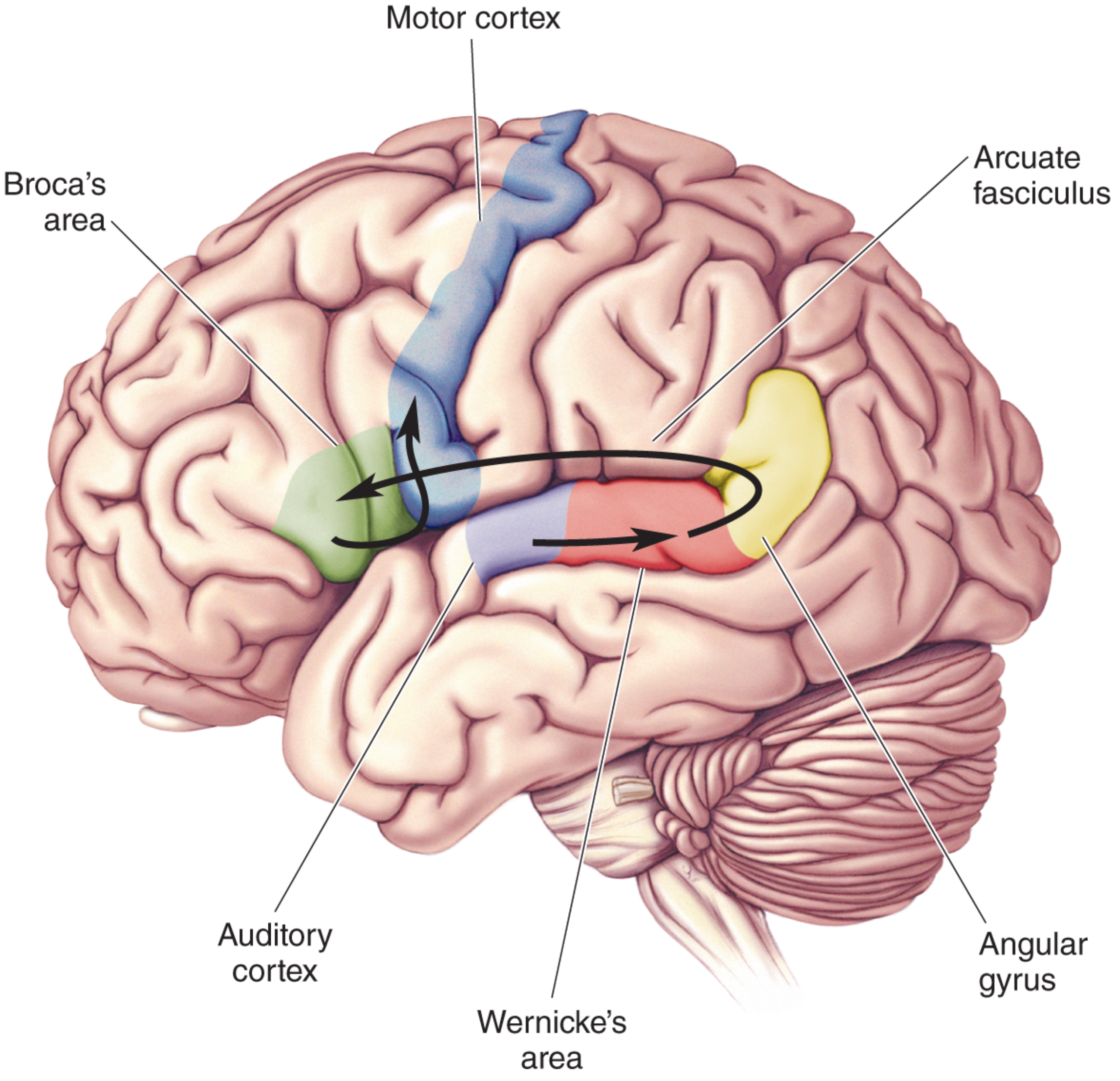
| Structure | Location | Function |
|---|---|---|
| Broca's area | Inferior frontal gyrus (BA 44/45), left | Motor speech programming, grammar production |
| Wernicke's area | Posterior superior temporal gyrus (BA 22), left | Auditory language comprehension, word meaning |
| Arcuate fasciculus | White matter arc, temporal → frontal | Connects Wernicke to Broca; underlies repetition |
| Angular gyrus | Inferior parietal lobule (BA 39) | Cross-modal integration (visual → language); reading, writing, calculation |
| Supramarginal gyrus | Inferior parietal lobule (BA 40) | Phonological processing |
| Insular cortex | Deep to Sylvian fissure | Articulatory coordination |
3. Bedside Language Examination
- Spontaneous speech - Is it fluent (>100 words/min, normal phrase length, effortless) or nonfluent (<50 words/min, short phrases, effortful/telegraphic)?
- Comprehension - Follow 1-, 2-, 3-step commands; yes/no questions
- Repetition - Repeat phrases like "No ifs, ands, or buts" or "The orchestra played and the audience applauded"
- Naming - Name objects, body parts, colors (confrontation naming)
- Reading and Writing - Read a sentence aloud and silently; write to dictation
4. The Major Aphasia Syndromes
4a. Broca's Aphasia (Non-fluent / Expressive / Motor Aphasia)
- Non-fluent speech - slow, labored, effortful, choppy ("telegraphic")
- Agrammatism - content words (nouns, verbs) produced, but function words (articles, conjunctions, prepositions) and grammatical morphemes are dropped. Classic example: "Wife... come... hospital."
- Apraxia of speech - inconsistent phonemic errors (p→b, t→d), groping, false starts
- Comprehension relatively preserved (impaired for complex syntax)
- Repetition impaired
- Reading often impaired ("third alexia"); writing markedly impaired (dysmorphic, dysgrammatic)
| Feature | Finding |
|---|---|
| Spontaneous speech | Nonfluent, mute, or telegraphic; sometimes dysarthric |
| Naming | Impaired (tip-of-tongue phenomenon, literal paraphasias) |
| Comprehension | Intact (mild difficulty with complex syntax) |
| Repetition | Impaired |
| Reading | Often impaired |
| Writing | Impaired (dysmorphic, dysgrammatic) |
| Associated | Right hemiparesis, hemisensory loss, ± limb apraxia |
Mini-Broca aphasia: Mildest form - slightly effortful, halting speech only. Good prognosis. Recovers quickly.
4b. Wernicke's Aphasia (Fluent / Receptive / Sensory Aphasia)
- Fluent speech - normal or even rapid output (logorrhea), normal phrase length and prosody
- Speech is empty of meaning - full of paraphasias:
- Verbal/semantic paraphasia: wrong word substitution ("The grass is blue")
- Literal/phonemic paraphasia: wrong phoneme/syllable ("The grass is greel")
- Neologisms: invented non-words ("The grass is grumps")
- In extremis: jargon aphasia - speech is entirely incomprehensible gibberish
- Comprehension impaired - cannot follow commands, fails to understand conversation
- Repetition impaired
- Reading comprehension impaired; writing produces well-formed letters but paragraphic/meaningless content
| Feature | Finding |
|---|---|
| Spontaneous speech | Fluent, paraphasic, sometimes logorrhoeic |
| Naming | Impaired (bizarre paraphasic misnaming) |
| Comprehension | Impaired |
| Repetition | Impaired |
| Reading | Impaired (comprehension and reading aloud) |
| Writing | Well-formed letters but paragraphic/meaningless |
| Associated | ± Right hemianopia; motor/sensory signs usually absent |
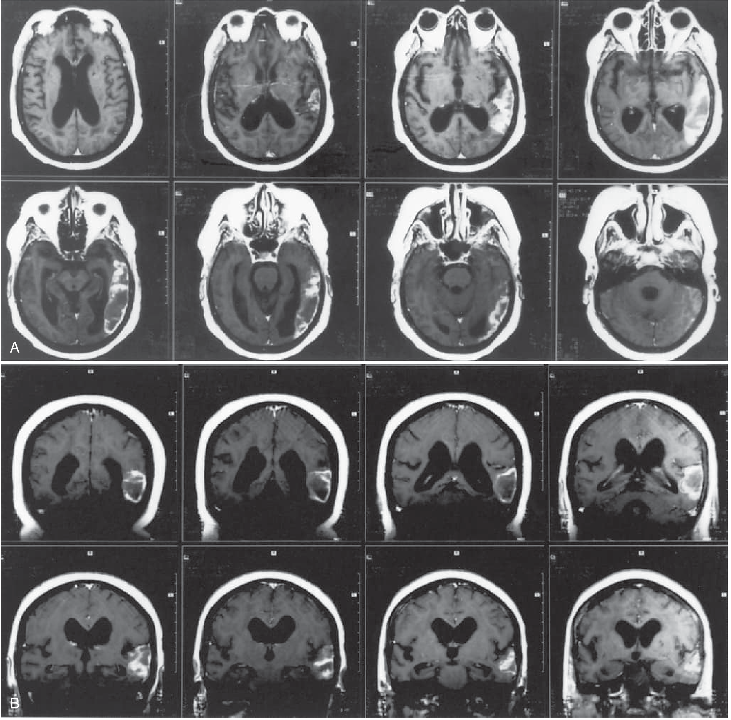
4c. Global Aphasia
- ALL language functions severely impaired: nonfluent/mute, comprehension impaired, repetition impaired, reading impaired, writing impaired
- Patient may utter only stereotyped sounds ("ah," "tah-tah"), a habitual phrase, or remain entirely mute
- May still participate in gestures of greeting and self-help activities
| Feature | Finding |
|---|---|
| All domains | Impaired |
| Associated | Right hemiplegia + hemisensory loss + hemianopia |
4d. Conduction Aphasia
- Fluent spontaneous speech with literal (phonemic) paraphasias - patient knows what they want to say and is aware of errors
- Comprehension preserved
- Repetition disproportionately impaired - the defining hallmark. A patient who can hold a conversation and follows commands cannot repeat a 3-word phrase. Classic: a patient could not repeat "boy" but said "I like girls better."
- Conduite d'approche: repeated self-correction attempts, circling around the target word (patient is aware of errors and tries to fix them)
- Reading aloud impaired; reading comprehension largely intact
| Feature | Finding |
|---|---|
| Spontaneous speech | Fluent, hesitant, literal paraphasias |
| Naming | Moderately impaired |
| Comprehension | Intact |
| Repetition | Markedly impaired (hallmark) |
| Reading aloud | Impaired; comprehension largely intact |
| Writing | Variable deficits |
4e. Anomic Aphasia
- Fluent speech with word-finding pauses and circumlocution (talking around the word)
- Comprehension, repetition, reading, writing all intact
- Naming is the sole deficit - patients know what the object is, can describe its use, but cannot produce its name
- Less common words give more trouble than common ones
- Inability to produce nouns → temporal lobe lesion
- Inability to produce verbs → frontal lobe lesion
- Angular gyrus lesions often add: Gerstmann syndrome (agraphia + acalculia + right-left disorientation + finger agnosia), plus alexia
- Anomia is seen non-specifically with any left hemisphere or even diffuse disease (Alzheimer's) - has limited localizing value in isolation
| Feature | Finding |
|---|---|
| Spontaneous speech | Fluent, word-finding pauses, circumlocution |
| Naming | Impaired |
| Comprehension | Intact |
| Repetition | Intact |
| Reading | Intact |
| Writing | Intact except for anomia |
4f. Transcortical Aphasias
| Type | Fluency | Comprehension | Repetition | Equivalent to | Lesion Location |
|---|---|---|---|---|---|
| Transcortical Motor (TCM) | Nonfluent, hesitant | Intact | Preserved | Broca (minus repetition deficit) | Anterior to Broca's area; supplementary motor area; ACA/MCA watershed |
| Transcortical Sensory (TCS) | Fluent, paraphasic | Impaired | Preserved | Wernicke (minus repetition deficit) | Posterior temporal-parietal, MCA/PCA watershed |
| Mixed Transcortical (Isolation syndrome) | Nonfluent/mute | Impaired | Preserved (echolalic) | Global (minus repetition) | Large watershed infarcts sparing perisylvian cortex; advanced dementia |
5. Classification Summary Table
| Aphasia | Fluency | Comprehension | Repetition | Naming | Key Feature | Lesion |
|---|---|---|---|---|---|---|
| Broca | Non-fluent | Intact* | Impaired | Impaired | Telegraphic/agrammatic speech | Inferior frontal (BA44/45) |
| Wernicke | Fluent | Impaired | Impaired | Impaired | Paraphasia, jargon, anosognosia | Post. superior temporal (BA22) |
| Global | Non-fluent | Impaired | Impaired | Impaired | All language impaired + hemiplegia | Entire perisylvian zone (proximal MCA) |
| Conduction | Fluent | Intact | Markedly impaired | Moderate | Disproportionate repetition deficit, conduite d'approche | Arcuate fasciculus / supramarginal gyrus |
| Anomic | Fluent | Intact | Intact | Impaired | Word-finding only; otherwise intact | Angular gyrus / temporal |
| Transcortical Motor | Non-fluent | Intact | Preserved | Impaired | Like Broca but can repeat | Anterior to Broca, SMA, ACA/MCA watershed |
| Transcortical Sensory | Fluent | Impaired | Preserved | Impaired | Like Wernicke but can repeat | MCA/PCA watershed |
| Mixed Transcortical | Non-fluent | Impaired | Preserved (echolalic) | Impaired | Like Global but echolalia | Bilateral watershed, advanced dementia |
6. Special / Less Common Aphasias
7. Primary Progressive Aphasia (PPA)
| Variant | Core deficit | Pathology |
|---|---|---|
| Non-fluent/Agrammatic PPA | Non-fluent, agrammatic (like Broca) | TDP-43, tau (CBD/PSP) |
| Semantic variant PPA (svPPA) | Word meaning lost (semantic memory) - fluent speech but cannot tell you what a "comb" is | TDP-43 (semantic dementia) |
| Logopenic variant PPA (lvPPA) | Word retrieval + sentence repetition impaired | Alzheimer pathology |
8. Language Lateralization
- ~96% of right-handers: Language dominant in the left hemisphere
- ~60-70% of left-handers: Still left-hemisphere dominant
- Prognosis from aphasia is better in left-handers than right-handers, due to greater bihemispheric language representation
- The right hemisphere contributes to prosody (emotional tone of speech) and pragmatics (context-appropriate language); right hemisphere lesions cause aprosodia (flat, robotic speech)
9. Paraphasia Types (Quick Reference)
| Type | Description | Example | Associated aphasia |
|---|---|---|---|
| Verbal/semantic | Wrong word, same category | "knife" → "fork" | Wernicke |
| Literal/phonemic | Wrong phoneme | "table" → "tabel" | Conduction, Wernicke |
| Neologism | Invented non-word | "table" → "trable" | Wernicke |
| Circumlocution | Talking around the word | "the thing you tell time with" | Anomic |
| Perseveration | Repeating a previous response | "clock... clock... clock" | Broca, global |
10. Assessment Tools
- Boston Diagnostic Aphasia Examination (BDAE) - gold standard research battery
- Western Aphasia Battery (WAB) - produces "Aphasia Quotient" score
- Bedside screening: The three-item language screen of fluency + comprehension + repetition classifies most aphasias
- Imaging: MRI DWI (acute stroke), FLAIR (subacute); PET/SPECT for PPA
11. Treatment and Prognosis
- Traumatic aphasia recovers faster and more completely than stroke-related aphasia
- Global aphasia - usually improves little
- Mini-Broca and dissociative syndromes - recover rapidly, often completely
- Better prognosis: left-handedness, younger age, smaller lesion, early onset of some comprehension, posterior (Wernicke) rather than anterior (Broca) lesion
- Recovery pattern: Global → severe Broca; Wernicke/conduction/transcortical → Anomic
- Spontaneous improvement occurs over months; active SLT accelerates word retrieval recovery
- Donepezil, memantine (for PPA with Alzheimer pathology)
- Bromocriptine (dopaminergic; some evidence for transcortical motor aphasia)
- Piracetam (mixed evidence)
- Transcranial magnetic stimulation (TMS) / tDCS - active research; may enhance SLT effects
Key Exam Mnemonics
- Frontal → Fails to speak (non-fluent) = Broca
- Temporal → Talks too much (fluent) = Wernicke
- Perisylvian lesion (Broca, Wernicke, Conduction, Global) → Repetition impaired
- Extra-perisylvian lesion (Transcortical variants) → Repetition preserved
- Frontal (Broca, TCM) → Comprehension intact
- Temporal (Wernicke, TCS, Global) → Comprehension impaired
- Parietal/arcuate (Conduction) → Comprehension intact, repetition gone
talk about cranial nerve functions and their palsy
cranial nerve palsy functions clinical features neurology 2024
Cranial Nerves: Functions and Palsies
Overview
Oh Oh Oh To Touch And Feel Very Good Velvet Ah Heaven (Olfactory, Optic, Oculomotor, Trochlear, Trigeminal, Abducens, Facial, Vestibulocochlear, Glossopharyngeal, Vagus, Accessory, Hypoglossal)
Some Say Marry Money But My Brother Says Big Brains Matter Most (S, S, M, M, B, M, B, S, B, B, M, M)
The 12 Cranial Nerves: Functions, Foramina, Palsies
CN I - Olfactory Nerve
| Cause | Mechanism |
|---|---|
| Head trauma (most common) | Shear injury to olfactory fili crossing the cribriform plate |
| Subfrontal meningioma | Compression of olfactory bulb/tract |
| Viral (COVID-19, post-URI) | Olfactory neuroepithelium damage |
| Neurodegenerative disease | Parkinson's, Alzheimer's - early sign, often precedes motor/cognitive symptoms by years |
| Zinc deficiency | Metabolic |
| Frontal lobe abscess/tumor | Mass effect |
CN II - Optic Nerve
| Location | Field Defect |
|---|---|
| Optic nerve (pre-chiasmal) | Monocular visual loss (ipsilateral) |
| Optic chiasm center (pituitary tumor) | Bitemporal hemianopia |
| Optic chiasm lateral (rare) | Binasal hemianopia |
| Optic tract (post-chiasmal) | Contralateral homonymous hemianopia |
| Optic radiation - temporal (Meyer's loop) | Contralateral superior quadrantanopia ("pie in the sky") |
| Optic radiation - parietal | Contralateral inferior quadrantanopia |
| Occipital cortex (V1) | Contralateral homonymous hemianopia with macular sparing |
- Optic neuritis: Painful monocular visual loss, RAPD (relative afferent pupillary defect), central scotoma. Common first presentation of MS. Treatment: IV methylprednisolone.
- Ischemic optic neuropathy (ION): Painless visual loss. Arteritic (GCA) vs. non-arteritic. ESR/CRP urgent if GCA suspected → immediate steroids.
- Papilledema: Bilateral optic disc swelling from raised ICP - visual fields initially normal; later concentric constriction. No pain.
- Optic atrophy: Pale disc - end result of any optic nerve damage.
CN III - Oculomotor Nerve
- Motor: Superior rectus (contralateral subnucleus!), inferior rectus, medial rectus, inferior oblique, levator palpebrae superioris
- Parasympathetic (Edinger-Westphal nucleus): Pupil constriction (sphincter pupillae) + lens accommodation (ciliary muscle)
- Superior Oblique = CN IV (trochlear)
- Lateral Rectus = CN VI (abducens)
- Everything else = CN III
- Complete ptosis (levator palpebrae failure)
- Eye deviated "down and out" (exotropia + hypotropia - unopposed action of lateral rectus (CN VI) and superior oblique (CN IV))
- Diplopia
- Fixed, dilated pupil (if parasympathetic fibers involved)
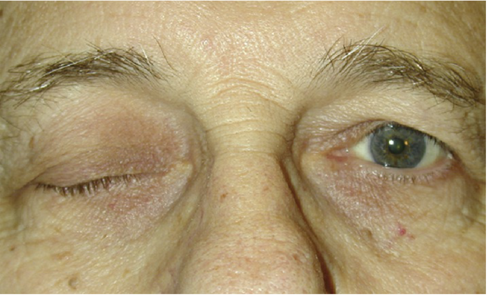
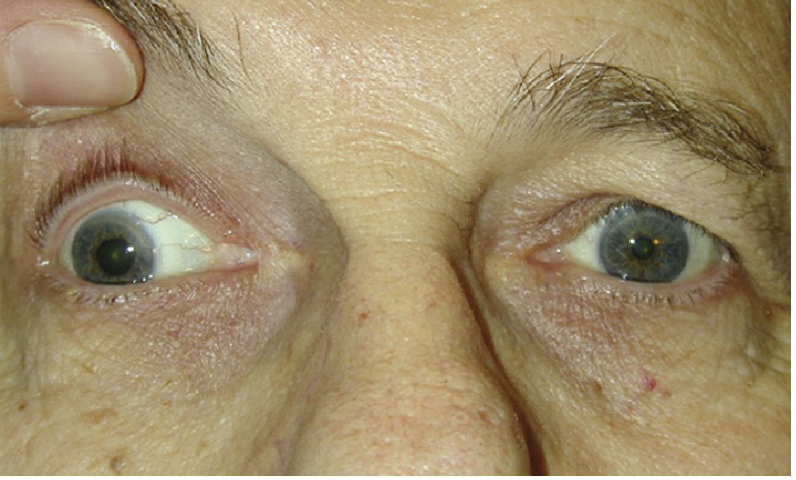
| Finding | Think | Urgency |
|---|---|---|
| Pupil-INVOLVING (dilated, fixed) | Posterior communicating artery aneurysm compressing outer parasympathetic fibers | EMERGENCY - CTA/MRA now |
| Pupil-SPARING (intact miosis) | Microvascular ischemia (DM, HTN) - inner axons spared, outer parasym fibers compressed by ischemia from within | Urgent work-up, less emergent |
- Pupil-involving: PCoA aneurysm (most serious), uncal herniation, cavernous sinus mass, pituitary apoplexy, tumor, trauma
- Pupil-sparing: Microvascular ischemia (DM most common), GCA, orbital disease
- In children: ophthalmoplegic migraine
| Location | Features |
|---|---|
| Nucleus (midbrain) | Ipsilateral CN III palsy + bilateral superior rectus weakness (superior rectus subnucleus crosses) + bilateral ptosis (midline levator nucleus) |
| Fascicle - through red nucleus | CN III palsy + contralateral tremor/ataxia = Benedikt syndrome |
| Fascicle - through cerebral peduncle | CN III palsy + contralateral hemiplegia = Weber syndrome |
| PCoA junction (subarachnoid) | CN III palsy, pupil-involving, painful |
| Cavernous sinus | CN III + IV + V1/V2 + VI + Horner's |
| Superior orbital fissure | CN III + IV + V1 + VI |
CN IV - Trochlear Nerve
- Vertical diplopia (worse looking down and in - e.g., reading, going downstairs)
- Head tilt to the opposite side (compensatory - tilting away from the affected eye reduces diplopia)
- Ipsilateral hypertropia (affected eye higher - superior oblique failure → unopposed inferior oblique lifts the eye)
- Extorsion of the affected eye
- Bielschowsky three-step test: confirms CN IV palsy
- Which eye is hypertropic in primary gaze? → the affected side
- Is hypertropia worse on gaze to the left or right? → worse on gaze to opposite side
- Is hypertropia worse on head tilt left or right? → worse tilting toward the affected eye
- Trauma (most common acquired) - the long intracranial course and dorsal exit make it vulnerable to contrecoup injury
- Microvascular ischemia (DM, HTN)
- Congenital (often decompensates in adulthood - look for childhood photographs showing old head tilt)
- Cavernous sinus, superior orbital fissure
- Tentorial meningioma
CN V - Trigeminal Nerve
- Motor nucleus: Pons
- Chief sensory nucleus: Pons (light touch, pressure)
- Spinal nucleus (descending): Pons → medulla → C2 (pain, temperature)
- Mesencephalic nucleus: Midbrain (proprioception from jaw muscles) Exit: Three divisions:
- V1 (Ophthalmic): Superior orbital fissure
- V2 (Maxillary): Foramen rotundum
- V3 (Mandibular): Foramen ovale
- Sensory: Facial sensation (V1: forehead/cornea/scalp, V2: cheek/upper lip/palate, V3: jaw/lower lip/chin/anterior 2/3 tongue somatosensation)
- Motor (V3 only): Muscles of mastication (masseter, temporalis, medial/lateral pterygoids), tensor tympani, tensor veli palatini, mylohyoid, anterior digastric
- Reflexes: Afferent limb of corneal reflex (V1) and jaw jerk reflex (V3)
- Parasympathetic distribution: CN V branches carry parasympathetic fibers from CN III (to ciliary ganglion → pupil/lens), VII (pterygopalatine ganglion → lacrimal/nasal; submandibular ganglion → salivary), IX (otic ganglion → parotid)
- Lancinating, electric-shock pain in V2/V3 distribution (rarely V1)
- Triggered by light touch - eating, talking, brushing teeth, cold wind
- Most common in women >50 years
- Cause: Most often vascular compression of CN V root at pons (superior cerebellar artery most common)
- Treatment: Carbamazepine (first-line), oxcarbazepine; if refractory → microvascular decompression (MVD), gamma knife
- Red flags for secondary causes: age <50, bilateral, onset in V1, sensory deficit (suggests tumor, MS)
- Numbness in trigeminal distribution
- Causes: Sjogren's syndrome (most common systemic cause), scleroderma, SLE, cancer (skull base)
- CN V3 motor lesion → jaw deviates TOWARD the side of the lesion (ipsilateral pterygoids weak → contralateral push unopposed)
- Afferent limb: CN V1 → both eyes blink (consensual)
- Efferent: CN VII (orbicularis oculi)
- If V1 lesion → ipsilateral corneal reflex lost, but consensual reflex (other eye blinks to corneal touch of normal eye) is intact
- V1: Forehead sensation, corneal reflex
- V2: Cheek sensation
- V3: Chin/jaw sensation + jaw strength (bite, jaw opening)
CN VI - Abducens Nerve
- Esotropia (eye deviated inward - unopposed medial rectus)
- Horizontal diplopia (worse on ipsilateral gaze)
- Inability to abduct the eye beyond midline
- Head turn toward affected side (compensatory)
- Raised ICP (most common false localizing sign - stretching of long intracranial course)
- Microvascular ischemia (DM, HTN)
- Gradenigo syndrome: Petrous apex lesion → CN VI palsy + CN V pain + ipsilateral otitis media (Petrous Apex Syndrome)
- Cavernous sinus pathology
- Wernicke's encephalopathy (thiamine deficiency - bilateral CN VI)
- Tumor (brainstem, clivus, NPC extending to skull base)
- MS (internuclear ophthalmoplegia is more common, but CN VI fascicle involvement possible)
- Foville syndrome (dorsal pons): Ipsilateral CN VI palsy + ipsilateral CN VII palsy + ipsilateral conjugate gaze palsy (PPRF damage) + contralateral hemiplegia
CN VII - Facial Nerve
- Motor: All muscles of facial expression (frontalis, orbicularis oculi, orbicularis oris, buccinator, platysma, stapedius)
- Special sensory (taste): Anterior 2/3 of tongue via chorda tympani → geniculate ganglion → NTS
- Parasympathetic:
- Greater petrosal nerve → pterygopalatine ganglion → lacrimal gland (tearing) + nasal/palatal glands
- Chorda tympani → submandibular ganglion → submandibular + sublingual salivary glands
- General sensory: Small area of skin around external auditory meatus (Ramsay Hunt zone)
- Greater petrosal nerve (at geniculate ganglion) - lacrimation
- Nerve to stapedius - dampens loud sounds
- Chorda tympani - taste anterior 2/3 tongue + submandibular/sublingual salivation
To Zanzibar By Motor Car Temporal, Zygomatic, Buccal, Marginal mandibular, Cervical
| Feature | UMN (central) lesion | LMN (peripheral) lesion |
|---|---|---|
| Forehead | SPARED (bilateral cortical input to upper face) | INVOLVED (whole face, including forehead) |
| Lower face | Contralateral weakness | Ipsilateral weakness |
| Eye closure | Usually intact | Impaired - lagophthalmos |
| Taste | Intact | Lost (if lesion proximal to chorda tympani) |
| Lacrimation | Intact | Lost (if proximal to greater petrosal nerve) |
| Cause | Contralateral MCA stroke, tumor | Bell's palsy, Ramsay Hunt, parotid tumor, otitis media, trauma |
| Level of lesion | Motor palsy | Taste lost | Hyperacusis | Lacrimation lost |
|---|---|---|---|---|
| Brainstem (CN VII nucleus) | Ipsilateral whole face | Yes | Yes | Yes |
| Geniculate ganglion | Ipsilateral whole face | Yes | Yes | Yes |
| After stapedius branch | Ipsilateral whole face | Yes | No | No |
| After chorda tympani | Ipsilateral whole face | No | No | No |
| Stylomastoid foramen | Ipsilateral whole face | No | No | No |
| Parotid (terminal branches) | Partial (branch-specific) | No | No | No |
- Most common cause of acute LMN CN VII palsy (70% of all facial palsies)
- Idiopathic; HSV-1 reactivation in geniculate ganglion strongly implicated
- Acute onset unilateral, complete facial weakness (whole face including forehead)
- ± Pain behind ear (prodrome), ± taste loss, ± hyperacusis, ± eye watering/dryness
- Management: Oral prednisolone (25-50mg/day × 10 days) within 72 hours + antiviral (acyclovir/valacyclovir) if severe. Eye protection (lubricants, tape at night) is mandatory to prevent corneal exposure.
- Prognosis: ~71% recover fully; majority improve within 3-6 months
- Herpes zoster reactivation in geniculate ganglion
- Triad: Ipsilateral LMN facial palsy + herpetic vesicles in external auditory canal/auricle + ipsilateral sensorineural hearing loss ± vertigo
- More severe than Bell's palsy - only 50% full recovery
- Treatment: Antiviral + steroids urgently
CN VIII - Vestibulocochlear Nerve
- Cochlear division: Hearing (frequency-specific hair cells of organ of Corti → spiral ganglion → cochlear nuclei → bilateral superior olivary nuclei → inferior colliculus → medial geniculate → auditory cortex)
- Vestibular division: Balance, head position, acceleration (utricle, saccule, 3 semicircular canals → Scarpa's ganglion → vestibular nuclei → cerebellum, spinal cord, extraocular motor nuclei)
- Conductive: External/middle ear problem (cerumen, otitis media, ossicular chain disruption)
- Sensorineural: Hair cell/CN VIII/central pathway problem
- Weber test: Tuning fork on vertex - sound lateralizes to WORSE ear in conductive loss; BETTER ear in sensorineural loss
- Rinne test: BC > AC = conductive loss; AC > BC = normal or sensorineural
- Presbycusis (age-related, high-frequency first)
- Noise-induced
- Vestibular schwannoma (acoustic neuroma): Progressive unilateral SNHL + tinnitus ± imbalance; MRI with gadolinium diagnostic; at CPA angle
- Ototoxic drugs (aminoglycosides, cisplatin, loop diuretics, quinine)
- Meniere's disease (episodic vertigo + fluctuating SNHL + tinnitus + aural fullness)
- Sudden SNHL (treat with steroids within 24-72 hours)
- BPPV, vestibular neuritis, labyrinthitis, Meniere's - covered in previous session
CN IX - Glossopharyngeal Nerve
- Motor: Stylopharyngeus (elevates pharynx) - only muscle CN IX innervates
- General sensory (afferent): Posterior 1/3 of tongue, pharynx, tonsils, middle ear (tympanic plexus), carotid sinus/body (chemoreceptors, baroreceptors)
- Special sensory (taste): Posterior 1/3 of tongue
- Parasympathetic: Inferior salivatory nucleus → lesser petrosal nerve → otic ganglion → parotid gland salivation
- Isolated CN IX lesion is rare
- Loss of gag reflex (afferent limb = CN IX, efferent = CN X)
- Dysgeusia (posterior tongue taste lost)
- Referred otalgia (ear pain from pharyngeal pathology - Jacobson's nerve)
- Glossopharyngeal neuralgia: Severe lancinating pain triggered by swallowing, talking, yawning - similar to trigeminal neuralgia but in throat/ear territory. Can cause cardiac syncope (vagal efferent activation from CN IX trigger). Treatment: carbamazepine; MVD if refractory.
CN X - Vagus Nerve
- Motor (nucleus ambiguus): Pharynx (levator veli palatini, most pharyngeal constrictors), larynx (all intrinsic laryngeal muscles via recurrent laryngeal nerve)
- Parasympathetic (dorsal motor nucleus): Heart (slows rate), bronchi, GI tract (esophagus to splenic flexure of colon)
- Sensory: Pharynx, larynx, external ear, dura of posterior fossa; aortic arch baroreceptors/chemoreceptors
- Special sensory (taste): Epiglottis/laryngeal inlet
- Hoarseness/dysphonia (laryngeal muscles - recurrent laryngeal nerve)
- Dysarthria (nasal speech - soft palate droop)
- Dysphagia (pharyngeal weakness - aspiration risk)
- Palatal deviation: Uvula deviates AWAY from the side of lesion (intact side pulls uvula toward itself)
- Loss of gag reflex (efferent limb)
- Tachycardia (unilateral vagal lesion - less relevant)
- Left RLN: Long course around aortic arch → vulnerable to mediastinal pathology (aortic aneurysm, lung cancer, mediastinal lymphadenopathy, hilar masses) - Ortner's syndrome
- Right RLN: Shorter, loops around subclavian artery
- Both: surgical injury (thyroidectomy, parathyroidectomy, neck dissection)
- Unilateral RLN palsy: Hoarseness, breathy voice, aspiration on thin liquids
- Bilateral RLN palsy: Inspiratory stridor, respiratory distress, possible asphyxia
- Dysarthria, dysphagia, hoarseness, tongue fasciculations, nasal regurgitation
- Tongue wasted and fasciculating, palate doesn't rise, absent gag
- Causes: motor neuron disease (ALS), brainstem infarct, Guillain-Barré, syringobulbia, nasopharyngeal carcinoma
CN XI - Spinal Accessory Nerve
- Sternocleidomastoid (SCM): Turns head to the OPPOSITE side, flexes neck
- Trapezius (upper): Elevates and retracts shoulder; shrugs
- SCM weakness: Inability to turn head to the opposite side against resistance
- Trapezius weakness: Shoulder drop (winging of scapula - medial), inability to shrug, shoulder droop, difficulty raising arm above 90°
- Ipsilateral weakness of both muscles
- Jugular foramen lesions (tumor, glomus jugulare, metastasis) - usually with CN IX and X also involved
- Neck surgery (lymph node biopsy, neck dissection)
- Trauma to posterior triangle of neck
- Radiation
- Neuromuscular (rare)
CN XII - Hypoglossal Nerve
-
LMN lesion (nucleus or nerve):
- Tongue deviates TOWARD the side of the lesion (weak side "falls" toward the lesion; the intact side's genioglossus pushes tongue to the weak side)
- Ipsilateral tongue atrophy and fasciculations
- Dysarthria (especially lingual consonants: L, R, N, T, D)
- Dysphagia
-
UMN lesion (corticobulbar tract):
- Tongue deviates AWAY from the lesion (toward the contralateral, weaker side)
- No atrophy
- Often part of contralateral hemiplegia
- Bilateral UMN lesion → pseudobulbar palsy
- Skull base (hypoglossal canal): Nasopharyngeal carcinoma, metastasis, meningioma, chordoma
- Medullary infarct (medial medullary syndrome = CN XII palsy + contralateral hemiplegia + contralateral proprioception loss)
- ALS (tongue fasciculations prominent)
- Neck mass, carotid dissection
Cranial Nerve Summary Table
| CN | Name | Modalities | Exit foramen | Key functions | Palsy sign(s) |
|---|---|---|---|---|---|
| I | Olfactory | SS | Cribriform plate | Smell | Anosmia |
| II | Optic | SS | Optic canal | Vision, pupil afferent | Visual field defect, RAPD |
| III | Oculomotor | SM + PS | Superior orbital fissure | All EOM except SO+LR; levator palpebrae; pupil constriction | Ptosis, down-and-out eye, mydriasis |
| IV | Trochlear | SM | Superior orbital fissure | Superior oblique (intortion, depression) | Vertical diplopia, head tilt, hypertropia |
| V | Trigeminal | SM + GS | SOF (V1), Rotundum (V2), Ovale (V3) | Face sensation; muscles of mastication; corneal reflex afferent | Facial numbness, jaw deviation, trigeminal neuralgia |
| VI | Abducens | SM | Superior orbital fissure | Lateral rectus (abduction) | Esotropia, horizontal diplopia, cannot abduct |
| VII | Facial | SM + SS + PS + GS | Stylomastoid foramen | Facial expression; taste ant 2/3 tongue; lacrimation; salivation | Bell's palsy, forehead sparing vs. whole face weakness |
| VIII | Vestibulocochlear | SS | Internal acoustic meatus | Hearing, balance | SNHL, vertigo, tinnitus |
| IX | Glossopharyngeal | SM + GS + SS + PS | Jugular foramen | Stylopharyngeus; taste post 1/3 tongue; gag afferent; parotid salivation | Loss of gag, dysphagia, glossopharyngeal neuralgia |
| X | Vagus | SM + GS + SS + PS | Jugular foramen | Pharynx/larynx; palate; parasympathetics thorax/abdomen | Hoarseness, dysphagia, uvula deviation away |
| XI | Accessory | SM | Jugular foramen | SCM, upper trapezius | Inability to shrug/turn head; shoulder drop |
| XII | Hypoglossal | SM | Hypoglossal canal | All tongue muscles | Tongue deviates toward lesion (LMN), atrophy, dysarthria |
Skull Foramina Quick Reference
| Foramen | Location | Contents |
|---|---|---|
| Cribriform plate | Ethmoid | CN I |
| Optic canal | Lesser wing sphenoid | CN II, ophthalmic artery |
| Superior orbital fissure | Between greater/lesser wings | CN III, IV, V1, VI, ophthalmic veins, sympathetics |
| Foramen rotundum | Greater wing sphenoid | CN V2 |
| Foramen ovale | Greater wing sphenoid | CN V3, accessory meningeal artery |
| Internal acoustic meatus | Temporal bone | CN VII, CN VIII |
| Jugular foramen | Temporal/occipital | CN IX, X, XI, jugular vein |
| Hypoglossal canal | Occipital bone | CN XII |
Multi-CN Syndromes
Cavernous Sinus Syndrome
- Findings: CN III/IV/VI palsies (ophthalmoplegia, ptosis, diplopia) + V1/V2 sensory loss + Horner's syndrome (sympathetic damage)
- Causes: Cavernous sinus thrombosis (septic - paranasal sinus/dental infection), carotid-cavernous fistula, pituitary apoplexy, meningioma, Tolosa-Hunt syndrome (painful idiopathic)
- Key: Proptosis + chemosis + ophthalmoplegia in a patient with sinusitis/dental infection = septic cavernous sinus thrombosis → urgent CT/MRI + antibiotics
Superior Orbital Fissure Syndrome
Orbital Apex Syndrome
Jugular Foramen Syndrome (Vernet syndrome)
- Dysphagia, hoarseness, uvula deviation, loss of gag, SCM/trapezius weakness
- Cause: Glomus jugulare tumor, NPC, metastases, meningioma
Posterior Fossa / Foramen Magnum
Cerebellopontine Angle (CPA) lesion
- Most common: Vestibular schwannoma (acoustic neuroma)
- Also: Meningioma, epidermoid cyst, facial neuroma
Medial Medullary Syndrome (Dejerine syndrome)
- CN XII palsy (ipsilateral tongue deviation)
- Contralateral hemiplegia (pyramid)
- Contralateral proprioception/vibration loss (medial lemniscus)
- Cause: Anterior spinal artery or vertebral artery occlusion
Lateral Medullary Syndrome (Wallenberg syndrome)
- Ipsilateral CN V (facial pain/numbness), CN IX/X (dysphagia/hoarseness), CN XI (shoulder drop), Horner's syndrome (descending sympathetics)
- Contralateral body pain/temperature loss
- Ipsilateral cerebellar signs (PICA territory)
- Cause: PICA or vertebral artery occlusion
Parasympathetic CN Summary
| CN | Ganglion | Target |
|---|---|---|
| CN III | Ciliary ganglion | Pupil constriction, lens accommodation |
| CN VII (greater petrosal) | Pterygopalatine ganglion | Lacrimal gland, nasal/palatal mucosa |
| CN VII (chorda tympani) | Submandibular ganglion | Submandibular + sublingual glands |
| CN IX | Otic ganglion | Parotid gland |
| CN X | Myenteric/submucosal ganglia in organs | Heart, lungs, esophagus → splenic flexure |
talk about stroke and predispositions like cardioembolic events
stroke cardioembolic causes risk factors AHA guidelines 2024 2025
stroke cardioembolic atrial fibrillation prevention anticoagulation