
Osseous surgeries in periodontics
osseous surgery periodontics types osteoplasty ostectomy bone surgery periodontology
osseous surgery periodontics resective regenerative
https://pocketdentistry.com/60-resective-osseous-surgery
osseous surgery periodontal bone defects osteoplasty ostectomy
Osseous Surgeries in Periodontics
Classification
1. Additive (Regenerative) Osseous Surgery
2. Subtractive (Resective) Osseous Surgery
Resective Osseous Surgery - Core Procedures
| Procedure | Definition |
|---|---|
| Osteoplasty | Reshaping bone WITHOUT removing tooth-supporting (alveolar) bone. Corrects bony contour. |
| Ostectomy (Osteoectomy) | REMOVAL of tooth-supporting alveolar bone. Used to eliminate pockets and achieve positive architecture. |
Bony Architecture Concepts
- Positive architecture - Normal/ideal; interproximal bone coronal to facial/lingual bone
- Negative (reverse) architecture - Facial/lingual bone coronal to interproximal bone; abnormal
- Flat architecture - Interproximal and facial/lingual bone at the same level
Classification of Bony (Osseous) Defects
| Type | Description | Treatment preference |
|---|---|---|
| 1-wall defect | One remaining wall | Resective surgery (osteoplasty/ostectomy) |
| 2-wall defect (crater) | Two remaining walls; most common interproximal defect | Either resective or regenerative depending on depth/width |
| 3-wall defect | Three remaining walls; narrow and deep | Regenerative surgery preferred (best prognosis) |
| Combined/hemiseptal | Mixed | Case-dependent approach |
Indications for Osseous Surgery
- Residual pocketing (>4 mm) after completion of non-surgical therapy (scaling and root planing)
- Shallow to moderate bone loss (2-3 mm)
- Infrabony pockets with 1 or 2 bony walls
- Moderate-length root trunks
- Furcation defects (Class I/II) amenable to resection
- Elimination of bony exostoses, ledges, and irregular contours
- Deep localized bone defects (>3 mm) - regenerative surgery preferred
- Anterior maxilla - avoided due to esthetic concerns (gingival recession)
- Advanced attachment loss with deep intrabony defects
- Patients where tooth extraction is the better option
Surgical Technique (Resective Osseous Surgery)
- Flap reflection - Full-thickness mucoperiosteal flap raised (alternatively, split-thickness flap in fiber retention osseous surgery)
- Root debridement - Thorough scaling and root planing
- Soft tissue removal from defects using curettes, ultrasonic scalers, or small burs
- Osteoplasty first - Bony ledges, shelves, and exostoses removed with round surgical burs
- Ostectomy - Using end-cutting burs, bone removed toward the lingual side to the depth of interproximal defect
- Contouring - Establish positive architecture on mid-facial and mid-lingual aspects
- Flap closure - Sutured at or slightly apical to the crest of bone
Variant: Fiber Retention Osseous Resective Surgery (FROSS)
- Similar pocket depth reduction compared to conventional osseous surgery
- Less gingival recession
- Less dentinal hypersensitivity
Additive Osseous Surgery - Regenerative Procedures
Bone Grafts
| Type | Source | Examples |
|---|---|---|
| Autograft | Patient's own bone | Healing socket, chin, ramus, iliac crest |
| Allograft | Human cadaveric bone | FDBA (freeze-dried bone allograft), DFDBA (demineralized) |
| Xenograft | Animal-derived | Bio-Oss (bovine), porcine |
| Alloplast | Synthetic | Hydroxyapatite, beta-TCP, bioactive glass |
Guided Tissue Regeneration (GTR)
- Uses a membrane (resorbable or non-resorbable) to exclude epithelium and connective tissue
- Allows selective repopulation of the root surface by periodontal ligament cells
- Best for 3-wall and combined defects
- Non-resorbable membranes (e.g., PTFE/ePTFE) require a second surgery for removal
- Resorbable membranes (collagen, polylactic acid) eliminate second surgery
Biologics
- Enamel matrix derivatives (EMD) - Emdogain; promotes cementum regeneration
- Platelet-rich plasma/fibrin (PRP/PRF) - Autologous growth factors
- rhPDGF-BB (GEM 21S) - Recombinant human PDGF with beta-TCP scaffold
Selection of Treatment Technique
- 1-wall angular defects - Resective recontouring
- 3-wall narrow deep defects - Regenerative (best outcome)
- 2-wall angular defects - Either method based on depth, width, configuration
- Interproximal craters (2-wall) - Resection effective for shallow defects; regeneration for deeper ones
- Wide shallow defects - Resective surgery
Expected Outcomes
- Significant pocket depth reduction in pockets >4 mm
- Mean attachment loss circumferentially: ~0.6 mm (properly applied)
- Gingival recession is an expected consequence
- High long-term predictability when patient selection is appropriate
- Variable bone fill (best in 3-wall defects)
- Clinical attachment level gain
- Improved long-term tooth prognosis in deep intrabony defects
Summary Table
| Feature | Resective Osseous Surgery | Regenerative Osseous Surgery |
|---|---|---|
| Goal | Eliminate pocket by removing bone | Regenerate lost attachment/bone |
| Defect type | 1-2 wall, shallow (2-3mm) | 3-wall, deep, narrow |
| Bone modification | Ostectomy + osteoplasty | Graft + GTR/biologics |
| Recession | Expected | Minimized |
| Predictability | High | Moderate-high (defect dependent) |
| Re-entry surgery | Not needed | Sometimes (non-resorbable membrane) |
What are the latest clinical guidelines for selecting resective vs. regenerative osseous surgery?
resective regenerative osseous surgery selection criteria periodontal defects
periodontal infrabony defect treatment guidelines 2023 2024 2025
EFP AAP 2024 2025 guidelines resective regenerative periodontal osseous surgery selection
PMID: 39058949
PMID: 33025619
https://pmc.ncbi.nlm.nih.gov/articles/PMC12476086
Latest Clinical Guidelines: Resective vs. Regenerative Osseous Surgery
1. The Overarching Framework (EFP S3-Level Guidelines)
- Step 1-2 (non-surgical therapy): Scaling and root planing + behavioral modification
- Re-evaluation at 8-12 weeks: Assess residual pocketing and bleeding on probing
- Surgical threshold: Sites with probing depth >5 mm with bleeding on probing after non-surgical therapy are candidates for surgery
- For residual deep pockets with complex defects, the guideline supports flap surgery + regenerative procedures for deep intrabony defects, with regenerative therapy (GTR or enamel matrix derivatives) recommended when an adequate RCT base exists
2. Primary Decision Driver: Defect Morphology
Defect Depth
- Greater initial defect depth predicts more radiographic bone gain after regenerative surgery
- Narrower defect angle and more bony walls both independently improve CAL gain and bone fill at 12 months
- These associations held irrespective of the biomaterial used - defect anatomy matters more than the choice of graft or membrane
Wall Count and Geometry
| Defect Type | First-Choice Strategy | Rationale |
|---|---|---|
| 3-wall intrabony, narrow, deep | Regenerative (GTR / EMD / graft) | Best blood supply, containment, predictable bone fill |
| 2-wall defect (crater), shallow <3 mm | Resective (osteoplasty ± ostectomy) | Too shallow for regeneration to be cost-effective |
| 2-wall defect, deep >3 mm | Regenerative preferred | Sufficient depth for meaningful attachment gain |
| 1-wall angular defect | Resective (recontouring) | Poor containment limits regenerative potential |
| Hemiseptal defect | Case-by-case; resective or combined | Anatomy limits true regeneration |
| Suprabony pocket (horizontal loss) | Resective | No bony walls to contain regenerative material |
- Number of walls
- Defect depth
- Extension to buccal/lingual surfaces
3. Defect Depth Threshold
- Intrabony component ≤3 mm - resective approach appropriate; small defect can be flattened by osteoplasty without unacceptable bone sacrifice
- Intrabony component >3 mm - resective surgery alone is not recommended; regenerative surgery or extraction should be considered
- Infrabony pockets with deep narrow defects - regenerative surgery achieves the best long-term tooth survival
4. Furcation Involvement
| Furcation Class | Strategy |
|---|---|
| Class I | Resective (odontoplasty, osteoplasty); good prognosis |
| Class II mandibular | Regenerative if anatomy permits (GTR/graft); tunnel preparation as alternative |
| Class II maxillary | More complex; regenerative if contained, resective if not; root resection in some cases |
| Class III | Resective (root resection, hemisection, extraction) - regeneration generally unpredictable |
5. Esthetic Zone Considerations
- Resective surgery in the anterior maxilla is generally avoided due to inevitable gingival recession exposing root surfaces with poor esthetic outcomes
- In esthetic zones, regenerative surgery or conservative flap debridement is strongly preferred
- The EFP guideline states regenerative procedures should be performed by clinicians with advanced training given technical complexity (MDPI Advances in Regenerative Therapies)
6. Patient-Level Factors
| Factor | Impact on Decision |
|---|---|
| Smoking | Significantly impairs regenerative outcomes (especially GTR); resective more predictable in heavy smokers |
| Diabetes control | Poor glycemic control reduces regenerative healing; resective may be safer |
| Compliance/oral hygiene | Poor plaque control negates regenerative gains; resective preferred until control achieved |
| Systemic risk profile | High-risk patients may benefit from simpler, faster resective procedures |
| Strategic value of tooth | Abutment or lone-standing tooth favors regenerative effort |
| Residual bone support | Minimal remaining bone - extraction may outweigh surgical benefit |
7. FibReORS - The Modern Hybrid Option (2025 Update)
- Uses a split-thickness flap to preserve supracrestal connective tissue fibers
- Allows more coronal positioning of the base of the defect
- Reduces supporting bone removal compared to traditional ostectomy
- Achieves similar pocket depth reduction with less recession and less dentinal hypersensitivity
- Represents a paradigm shift treating the mineralized and unmineralized connective tissue complex as a unit
8. Synthesized Decision Algorithm (Current Evidence)
Residual pocket after non-surgical therapy
            │
    ┌───────▼────────┐
    │ Defect type?   │
    └───────┬────────┘
            │
    ┌───────▼──────────────────────────────────┐
    │ Suprabony (horizontal bone loss)?         │──► Resective (osteoplasty/ostectomy)
    └───────┬──────────────────────────────────┘
            │ Infrabony
    ┌───────▼────────────────────────────┐
    │ Wall count and depth?              │
    └───────┬────────────────────────────┘
            │
    1-wall or 2-wall shallow (<3 mm) ──────────► Resective
    2-wall deep (>3 mm) + narrow angle ────────► Regenerative preferred
    3-wall narrow deep ────────────────────────► Regenerative (best outcomes)
    Furcation Class I-II (accessible) ─────────► Resective or regenerative
    Furcation Class III ────────────────────────► Resective/extraction
            │
    + Overlay patient factors (smoking, compliance, esthetics, strategic value)
Key Evidence Summary
| Source | Type | Key Finding |
|---|---|---|
| Nibali et al. 2021 (PMID 33025619) | Systematic Review + Meta-analysis | Deeper, narrower, more-walled defects = better regenerative outcomes regardless of biomaterial |
| Nibali & Cortellini 2025 (PMID 39058949) | Review | New classification linking defect anatomy to regenerative treatment planning |
| Carnevale et al. 2025 (PMID 40757958) | Review | Resective surgery validated for residual pockets and furcation defects; long-term data strong |
| EFP S3 Guideline 2025 (PMC 12839767) | Practice Guideline methodology | GTR/EMD for deep intrabony; resective/regenerative for furcations based on class and strategic value |
| Osseous Resective Surgery Review 2025 (PMC 12476086) | Review | Three histological outcomes framework; FibReORS as hybrid; defect anatomy > biomaterial choice |
Peri implantitis management strategies
peri-implantitis treatment management
peri-implantitis management 2024 2025 EFP AAP guidelines non-surgical surgical resective regenerative
PMID: 40501397
PMID: 39726090
PMID: 38789758
https://www.perio.org/press-release/academy-of-osseointegrat…
PMID: 39572812
PMID: 35476853
implantoplasty peri-implantitis resective surgery outcomes
peri-implantitis bone defect resective regenerative surgery implant
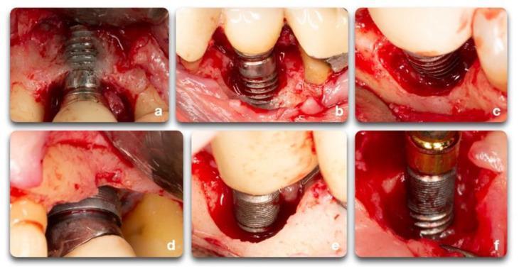
This composite of six intraoperative clinical photographs (a-f) illustrates various morphological classifications of peri-implant bone defects encountered during surgical treatment of peri-implantitis. (a) Displays a facial dehiscence where the buccal bone plate is lost, exposing the implant threads. (b, c) Show a combination of facial dehiscence and a semi-circumferential defect, indicating bone loss around a portion of the implant perimeter. (d) Illustrates a circumferential defect characterized by bone loss surrounding the entire implant diameter, combined with supra-crestal bone resorption where the bone level has receded below the original implant crest. (e) Features a deep circumferential defect with multiple remaining bony walls. (f) Demonstrates a semi-circumferential defect associated with supra-crestal bone loss. The images highlight key diagnostic features including altered tissue contours, varying degrees of implant thread exposure, and the presence of inflammatory granulation tissue. These visual examples categorize defects essential for determining the feasibility of regenerative procedures versus resective surgery in periodontology and oral implantology.
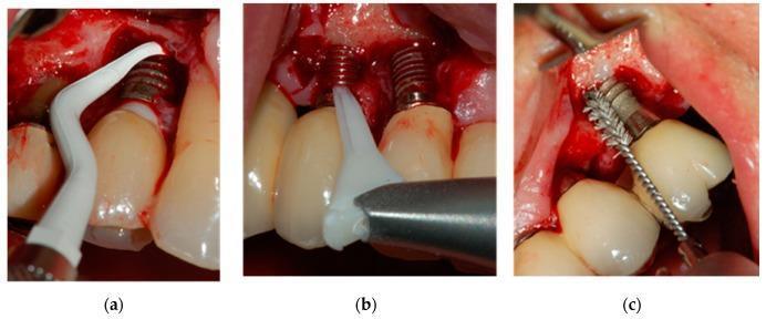
This composite of three clinical intraoral photographs (a, b, and c) demonstrates different mechanical debridement techniques for the surgical treatment of peri-implantitis. Each image shows an exposed dental implant within an osseous defect following the elevation of a full-thickness mucoperiosteal flap. In image (a), a white, curved plastic Gracey-type curette is used to manually scale the implant surface to remove biofilm and granulation tissue. Image (b) shows the use of a Perio-Flow® air-polishing device, featuring a flexible white nozzle designed to deliver glycine powder under pressure for non-contact surface decontamination. Image (c) illustrates a Ti-Brush® consisting of a stainless steel shaft with titanium bristles, which is used for mechanical scrubbing and polishing of the implant threads. The images highlight the variations in instrument morphology—from manual scraping tools to pressurized air nozzles and rotating brushes—used in regenerative or resective periodontal surgery to manage peri-implant bone loss.
Peri-Implantitis: Management Strategies
2025 Landmark Guideline: AO/AAP Consensus
Risk Factors and Etiology
- History of periodontitis (strongest risk factor)
- Smoking
- Uncontrolled diabetes
- Poor biofilm/plaque control
- Obesity
- Implant malposition
- Unfavorable prosthetic factors (overhangs, subgingival cement excess)
- Suboptimal peri-implant soft tissue phenotype (thin/deficient keratinized mucosa)
- Lack of regular maintenance
Classification of Peri-Implant Bone Defects
| Defect Type | Description | Surgery Preferred |
|---|---|---|
| Suprabony / supra-crestal | Bone loss at or above crestal level; horizontal | Resective |
| Facial dehiscence | Buccal bone plate lost, threads exposed | Resective or combined |
| Semi-circumferential | Bone loss around part of implant perimeter | Regenerative possible |
| Circumferential (contained) | Bone loss around entire implant; walls present | Regenerative |
| Deep circumferential with multiple walls | Deep contained defect; best regenerative candidate | Regenerative |
Stepwise Management Framework
STEP 1 - Risk Factor Modification and Patient Education
- Smoking cessation counseling
- Glycemic control in diabetics
- Oral hygiene instruction and reinforcement
- Correction of prosthetic risk factors (removal of subgingival cement, prosthesis redesign if needed)
STEP 2 - Non-Surgical Treatment
- Titanium or carbon fiber curettes (avoid scratching the implant surface with steel instruments)
- Ultrasonic devices with plastic/PEEK tips
- Air-polishing with glycine or erythritol powder (soft, non-abrasive)
- Ti-brushes for mechanical scrubbing of implant threads

- Local antiseptics: chlorhexidine chips/gel placed subgingivally
- Photodynamic therapy (PDT) as an adjunct to mechanical debridement
- Local antibiotic delivery (minocycline microspheres)
- A systematic review and meta-analysis (Wang et al., 2022, PMID: 35476853) found adjunctive systemic antibiotics yield statistically greater probing depth reduction (0.56 mm at 3 months, 0.92 mm at 12 months) and radiographic bone gain vs. debridement alone
- However, the Frontiers of Oral and Maxillofacial Medicine 2025 review notes two meta-analyses and an RCT show NO consistent benefit for systemic antibiotics post-operatively
- Current position: Antibiotics considered for acute inflammatory presentations or peri-implant abscesses; routine systemic use is not recommended
- Disease stabilization achieved in only ~40% of peri-implantitis cases (Meyle & Fischer-Wasels, BDJ, 2024, PMID: 39572812)
- The inaccessibility of implant threads and fixture geometry limits thoroughness
- Non-surgical therapy is an essential preparatory step but rarely definitive for peri-implantitis
STEP 3 - Surgical Treatment
A. Flap for Access (Open Flap Debridement - OFD)
- Full-thickness flap reflected for direct visualization and debridement
- Thorough root/implant surface decontamination
- No bone modification
- Indicated when pockets are accessible and bone loss is minimal
- Represents the baseline surgical approach
B. Resective Surgery
- Suprabony defects / horizontal bone loss
- Facial dehiscence-type defects
- Non-contained defects with 1-2 walls
- Areas where esthetics are not a primary concern (posterior regions)
- Shallow, wide defects unfavorable for regeneration
- Bone recontouring (osteoplasty) to eliminate ledges and create physiologic architecture
- Implantoplasty: Mechanical reduction of exposed implant threads using rotary instruments (carbide burs, diamond burs). This smooths the rough implant surface to reduce bacterial adhesion
- A 2023 systematic review (Dasgupta et al., PMID: 37929359) supports implantoplasty's efficacy in peri-implantitis management
- A 2025 RCT (Cruz et al., PMID: 40419076) confirms benefits at 24 months
- Concerns: Generates titanium particles, reduces implant wall thickness and fracture resistance - should be reserved for appropriate cases
- Apical repositioning of flap to eliminate residual pockets
- Expected outcome: Effective pocket elimination; post-operative mucosal recession is expected and should be discussed with patients pre-operatively
C. Reconstructive (Regenerative) Surgery
- Contained circumferential defects (3-wall or deep semi-circumferential)
- Deep defects with multiple remaining bony walls
- Cases where implant re-osseointegration is the goal
- Esthetic zones where recession must be minimized
- Bone grafts: Autogenous bone, allografts (DFDBA/FDBA), xenografts (bovine hydroxyapatite/Bio-Oss), alloplasts (beta-TCP, bioactive glass)
- Barrier membranes (GTR): Resorbable (collagen) or non-resorbable (ePTFE/titanium-reinforced) to exclude epithelium and guide bone regeneration
- Biologics: Enamel matrix derivatives, PRP/PRF, rhPDGF-BB
- Chemical: citric acid, EDTA, H₂O₂, tetracycline
- Mechanical: Ti-brushes, air-polishing, ultrasonic
- Laser-assisted (Er:YAG): evidence supporting decontamination without thermal damage
D. Soft Tissue Augmentation
- Indicated when thin/deficient keratinized mucosa contributes to peri-implant instability
- Free gingival graft or connective tissue graft to increase soft tissue thickness and keratinized band
- Improves peri-implant phenotype and resistance to disease recurrence
Surgical Decision Algorithm
Peri-implantitis confirmed (BOP + bone loss)
           │
     ┌─────▼──────────────────────────────────────┐
     │ Step 1: Risk factor control + hygiene       │
     └─────┬──────────────────────────────────────┘
           │
     ┌─────▼──────────────────────────────────────┐
     │ Step 2: Non-surgical debridement ± adjuncts │
     └─────┬──────────────────────────────────────┘
           │ Re-evaluate 6-12 weeks
     ┌─────▼───────────────────┐
     │ Disease resolved?        │──► YES → Supportive maintenance
     └─────┬───────────────────┘
           │ NO
     ┌─────▼──────────────────────────────────────────────────┐
     │ Assess defect morphology (CBCT/clinical/radiographic)   │
     └─────┬──────────────────────────────────────────────────┘
           │
    Suprabony / horizontal  ──────────────────► Resective ± implantoplasty
    Semi-circumferential    ──────────────────► Resective or combined
    Contained circumferential (≥3 walls)  ────► Regenerative
    Thin keratinized mucosa  ─────────────────► Soft tissue augmentation
    Disease uncontrollable / failed     ──────► Implant removal
Supportive Peri-Implant Maintenance (SPIM)
- 3-month recall intervals initially; extend based on stability
- Probing, radiographic monitoring, BOP assessment at every visit
- Repeated professional debridement
- Reinforced home care with implant-specific brushes and interdental aids
- Adjunctive use of chlorhexidine rinse in high-risk patients
Summary: Treatment Selection at a Glance
| Scenario | Recommended Strategy |
|---|---|
| Mucositis only (no bone loss) | Non-surgical debridement + risk control |
| Early peri-implantitis, shallow defects | Non-surgical + re-evaluate; OFD if needed |
| Suprabony / horizontal bone loss | Resective surgery ± implantoplasty |
| Deep contained circumferential defect | Regenerative surgery |
| Thin/absent keratinized mucosa | Soft tissue augmentation |
| Uncontrolled, progressive, or failed | Implant removal |
| All cases post-treatment | Supportive peri-implant maintenance (3-month intervals) |
Bone graft amd bone substitutes
bone graft substitutes periodontal regeneration classification
bone grafts bone substitutes periodontics classification autograft allograft xenograft alloplast mechanisms 2024 2025
https://pmc.ncbi.nlm.nih.gov/articles/PMC12833343
PMID: 41602459
bone graft classification types autograft allograft xenograft alloplast periodontal
Bone Grafts and Bone Substitutes in Periodontics
Biological Mechanisms: The Triad of Bone Regeneration
| Property | Definition | Which Grafts Possess It |
|---|---|---|
| Osteogenesis | New bone formation by viable osteoblasts or osteoprogenitor cells within the graft itself | Autograft only |
| Osteoinduction | Stimulation of undifferentiated host mesenchymal cells to differentiate into bone-forming cells via BMPs and growth factors | Autograft (high), DFDBA (variable), some growth factor-based materials |
| Osteoconduction | Provides a scaffold/framework for ingrowth of blood vessels and bone-forming cells from the host | All graft types possess this |
| Osseointegration | Direct structural and functional connection between living bone and the implant/graft surface | Goal of all grafting procedures |
- Osteopromotion - Enhancement of osteoinduction without independent osteoinductive property (e.g., enamel matrix derivatives enhance the osteoinductive effect of DFDBA but cannot stimulate bone alone)
Classification of Bone Grafts
Based on Source (Primary Classification)
BONE GRAFTS
├── 1. AUTOGRAFT (Autogenous)
├── 2. ALLOGRAFT
├── 3. XENOGRAFT
└── 4. ALLOPLAST (Synthetic)
1. Autograft (Autogenous Bone Graft)
- Healing extraction sockets
- Edentulous ridges
- Mandibular symphysis (chin)
- Mandibular ramus / retromolar area
- Maxillary tuberosity
- Tori (when present)
- Iliac crest (most common extraoral)
- Calvarium
- Tibia
- Radius
- Cancellous: High surface area, rapid revascularization, high osteogenic/osteoinductive/osteoconductive potential. Poor mechanical strength. Remodels faster.
- Cortical: Provides structural support and mechanical stability. Lower biological activity. Takes longer to remodel.
- Corticocancellous: Combination - provides both structural support and biological activity.
- All three bone-regenerative properties
- Fully biocompatible, no immune reaction
- No disease transmission risk
- Fastest incorporation
- Requires a second surgical site (donor site morbidity)
- Limited quantity available
- Donor site pain, scarring, bleeding
- Unpredictable and sometimes rapid resorption
- Rapid reappearance of defects reported
2. Allograft
| Form | Description | Properties |
|---|---|---|
| Fresh-frozen allograft | Stored at -70°C; retains some proteins | Some osteoinductive potential; disease transmission risk higher |
| Freeze-dried bone allograft (FDBA) | Freeze-dried, vacuum-sealed; long shelf life | Primarily osteoconductive; some osteoinduction |
| Demineralized freeze-dried bone allograft (DFDBA) | Acid-treated to remove mineral component; exposes BMPs | Osteoconductive + osteoinductive; no osteogenesis |
| Irradiated allograft | Gamma/electron beam sterilization | Reduces infection risk but decreases osteoinductive activity |
- Most widely used allograft in periodontics
- Demineralization exposes bone morphogenetic proteins (BMPs), particularly BMP-2 and BMP-7
- Osteoinductive potential is highly variable between products and donors
- Available as putty, injectable gel, paste, powder, strips
- Can be mixed with carriers: glycerol, calcium sulfate, sodium hyaluronate, gelatin
- Significant inter-batch variability is a clinical limitation
- No donor site morbidity
- Available in larger quantities
- Long shelf life (FDBA/DFDBA)
- Well-documented clinical use in periodontics
- No viable cells (no osteogenesis)
- Variable osteoinductive strength
- Risk of immune response (minimal but present)
- Rare risk of disease transmission
- Compromised mechanical strength vs. autograft
3. Xenograft
- Bovine (most common): Deproteinized bovine bone matrix (DBBM) - e.g., Bio-Oss® (Geistlich, Switzerland) - the most widely used and documented xenograft in clinical dentistry
- Porcine: Processed similarly; gaining use
- Equine
- Coralline hydroxyapatite: Derived from coral skeleton; naturally porous
- Osteoconductive only - provides scaffold for bone ingrowth
- No osteogenesis, no osteoinduction
- Excellent biocompatibility
- Structural similarity to native bone mineral
- Very slow resorption (long-term volume maintenance - a clinical advantage for ridge preservation)
- Excellent track record in socket preservation and sinus floor elevation
- Alveolar socket preservation post-extraction
- Sinus floor elevation (sinus lift)
- Ridge augmentation
- Combined with membranes (GBR) for contained defects
- No donor site surgery
- Abundant supply
- Excellent dimensional stability
- Long-term volume maintenance
- Well-documented safety and efficacy
- Osteoconductive only (no osteoinduction or osteogenesis)
- Slow resorption (sometimes persists long-term in tissues)
- Theoretical (very low) infection/prion risk
- Religious/cultural concerns for bovine/porcine sources
4. Alloplast (Synthetic Bone Substitutes)
A. Calcium Phosphate Ceramics
| Material | Key Properties | Examples |
|---|---|---|
| Hydroxyapatite (HA) | Chemically identical to bone mineral; osteoconductive; very slow resorption; excellent biocompatibility | Calcitite®, OsteoGraf/N® |
| Beta-tricalcium phosphate (β-TCP) | Osteoconductive; faster resorbable than HA; replaced by new bone | Cerasorb®, Vitoss® |
| Biphasic calcium phosphate (BCP) | Mixture of HA + β-TCP; tunable resorption rate; combines stability of HA with resorbability of β-TCP | Straumann® BoneCeramic, MBCP® |
| Nanocrystalline HA | Enhanced surface area; improved cell attachment; better bone fill in periodontal defects per 2022 systematic review (Shaheen, PMID: 36570589) | NanoBone® |
B. Bioactive Glass (Bioglass)
- Silica-based material; forms a biologically active carbonate apatite layer on contact with body fluids
- Bonds directly to both bone AND soft tissue
- Osteoconductive and somewhat osteostimulative
- Releases ions (Si, Ca, P, Na) that upregulate osteogenic genes
- Example: PerioGlas® (45S5 Bioglass) - specifically designed for periodontal defects
C. Calcium Sulfate
- Rapidly resorbing osteoconductive scaffold
- Used as a space maintainer and carrier for other materials
- Often used with DFDBA to enhance handling characteristics
- Example: Capset®, OsteoSet®
D. Polymer-based Substitutes
- Biodegradable polymers (PLA, PGA, PLGA) used as scaffolds
- Can be combined with growth factors or cells
- Mostly experimental/emerging in periodontics
- Osteoconductive only (no osteogenesis or osteoinduction)
- No risk of disease transmission
- Unlimited supply, consistent composition
- Controlled manufacturing allows customization of porosity, particle size, resorption rate
- Good dimensional stability
- Lower biological activity than biological grafts
Classification by Laurencin et al. (Bone Graft Substitute System)
| Category | Materials |
|---|---|
| Allograft-based | FDBA, DFDBA, DBM - structural or filler grafts |
| Factor-based | BMPs (rhBMP-2, rhBMP-7), PDGF, FGF, VEGF - natural or recombinant growth factors |
| Cell-based | Bone marrow aspirate concentrate (BMAC), platelet-rich plasma (PRP), platelet-rich fibrin (PRF) |
| Ceramic-based | HA, β-TCP, BCP, bioactive glass - scaffolds for bone ingrowth |
| Polymer-based | PLA, PGA, PLGA scaffolds |
| Miscellaneous | Coral-derived materials, marine sources |
Biologics and Growth Factors (Adjuncts to Bone Grafts)
| Agent | Mechanism | Clinical Use |
|---|---|---|
| Platelet-Rich Plasma (PRP) | Concentrated autologous platelets; releases PDGF, TGF-β, VEGF | Combined with bone grafts to enhance healing |
| Platelet-Rich Fibrin (PRF) | Second-generation platelet concentrate; fibrin scaffold with growth factors | Used as membrane + graft adjunct |
| Enamel Matrix Derivatives (EMD / Emdogain®) | Amelogenin proteins; promotes cementum formation and periodontal ligament regeneration; enhances DFDBA osteoinduction | Infrabony defects; root coverage |
| rhBMP-2 (GEM 21S®, INFUSE®) | Recombinant human bone morphogenetic protein-2; potent osteoinduction | Ridge augmentation, sinus lift |
| rhPDGF-BB (GEM 21S®) | Recombinant platelet-derived growth factor-BB combined with β-TCP scaffold | Periodontal osseous defects; FDA approved |
| FGF-2 | Fibroblast growth factor; promotes angiogenesis and cell proliferation | Investigational in periodontics |
Comparison Summary Table
| Property | Autograft | Allograft (FDBA) | Allograft (DFDBA) | Xenograft | Alloplast |
|---|---|---|---|---|---|
| Osteogenesis | ✅ High | ❌ | ❌ | ❌ | ❌ |
| Osteoinduction | ✅ High | ⚠️ Low | ✅ Variable | ❌ | ❌ |
| Osteoconduction | ✅ | ✅ | ✅ | ✅ | ✅ |
| Disease transmission | ❌ None | ⚠️ Rare | ⚠️ Rare | ⚠️ Very rare | ❌ None |
| Immune reaction | ❌ None | ⚠️ Possible | ⚠️ Possible | ⚠️ Low | ❌ None |
| Donor site morbidity | ⚠️ Yes | ❌ | ❌ | ❌ | ❌ |
| Availability | Limited | Moderate | Moderate | High | Unlimited |
| Resorption rate | Fast | Moderate | Moderate | Very slow | Variable |
| Cost | Low | Moderate | Moderate | Moderate | Low-moderate |
| Gold standard status | ✅ Yes | - | - | - | - |
Ideal Bone Graft Material Properties (Criteria)
- Abundant supply without donor site harm
- Osteogenic, osteoinductive, and osteoconductive potential
- No immune reaction from the host
- Rapid revascularization capacity
- Resorbable and replaceable by native bone
- Mechanical properties comparable to native bone
- No risk of infection or disease transmission
- Easy handling and application
Emerging Technologies (2024-2025)
- 3D-printed patient-specific ceramic scaffolds - customized geometry matching exact defect anatomy; an RCT in 2024 (Kim et al.) showed favorable outcomes
- Nanotechnology-enabled delivery systems - nanocrystalline HA and nano-HA/collagen composites with superior cell adhesion
- Growth factor-loaded scaffolds - controlled release of BMPs, PDGF embedded within scaffold architecture
- Polydopamine-modified surfaces - enhances osseointegration and antibacterial performance
- Cell-sheet engineering - periodontal ligament cell sheets for true periodontal regeneration
Clinical Applications in Periodontics
| Clinical Indication | Preferred Graft(s) |
|---|---|
| Intrabony periodontal defect | DFDBA ± EMD, autograft, xenograft + GTR membrane |
| Furcation defect | DFDBA, xenograft + membrane |
| Ridge preservation (socket) | Xenograft (Bio-Oss), allograft, alloplast |
| Ridge augmentation | Autograft (gold standard) ± xenograft, GBR membrane |
| Sinus floor elevation | Xenograft (Bio-Oss), autograft, BCP |
| Peri-implantitis regeneration | Xenograft, autograft ± resorbable membrane |
| Horizontal bone augmentation | Autograft + xenograft block (shell technique), GBR |
Search for latest clinical guidelines on bone graft selection for peri-implantitis
bone graft selection peri-implantitis reconstructive surgery
peri-implantitis regenerative therapy bone substitute outcomes
bone graft selection peri-implantitis guidelines 2024 2025 xenograft allograft autograft alloplast reconstructive
PMID: 40344243
PMID: 37728141
PMID: 36635029
PMID: 39704200
PMID: 40539762
Bone Graft Selection for Peri-Implantitis: Latest Clinical Guidelines
Foundational Principle: Is Reconstructive Surgery Even Indicated?
Reconstructive/regenerative therapy is indicated only for CONTAINED peri-implant bone defects - specifically intrabony (angular) defects ≥3 mm deep with at least 2-3 remaining bony walls.
| Defect Type | Recommended Surgery | Role of Bone Graft |
|---|---|---|
| Horizontal / non-contained | Access flap ± resective | No graft indicated |
| Intrabony, contained, <3 mm | Access flap | Graft questionable |
| Intrabony, contained, ≥3 mm, 2-4 walls | Reconstructive (graft ± membrane) | Primary indication |
| Mixed morphology | Judgment-based | Case-by-case |
The Critical 2025 Evidence: Does Adding a Bone Graft Help?
Landmark 3-Year RCT (Alibegovic et al., 2025)
- Control: Access flap surgery alone
- Test: Access flap surgery + bone substitute material
- Both groups showed 3.2-3.5 mm probing depth reduction
- Marginal bone level gain of 1.1-1.3 mm in both groups
- Primary composite outcome achieved at only 14% of implants in both groups
- Disease resolution in only 39% overall
- Conclusion: Bone substitute did NOT show clear benefit over access flap alone at 3 years
7-Year RCT Follow-up (Isler et al., 2025)
- Xenogenic bone graft + either concentrated growth factor (CGF) or collagen membrane (CM)
- Treatment success at 7 years: only 23-31% of patients
- Disease recurrence in 31-35%
- Collagen membrane group had more favorable outcomes for probing depth and defect depth than CGF at 7 years
- Baseline suppuration was the only significant predictor of treatment failure (OR = 15.45)
- Conclusion: Long-term success is limited and failure rates increase over time regardless of biologic adjunct used
Systematic Review + Meta-Analysis (Donos et al., 2023)
- 7 RCTs comparing reconstructive surgery vs. access flap alone
- No significant difference in PPD change between groups at 12 months (-0.387 mm, p=0.325)
- No clear hierarchy of efficacy established among different biomaterials
- Conclusion: It was not possible to establish one biomaterial as superior over others for peri-implantitis
Material-by-Material Evidence Summary (2024-2025)
1. Xenografts (e.g., Bio-Oss® - Deproteinized Bovine Bone)
- Low resorption rate is an advantage in peri-implantitis - maintains volume longer while re-osseointegration is attempted
- Current evidence indicates bone substitutes with LOW resorption rates tend to yield more favorable outcomes compared to rapidly-resorbing materials and autogenous bone
- Most of the published RCTs on reconstructive peri-implantitis therapy have used xenografts (especially bovine-derived DBBM)
- Bio-Oss granules remain the most documented graft for this indication
2. Autogenous Bone
- Despite being the gold standard in other settings, rapidly resorbing behavior is a disadvantage in peri-implantitis
- Ramanauskaite et al. 2025 explicitly state that autogenous bone yields less favorable outcomes compared to bone substitutes with low resorption rates
- Mechanism: autograft resorbs before adequate re-osseointegration can be established on the contaminated implant surface
- May still have a role as a supplemental component in a "sandwich" or combined technique
3. Alloplasts (β-TCP, BCP, Bioactive Glass)
- β-TCP and BCP are resorbable, which may be disadvantageous for the same reasons as autograft
- Bioactive glass (e.g., PerioGlas) has some supporting case series data but limited RCT-level evidence for peri-implantitis specifically
- BCP (HA + β-TCP) represents a middle ground - tunable resorption rate by adjusting HA:TCP ratio
- Evidence base is weaker than for xenografts in this specific indication
4. Allografts (FDBA / DFDBA)
- Moderate resorption rate
- Some osteoinductive potential (DFDBA)
- Limited RCT data specifically for peri-implantitis reconstruction
- More commonly used in periodontal intrabony defects than peri-implantitis
- May serve as an alternative when xenograft is not available or acceptable to patient
5. "Sandwich" / Combination Technique
- Layers different graft materials to mimic natural bone composition
- E.g., autograft (inner layer for osteogenesis) + xenograft (outer layer for volume maintenance)
- Recent human histological studies show promising outcomes
- Leverages unique properties of each material
- Currently investigational; requires further RCT validation
Barrier Membranes: Do They Add Value?
"Comparative studies have not demonstrated clear clinical advantages from the adjunctive use of barrier membranes."
- Collagen membrane (CM) produced more favorable 7-year outcomes than CGF alone for probing depth and defect depth
- This suggests membranes may have a role, especially for long-term stability
Biologics as Adjuncts
"Current evidence does not support definitive conclusions regarding the benefits of adding biologics to reconstructive treatments."
| Biologic | Evidence in Peri-Implantitis |
|---|---|
| EMD (Emdogain) | Insufficient evidence |
| PRP / PRF / CGF | No clear advantage over membrane alone at 7 years |
| rhBMP-2 | Not established for this indication |
| rhPDGF-BB | Investigational |
Implant Surface Decontamination: The Real Non-Negotiable
- Mechanical: titanium brushes, curettes, air-polishing (glycine/erythritol)
- Chemical: citric acid, EDTA, saline irrigation, H₂O₂
- Laser-assisted (Er:YAG): decontamination without thermal damage
Graft Selection Decision Algorithm (Current Best Evidence)
Peri-implantitis confirmed → Non-surgical therapy → Re-evaluate
                                      │
              Residual pocketing / bone loss persists
                                      │
                         ASSESS DEFECT MORPHOLOGY
                                      │
    ┌─────────────────────────────────┼──────────────────────────────────┐
    │                                 │                                  │
Suprabony /              Intrabony contained               Mixed /
horizontal               ≥3 mm, ≥2 walls                 borderline
    │                                 │                                  │
Access flap            RECONSTRUCTIVE SURGERY               Access flap
± resective         Thorough decontamination first          (consider
  surgery               ↓                                   case-by-case)
No graft          GRAFT SELECTION:
                  1st choice: Xenograft (low-resorption DBBM)
                  Alternative: Allograft (FDBA/DFDBA)
                  Avoid: Rapidly resorbing materials
                  Add membrane? Resorbable collagen preferred
                  Biologics: No strong evidence to mandate
                       │
               PREDICTORS OF FAILURE:
               Baseline suppuration (strongest)
               Poor plaque control
               Smoking
               Shallow defects (<3 mm)
                       │
              Supportive peri-implant maintenance
              (3-month intervals - MANDATORY)
Summary: What the Guidelines Currently Say
| Question | Current Guideline Answer (2025) |
|---|---|
| When to graft? | Contained intrabony defects ≥3 mm with ≥2 walls |
| Best single material? | Xenograft (low-resorption DBBM) - most evidence, best volume maintenance |
| Autograft for PI? | Not favored - too rapidly resorbing |
| Membrane needed? | Not definitively proven, but collagen membrane preferred when used |
| Biologics needed? | No clear evidence of benefit |
| Does grafting beat flap alone? | RCTs show no significant clinical difference at 12 months; some radiographic benefit |
| Long-term success? | Limited - only 23-39% at 7 years regardless of material |
| Most important factor? | Complete implant surface decontamination + lifelong maintenance |
Key Evidence References
| Paper | Type | Key Finding |
|---|---|---|
| Alibegovic et al. 2025 (PMID 39704200) | 3-year multicenter RCT | Bone substitute showed no benefit over access flap alone at 3 years |
| Isler et al. 2025 (PMID 40539762) | 7-year RCT | Only 23-31% success at 7 years; suppuration predicts failure; CM > CGF |
| Ramanauskaite et al. 2025 (PMID 40344243) | 2025 Review | Low-resorption substitutes favored; no clear membrane or biologic advantage |
| Monje et al. 2024 (PMID 37728141) | Periodontology 2000 Review | Comprehensive biomaterial framework for peri-implantitis reconstruction |
| Donos et al. 2023 (PMID 36635029) | Systematic review + Meta-analysis | No hierarchy of efficacy among biomaterials; no clear advantage of reconstruction vs. flap alone |
| AO/AAP Consensus 2025 (PMID 40501397) | Consensus guideline | Stepwise approach; surgical decision by defect morphology; maintenance essential |