
Structures forming angle of anterior chamber
Reading File
The angle of the anterior chamber (iridocorneal angle) is formed by structures at the junction of the cornea, sclera, and iris root. Key structures from front to back include: Schwalbe's line, the trabecular meshwork, Schlemm's canal, the scleral spur, the ciliary body band, and the iris root.
Searching sources...
Finding Sources
Finding Sources
Reading File
Reading File
Now let me get the dedicated Kanski section on the anterior chamber angle, which will have the most detailed gonioscopic anatomy:
Finding Sources
Reading File
Now let me find the dedicated gonioscopy/angle anatomy section in Kanski:
Finding Sources
Reading File
Reading File
Reading File
Excellent - I found the key content. Now let me fetch the critical anatomy diagram images:
Loading Image
Loading Image
I now have comprehensive, authoritative content from Kanski's Clinical Ophthalmology. Let me compile the full answer.
Structures Forming the Angle of the Anterior Chamber (Iridocorneal Angle)
The angle of the anterior chamber (AC) - also called the iridocorneal angle or filtration angle - is located at the junction of the peripheral cornea, sclera, iris root, and ciliary body. It is the site of aqueous humor drainage and is evaluated clinically by gonioscopy.
The structures are classically described from anterior (corneal side) to posterior (iris/ciliary body side):
1. Schwalbe's Line
- The most anterior structure seen on gonioscopy
- Marks the termination of Descemet's membrane and the peripheral end of the corneal endothelium
- Appears as a fine glistening ridge or white line
- Clinically identified by the corneal wedge (two slit-lamp beams converge here)
- A prominent, anteriorly displaced Schwalbe's line is called posterior embryotoxon (seen in Axenfeld-Rieger syndrome)
2. Trabecular Meshwork (TM)
Extends from Schwalbe's line posteriorly to the scleral spur. It has two portions:
| Zone | Appearance | Location |
|---|---|---|
| Anterior (non-pigmented) TM | Pale, non-pigmented | Overlies the non-functional part of the meshwork |
| Posterior (pigmented) TM | Pigmented band (variable) | Overlies Schlemm's canal - the functional filtration zone |
Structurally, the TM has three layers (from inner to outer):
- (A) Uveal meshwork - innermost, rope-like trabeculae, faces the AC
- (B) Corneoscleral meshwork - middle layer, sheet-like lamellae with elliptical holes
- Juxtacanalicular (cribriform) meshwork - outermost layer, adjacent to Schlemm's canal; the main site of aqueous outflow resistance
The TM is the major aqueous outflow pathway (~90% of drainage).
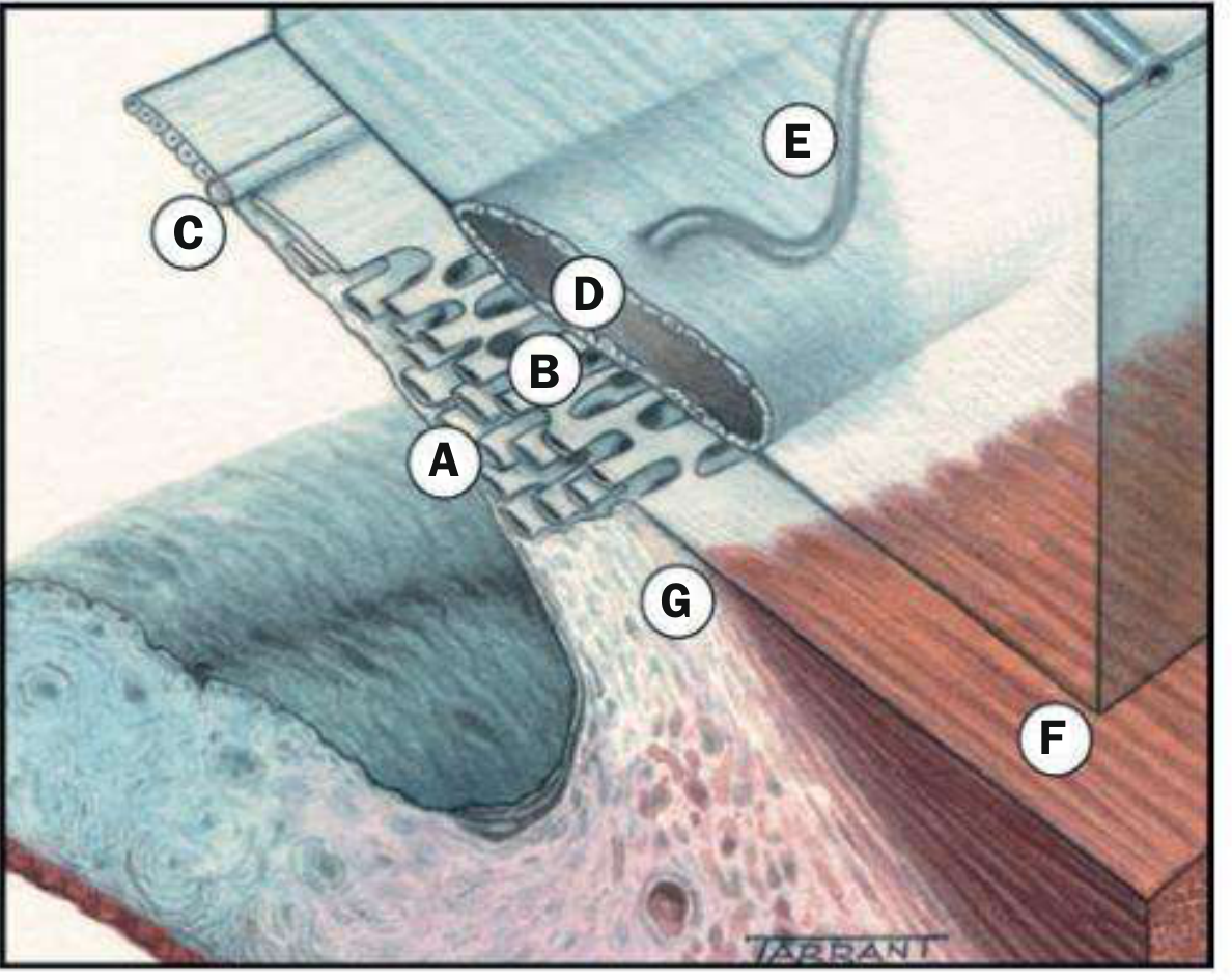
3. Schlemm's Canal (Scleral Venous Sinus)
- A circumferential endothelium-lined channel that encircles the eye at the corneoscleral junction
- Located just deep to the posterior pigmented trabecular meshwork
- Aqueous flows from the AC through the trabecular meshwork into Schlemm's canal
- Exits via collector channels (aqueous veins of Ascher) into episcleral veins
- Not directly visible gonioscopically, but the pigmented TM overlying it acts as a landmark
- Can be identified by pressing on the perilimbal episclera to reflux blood into the canal (turns red)
4. Scleral Spur
- A white/light-colored ridge projecting inward from the inner sclera
- The most important surgical landmark in the angle
- Represents the posterior edge of Schlemm's canal
- Anteriorly: posterior trabecular meshwork inserts here
- Posteriorly: longitudinal ciliary muscle fibers attach here
- Separates canal-based surgery (trabecular - anterior) from suprachoroidal surgery (ciliary body - posterior)
5. Ciliary Body Band
- A dark gray/brown band immediately posterior to the scleral spur
- Width varies and reflects how far the iris inserts posteriorly - a wide band suggests posterior iris insertion
- In angle-closure glaucoma, this may be obscured or absent
- Contains the longitudinal muscle of the ciliary body
6. Iris Root / Peripheral Iris
- The most posterior structure seen on gonioscopy
- Its insertion level onto the ciliary body determines the angle width
- High insertion (on scleral spur or TM) = narrow/closed angle - risk of angle-closure glaucoma
- Low insertion (on ciliary body band) = wide/open angle
Mnemonic (Anterior to Posterior)
"Some Trees Stand Straight, Cells Iris" Schwalbe's line → Trabecular meshwork → Schlemm's canal → Scleral spur → Ciliary body band → Iris root
Or the classic: SCSTCI (Schwalbe, Corneoscleral TM, Scleral Spur, Trabecular, Ciliary body, Iris)
Gonioscopic View
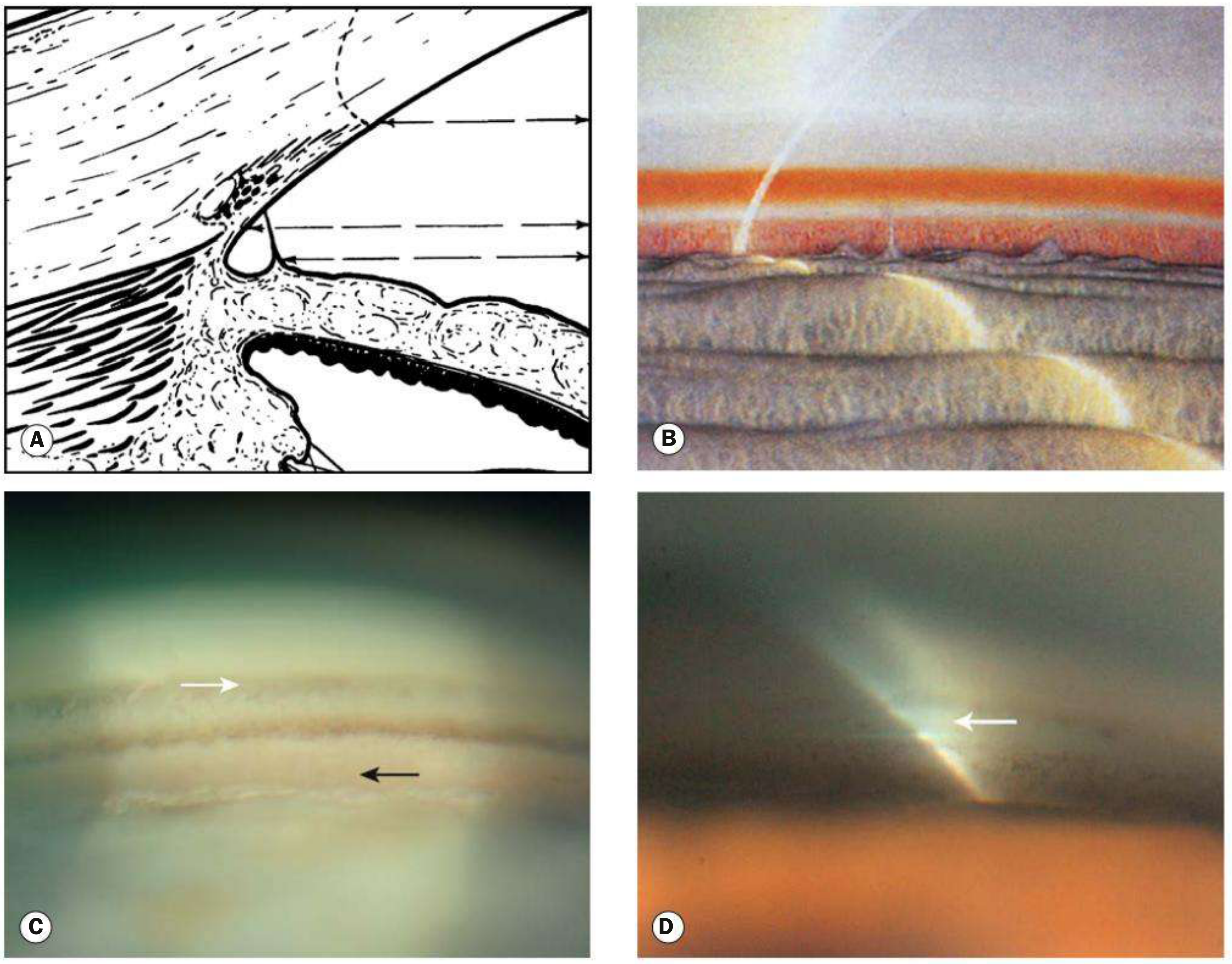
In the goniophotograph (C above), from superior to inferior: a broad Schwalbe's line (white arrow) → pale non-pigmented TM → pigmented TM → scleral spur → ciliary body band (black arrow).
Aqueous Outflow Pathway Summary
Ciliary body (production) → posterior chamber → pupil → anterior chamber → trabecular meshwork (90%) or uveoscleral route (10%) → Schlemm's canal → collector channels → aqueous veins of Ascher → episcleral veins → systemic venous circulation
Sources: Kanski's Clinical Ophthalmology: A Systematic Approach, 10th ed. (Figs. 1.52, 11.2, 11.3); Histology: A Text and Atlas with Correlated Cell and Molecular Biology (Figs. 24.6, 24.7)
2 pathological features and 2 abnormal contents of it ...write it crisp short
Finding Sources
Finding Sources
Finding Sources
Finding Sources
Finding Sources
Reading File
I have all the content needed. Here's the crisp answer:
Anterior Chamber Angle - Pathological Features & Abnormal Contents
2 Pathological Features
| # | Feature | Details |
|---|---|---|
| 1 | Peripheral Anterior Synechiae (PAS) | Adhesions between iris root and trabecular meshwork/cornea; seen in angle-closure glaucoma, uveitis, neovascular glaucoma - causes permanent angle closure |
| 2 | Angle Recession | Tear between the longitudinal and circular ciliary muscle after blunt trauma; appears as a deepened, irregular ciliary body band on gonioscopy - risk of late glaucoma |
2 Abnormal Contents
| # | Content | Details |
|---|---|---|
| 1 | Hyphema | Blood in the angle/anterior chamber - from trauma or neovascularization; blocks trabecular outflow → raised IOP |
| 2 | Rubeosis Iridis (NVA) | New vessels growing over the angle (neovascularization of angle) - seen in CRVO, PDR, ocular ischemia; leads to neovascular glaucoma |
Kanski's Clinical Ophthalmology, 10th ed.
This is a shared conversation. Sign in to Orris to start your own chat.