
Female pelvic bone
Reading File
Now let me search the medical library for female pelvic bone anatomy.
Finding Sources
Finding Sources
Reading File
Reading File
Finding Sources
Reading File
Loading Image
Loading Image
Loading Image
Loading Image
Reading File
Here is a comprehensive overview of the female pelvic bone based on authoritative anatomy textbooks.
Female Pelvic Bone
Overview
The skeleton of the female pelvis is formed by four bones: the sacrum, the coccyx, and the paired hip bones (os coxae / innominate bones). These unite at four joints to form a bony ring that supports the abdominal contents, transmits body weight to the lower limbs, and - uniquely in women - forms the birth canal.
- Berek & Novak's Gynecology, p. 148
- THIEME Atlas of Anatomy - General Anatomy and Musculoskeletal System
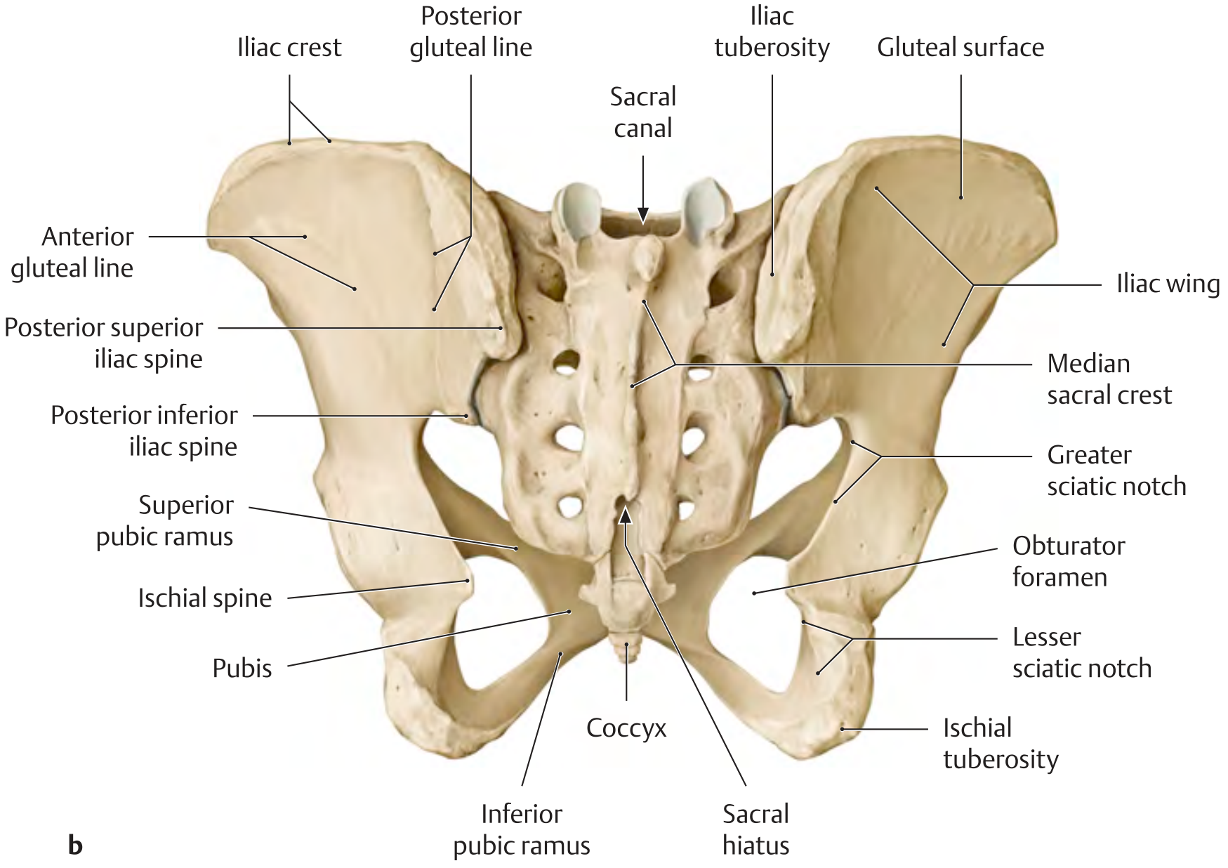
Posterior View of the Female Pelvis
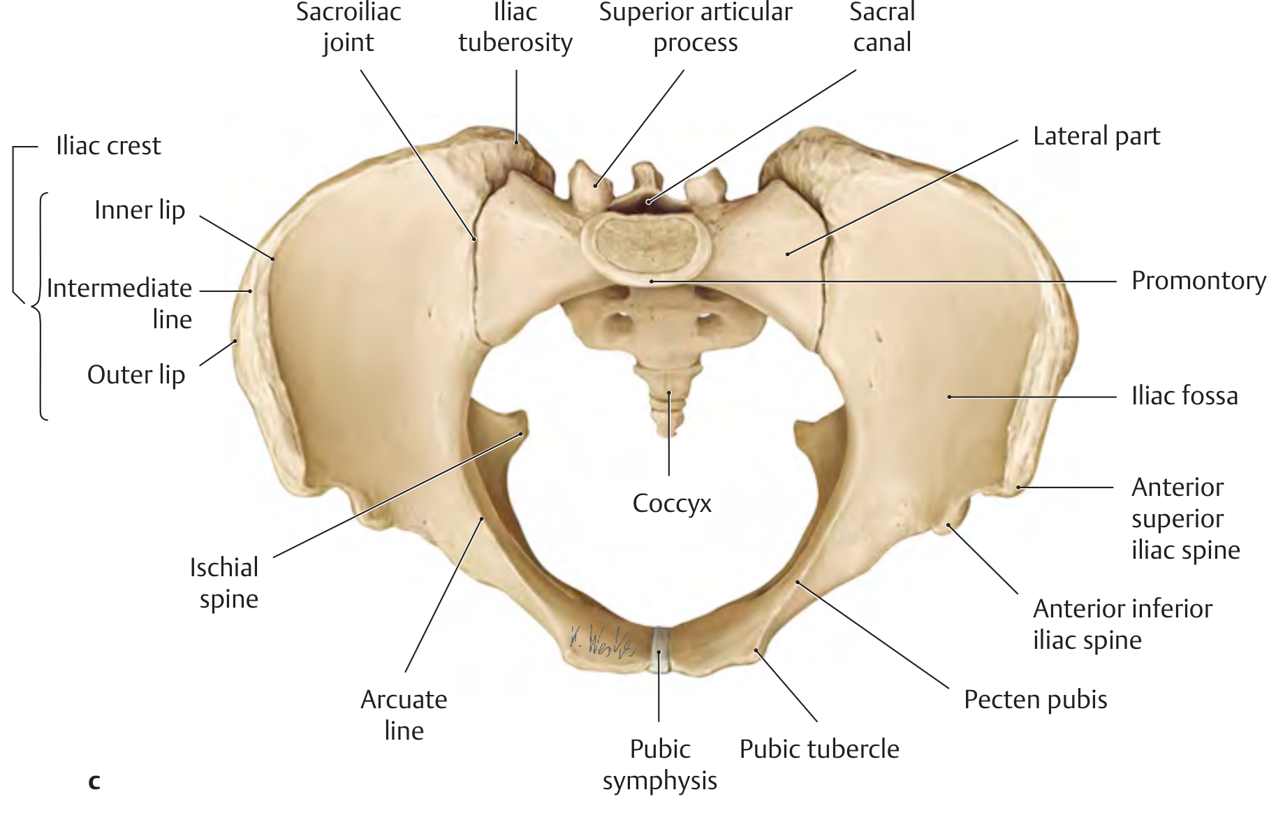
Superior (Inlet) View of the Female Pelvis
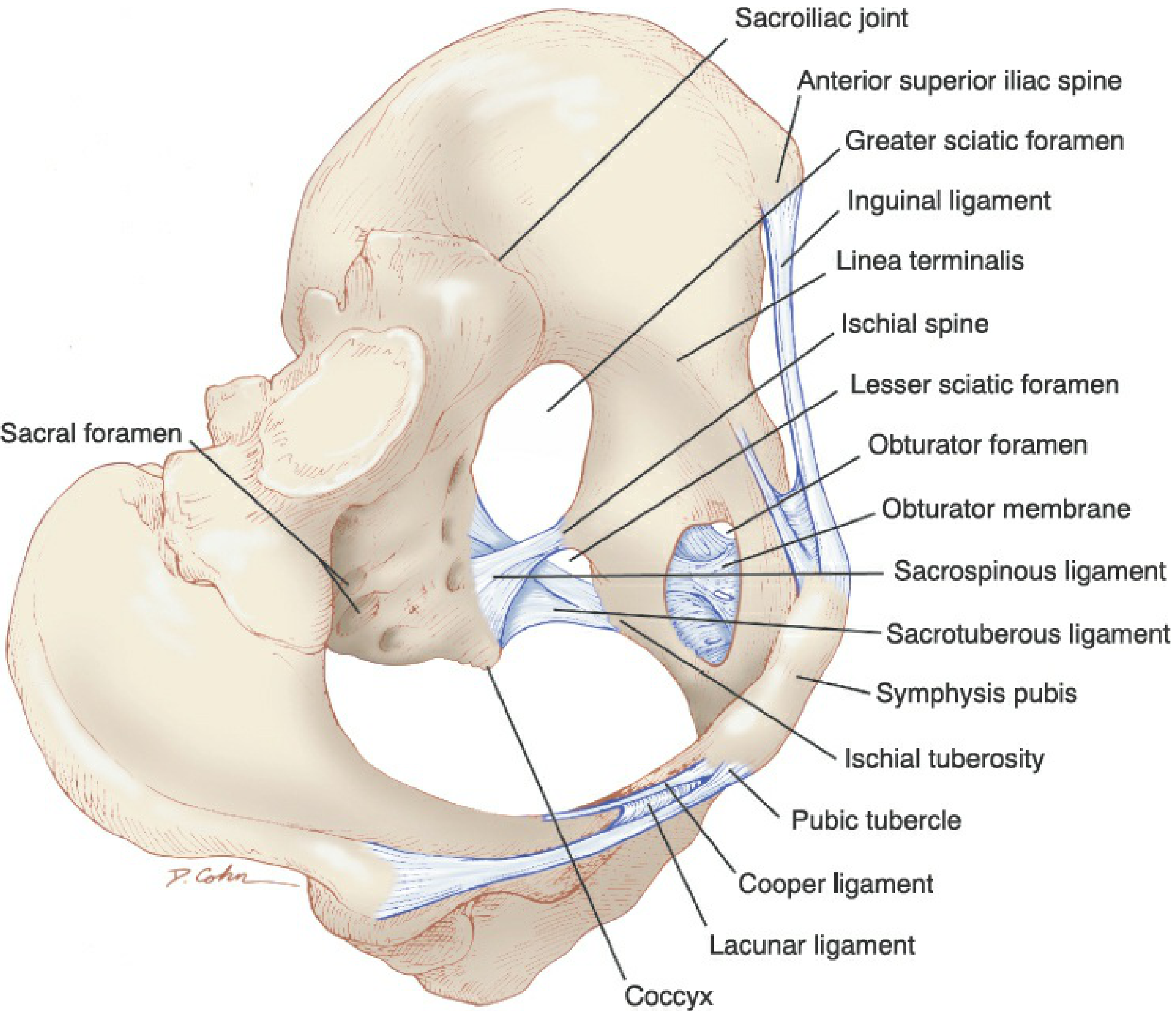
Female Pelvis with Ligaments and Foramina
Components of the Hip Bone (Os Coxae)
Each hip bone has three fused components - the ilium, ischium, and pubis - that converge at the acetabulum.
1. Ilium
| Landmark | Significance |
|---|---|
| Iliac crest | Attachment for abdominal muscles, iliac fascia, fascia lata |
| Anterior superior iliac spine (ASIS) | Attachment for inguinal ligament; lateral landmark for laparoscopic entry |
| Anterior inferior iliac spine (AIIS) | Attachment for rectus femoris |
| Posterior superior iliac spine (PSIS) | Attachment for sacrotuberous and posterior sacroiliac ligaments |
| Arcuate line | Marks the pelvic brim |
| Iliopectineal eminence (linea terminalis) | Junction of ilium and pubis |
| Iliac fossa | Smooth concavity covered by iliacus muscle |
2. Ischium
| Landmark | Significance |
|---|---|
| Ischial spine | Delineates greater and lesser sciatic notches; attachment for sacrospinous ligament; landmark for pudendal nerve block and fetal station assessment in labor |
| Ischial ramus | Encircles the obturator foramen with pubic rami; attachment for urogenital diaphragm |
| Ischial tuberosity | Weight-bearing bony prominence when sitting; landmark for anterior vaginal mesh placement |
3. Pubis
| Landmark | Significance |
|---|---|
| Body | Formed by midline fusion of superior and inferior pubic rami |
| Symphysis pubis | Fibrocartilaginous joint; allows flexibility during parturition |
| Superior and inferior pubic rami | Encircle the obturator foramen; landmark for transobturator incontinence sling passage |
| Pubic tubercle | Lateral projection; attachment for inguinal ligament and rectus abdominis |
Sacrum and Coccyx
| Feature | Detail |
|---|---|
| Sacral promontory | Most anterior projection of sacrum; landmark for laparoscopy and sacrocolpopexy; located just below the bifurcation of the common iliac arteries |
| Sacral foramina (4 pairs) | Exit sites for anterior and posterior sacral nerve rami and lateral sacral vessels |
| Sacral hiatus | Incomplete fusion of S5 posterior lamina; access point for caudal anesthesia |
| Auricular surfaces | Articulate with iliac bones to form synovial sacroiliac joints |
| Coccyx | 4 fused coccygeal vertebrae; joined to sacrum by the sacrococcygeal joint |
Pelvic Joints (Articulations)
The four pelvic articulations are:
- Symphysis pubis - fibrocartilaginous (symphyseal); loosened by relaxin in pregnancy to facilitate delivery
- Sacrococcygeal joint - fibrocartilaginous (symphyseal); allows some movement
- Two sacroiliac joints - synovial; stabilized by sacroiliac, iliolumbar, lumbosacral, sacrotuberous, and sacrospinous ligaments
Key Ligaments
| Ligament | Course | Clinical Relevance |
|---|---|---|
| Inguinal ligament | Lower border of external oblique aponeurosis folded on itself | Inguinal hernia repair |
| Cooper (pectineal) ligament | Along the pectineal line | Bladder suspension procedures (Burch colposuspension) |
| Sacrospinous ligament | Ischial spine to lateral sacrum | Vaginal vault suspension (sacrospinous fixation) |
| Sacrotuberous ligament | Sacrum to ischial tuberosity | Defines lesser sciatic foramen |
Pelvic Inlet and Outlet
The pelvis is divided into the greater pelvis (false pelvis, above the brim) and lesser pelvis (true pelvis, below the brim) by an oblique plane through:
- Sacral promontory
- Linea terminalis (arcuate line of ilium)
- Pectineal line of pubis
- Pubic crest
- Upper margin of symphysis pubis
The pelvic outlet is bounded by: tip of coccyx, symphysis pubis, and ischial tuberosities.
Female vs. Male Pelvis - Key Differences
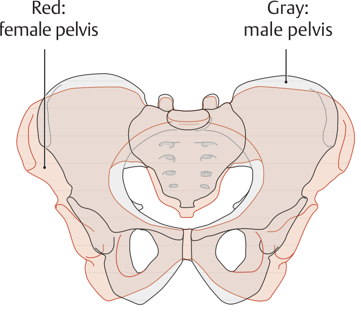
| Feature | Female | Male |
|---|---|---|
| Overall shape | Wider, broader | Taller, narrower, more massive |
| Pelvic inlet | Larger, nearly oval | Smaller, heart-shaped (sacral promontory more prominent) |
| Subpubic angle | ~90-100° | ~70° (acute) |
| Sacral curvature | Angulated at S3-S4 | Uniform curvature |
| Obturator foramen | Triangular | Oval |
These differences are directly adapted for childbirth. The wider oval inlet, larger pelvic outlet, and wider subpubic angle all allow passage of the fetal head.
Clinical Implications
MRI and CT pelvimetry studies have linked bony pelvic architecture to pelvic floor disorders. A wider transverse inlet, shorter obstetric conjugate (sacral promontory to pubic symphysis), and larger pelvic floor area are associated with higher risk of pelvic organ prolapse and pelvic neuropathy during childbirth. A less vertical orientation of the pelvic inlet directs intra-abdominal forces toward pelvic viscera rather than toward the pubic symphysis, potentially contributing to genital prolapse.
- Berek & Novak's Gynecology, p. 149
This is a shared conversation. Sign in to Orris to start your own chat.