
indication for proximal row carpelectomy
proximal row carpectomy indications outcomes
proximal row carpectomy wrist SLAC SNAC X-ray
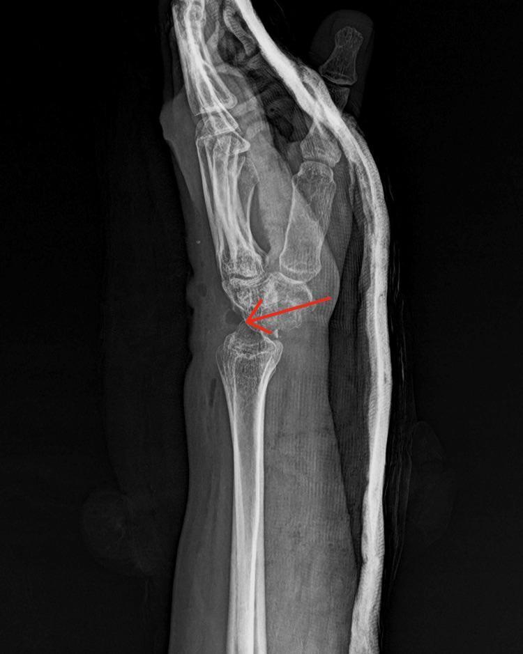
This lateral view X-ray of the wrist demonstrates post-operative findings following a proximal row carpectomy (PRC). The imaging reveals the surgical absence of the proximal carpal row (scaphoid, lunate, and triquetrum). The head of the capitate is shown articulating directly with the lunate fossa of the distal radius, a hallmark of this motion-preserving procedure used to treat advanced Kienbock's disease or SLAC/SNAC wrist. A red arrow highlights the surgical site and the new radiocapitate articulation. Encasing the forearm and wrist is a radiopaque cast or splint, which creates a hazy silhouette around the anatomical structures and confirms the immediate post-operative phase. The distal radius and distal carpal row (trapezium, trapezoid, capitate, and hamate) remain visible, though their spatial relationship is altered to accommodate the shortened carpus. This diagnostic image is significant for orthopedic training in identifying salvaging procedures for chronic wrist instability and carpal collapse.
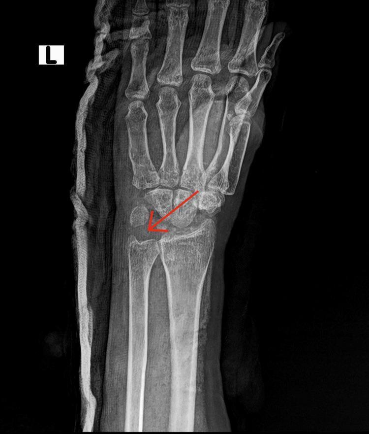
This diagnostic image is a post-operative anteroposterior (AP) view X-ray of the left wrist. The radiograph reveals significant surgical changes, specifically the absence of the proximal row carpal bones (scaphoid, lunate, and triquetrum), consistent with a proximal row carpectomy. A red arrow points to the radiolucent void created by this excision. The distal carpal row remains intact and articulates with the radius. The distal radius and ulna are visualized, as are the metacarpals and phalanges of the hand. Along the lateral aspect of the wrist, a dense, radiopaque, undulating material is visible, which represents post-surgical immobilization—likely a plaster or fiberglass cast or splint. This procedure is frequently performed as a motion-preserving salvage operation for conditions such as advanced Kienbock's disease or scaphoid nonunion advanced collapse (SNAC). The educational focus of the image is the radiological presentation of a partial carpectomy used in orthopedic surgery for chronic wrist pathology.
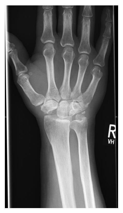
This posteroanterior (PA) X-ray of the right wrist demonstrates the postoperative appearance of a proximal row carpectomy (PRC). The primary diagnostic finding is the surgical absence of the scaphoid, lunate, and triquetrum. Consequently, the proximal carpal row is no longer present, and the capitate has migrated proximally to articulate directly with the lunate fossa of the distal radius. The newly formed radiocapitate articulation shows preserved joint space without significant degenerative changes or subchondral sclerosis. The distal radius and ulna remain intact, showing normal alignment and neutral ulnar variance. The distal carpal row (trapezium, trapezoid, capitate, and hamate) and the metacarpals appear structurally normal with maintained cortical thickness and trabecular density. This imaging is characteristic of a motion-preserving salvage procedure typically used for conditions like Stage III Kienböck's disease or scaphoid nonunion advanced collapse (SNAC), where the goal is to alleviate pain while maintaining functional wrist range of motion.
Proximal Row Carpectomy - Indications
Primary Indications
| Condition | Notes |
|---|---|
| SLAC wrist (Scapholunate Advanced Collapse) | Most common indication; late-stage radioscaphoid arthritis following scapholunate ligament injury |
| SNAC wrist (Scaphoid Nonunion Advanced Collapse) | Long-standing scaphoid nonunion with arthritis, osteonecrosis, and collapse of the proximal pole |
| Kienbock's disease (avascular necrosis of the lunate) | Used as a salvage option in advanced stages |
| Acute severe open carpal fracture-dislocations | Primary PRC indicated when there is significant bony disruption, comminuted scaphoid/lunate fractures, and disruption of blood supply to these bones |
| Failed prior wrist procedures | PRC can serve as a salvage when other procedures have failed |
Ideal Patient Profile
- Patients with limited functional demands (non-manual laborers)
- Those who desire some wrist mobility (PRC preserves ~50-60% of motion)
- Patients who accept the possibility of minimal persistent pain
- Healthy articular surfaces must be present at:
- Lunate fossa of the radius
- Proximal articular surface of the capitate
Contraindications
- Significant arthritis at the capitolunate joint or lunate fossa of the radius (relative contraindication - the proximal capitate pole can be excised with dorsal capsular flap interposition in selected cases)
- Manual laborers with heavy demands - wrist arthrodesis is generally preferred for these patients
PRC vs. Four-Corner Fusion
Post-PRC Radiograph (characteristic appearance)
Bones Excised
- Scaphoid, lunate, and triquetrum (entire proximal row)
- Pisiform is NOT excised (it is a sesamoid within the flexor carpi ulnaris tendon)
- The distal scaphoid pole may optionally be retained to provide a stable thumb base (but requires radial styloidectomy to prevent impingement)