
Dermetomes Of full body explain
Reading File
Finding Sources
Searching PubMed
dermatomes AND anatomy
Searching the Web
dermatomes clinical examination ASIA key sensory points spinal nerve roots reference
Reading File
Reading File
Searching Images
labeled full body dermatome map anterior posterior spinal nerve roots
Dermatomes of the full body
A dermatome is an area of skin whose sensation is supplied mainly by sensory fibers from one spinal nerve root.
There are 31 pairs of spinal nerves:
- 8 cervical: C1-C8
- 12 thoracic: T1-T12
- 5 lumbar: L1-L5
- 5 sacral: S1-S5
- 1 coccygeal: Co1
C1 has no dermatome because it has no significant sensory cutaneous distribution. Neighboring dermatomes overlap, so a single root lesion may reduce sensation rather than cause a perfectly sharp numb area. Guyton and Hall Textbook of Medical Physiology, p. 606.
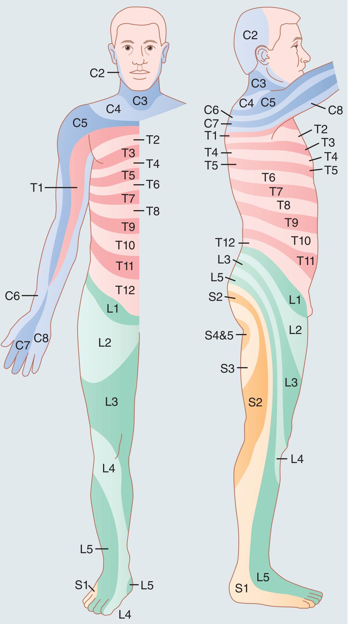
Key dermatome landmarks to memorize
| Spinal root | Main sensory area / landmark |
|---|---|
| C2 | Back of head, occiput |
| C3 | Lower neck |
| C4 | Shoulder tip, over acromioclavicular area |
| C5 | Lateral upper arm, deltoid region |
| C6 | Lateral forearm, thumb |
| C7 | Middle finger |
| C8 | Ring and little finger, medial forearm |
| T1 | Medial forearm and lower arm |
| T2 | Upper medial arm and axilla |
| T3 | Upper chest |
| T4 | Nipple line |
| T5-T6 | Mid-chest and xiphisternum region |
| T7-T9 | Upper abdominal wall |
| T10 | Umbilicus |
| T11 | Below umbilicus |
| T12 | Suprapubic / lower abdominal region |
| L1 | Groin, inguinal ligament |
| L2 | Upper anterior thigh |
| L3 | Lower anterior thigh and medial knee |
| L4 | Medial leg, medial ankle, medial foot |
| L5 | Lateral leg, dorsum of foot, great toe |
| S1 | Lateral foot, little toe, posterior calf |
| S2 | Posterior thigh and upper calf |
| S3 | Buttock / saddle region |
| S4-S5 | Perianal region, skin immediately around anus |
Distribution by body region
1. Head and neck
The face is mainly supplied by the trigeminal nerve (cranial nerve V), not cervical spinal dermatomes:
- V1 ophthalmic: forehead, upper eyelid, bridge of nose
- V2 maxillary: cheek, lateral nose, upper lip
- V3 mandibular: lower jaw, lower lip, chin
Cervical dermatomes begin at the posterior head and neck:
- C2: occiput
- C3: neck
- C4: shoulder top
2. Upper limb
Remember this sequence from the outer to inner side of the hand:
- C5: lateral upper arm
- C6: thumb
- C7: middle finger
- C8: little finger
- T1: medial forearm
A simple memory aid: C6 thumb, C7 middle, C8 little finger.
3. Trunk
Thoracic dermatomes are mostly horizontal bands around the chest and abdomen:
- T4: nipple
- T6: xiphisternum
- T10: umbilicus
- T12: just above pubic region
Below the umbilicus, these bands angle slightly downward toward the midline. General Anatomy and Musculoskeletal System, p. 103.
4. Lower limb
Because of limb rotation during development, leg dermatomes have a spiral pattern rather than simple horizontal bands:
- L1: groin
- L2: upper anterior thigh
- L3: medial knee
- L4: medial leg and medial malleolus
- L5: top of foot and great toe
- S1: outer foot and little toe
- S2: posterior thigh/calf
- S3-S5: saddle and perianal areas
A useful memory aid: L4 medial malleolus, L5 great toe, S1 little toe.
Clinical importance
Dermatomes help localize a problem in a nerve root, spinal cord, or sometimes a peripheral nerve.
Examples:
- Pain or numbness traveling from the low back to the great toe can suggest an L5 radiculopathy.
- Symptoms reaching the lateral foot or little toe can suggest S1 radiculopathy.
- Sensory loss around the umbilicus indicates approximately the T10 level.
- New numbness in the saddle/perianal area with bladder or bowel dysfunction is an emergency warning sign for possible cauda equina compression and needs immediate medical assessment.
- In spinal cord injury, testing sensation at standardized key points helps identify the neurological sensory level. The ASIA examination worksheet defines the sensory level as the lowest segment with normal light touch and pinprick sensation.
Do not interpret a dermatome map as exact borders in every person. Adjacent root territories substantially overlap, and maps differ slightly between anatomy references.
Nerve supply of full body explain
Reading File
“Nerve supply of the full body” is a large topic. It includes both the central nervous system and the peripheral nervous system.
Topic map
-
Basic organization
- Brain, spinal cord, spinal nerves, cranial nerves
- Sensory, motor, and autonomic nerve functions
-
Head and neck
- Cranial nerves I-XII
- Facial sensation, facial movements, eye movements, swallowing, tongue, hearing
-
Trunk
- Thoracic spinal nerves and intercostal nerves
- Abdominal wall nerves
- Diaphragm and phrenic nerve
-
Upper limb
- Brachial plexus
- Axillary, musculocutaneous, median, ulnar, radial nerves
- Main muscle actions and sensory areas
-
Lower limb
- Lumbar and sacral plexuses
- Femoral, obturator, sciatic, tibial, common fibular nerves
- Main muscle actions and sensory areas
-
Autonomic nerves
- Sympathetic system: “fight or flight”
- Parasympathetic system: “rest and digest”
- Control of heart, lungs, gut, bladder, glands, and blood vessels
-
Dermatomes and myotomes
- Skin areas and muscle actions linked to each spinal nerve root
- Useful for locating spinal or nerve-root injury
Basic overview
Nerves carry three major types of signals:
- Sensory (afferent): from skin, joints, muscles, and organs to the brain/spinal cord
- Motor (efferent): from brain/spinal cord to skeletal muscles
- Autonomic: involuntary control of organs, smooth muscle, cardiac muscle, and glands
The body has 12 pairs of cranial nerves arising from the brain and 31 pairs of spinal nerves arising from the spinal cord. Most peripheral nerves contain mixed sensory and motor fibers. Major limb nerves arise by mixing of spinal nerve roots in nerve plexuses, especially the brachial plexus for the upper limb and lumbosacral plexus for the lower limb.
Which section should I explain in detail first?
This is a shared conversation. Sign in to Orris to start your own chat.