
History taking and physical examination: Occupational history techniques. Cultural considerations in patient assessment.
Reading File
Finding Sources
Finding Sources
Searching PubMed
"cultural competency" AND "patient assessment"
Searching the Web
CDC taking an occupational history work related exposures cultural patient assessment CLAS standards
Reading File
Reading File
Reading Web Page
https://www.cdc.gov/niosh/bulletin/2024/primary-care-oh.html
1. Occupational history techniques
An occupational history identifies whether work may cause, worsen, or complicate illness. It should be part of every adult assessment, especially for respiratory, skin, neurologic, musculoskeletal, reproductive, hearing, and mental-health symptoms.
A. Start with broad screening questions
Use open, nonjudgmental questions:
- “What kind of work do you do?”
- “Please describe a usual workday.”
- “Have you ever thought your symptoms might be related to work?”
- “Do symptoms improve on days off, weekends, holidays, or vacation?”
- “Do coworkers have similar symptoms?”
- “Do you have unpaid work, hobbies, or home exposures such as renovations, farming, crafts, or vehicle repair?”
A useful prompt is WHACS:
| Letter | Question |
|---|---|
| W | What do you do? |
| H | How do you do it? |
| A | Are you concerned about exposures at work or outside work? |
| C | Are coworkers or household members exposed or affected? |
| S | Are you satisfied with the job? |
The CDC also advises asking about schedule, long shifts, multiple jobs, stress, and control over tasks because these affect health and self-management of chronic illness. CDC occupational-history guidance
B. Take a chronological job history
Record every important job, beginning with the current job and working backward:
- Job title, industry, employer, and location
- Dates of employment and duration
- Main duties rather than job title alone
- Training, job changes, unemployment, and military service
- Work relationships, job satisfaction, conflict, responsibility, and current feelings about work
Psychiatric history-taking also includes occupation chosen, training, ambitions, conflicts, relationships with supervisors/peers/subordinates, number and duration of jobs, and job-status changes. Kaplan and Sadock's Synopsis of Psychiatry, p. 110.
C. Characterize workplace exposures
Ask specifically about:
- Inhalational: dust, silica, asbestos, fumes, smoke, welding gases, vapors, aerosols, molds, animal proteins
- Chemical: solvents, pesticides, metals, dyes, disinfectants, acids/alkalis, pharmaceuticals
- Physical: noise, vibration, heat/cold, radiation, poor lighting
- Ergonomic: repetitive movements, lifting, awkward posture, prolonged sitting/standing
- Biological: blood/body fluids, animals, infectious patients, waste
- Psychosocial: workload, shift work, harassment, job insecurity, violence, low job control
Clarify the agent, intensity, frequency, route of exposure, ventilation, spills/accidents, and whether personal protective equipment is available, suitable, and consistently used.
D. Establish a work-symptom time relationship
Ask:
- When did symptoms begin relative to starting the job or a new task?
- Do they occur during a shift, after work, or at night?
- Do they improve away from work?
- Was there an acute high-level exposure?
- Have similar symptoms occurred in previous jobs?
A temporal link is important but does not by itself prove causation. Work can be the cause, a trigger, or an aggravating factor for pre-existing disease.
E. Assess consequences and safety
Determine:
- Work restrictions or sickness absence
- Ability to perform duties safely
- Risk to coworkers, family members, or the public
- Need for workplace modification, occupational-health referral, reporting, or compensation documentation
- Patient consent before contacting an employer
Document the occupational history clearly in the chart and update it when job or duties change.
2. Cultural considerations in patient assessment
Culture affects how patients describe symptoms, understand illness, decide whom to consult, communicate with clinicians, and accept treatment. It should be explored with humility rather than assumptions.
Principles
- Ask the person how they identify themselves. Do not assign identity based on appearance, surname, or language.
- Treat culture as broader than ethnicity or religion. It may include language, migration history, gender identity, sexual orientation, age, socioeconomic position, occupation, disability, caste, spirituality, and community.
- Recognize that one person may hold several intersecting identities.
- Avoid stereotyping. Cultural information should guide individualized care, not replace clinical reasoning.
The DSM-5-TR cultural formulation framework identifies five areas: cultural identity; cultural concepts of distress; psychosocial stressors and resilience; cultural features of the patient-clinician relationship; and an overall cultural assessment. Kaplan & Sadock’s Comprehensive Textbook of Psychiatry, p. 2567.
A. Cultural identity and communication
Assess:
- Preferred language for speaking and reading
- Health literacy and numeracy
- Preferred name, pronouns, and family role
- Country/place of origin, migration or displacement experience
- Religion, spiritual practices, and community supports
- Need for an interpreter
Use a qualified medical interpreter for significant language discordance. Do not routinely rely on children, relatives, or untrained staff for sensitive, complex, or consent-related discussions. Speak directly to the patient, use short sentences, avoid jargon, and confirm understanding using teach-back.
B. Patient’s explanatory model of illness
Explore the patient’s own understanding before imposing a biomedical explanation:
- “What do you think is causing this problem?”
- “What worries you most about it?”
- “What name do you give this illness?”
- “What treatments do you think would help?”
- “Who should be involved in decisions about your care?”
Patients may use culturally familiar expressions or bodily symptoms to communicate emotional distress. These are not automatically inaccurate, exaggerated, or evidence of psychiatric illness. Cultural concepts of distress can include idioms of distress, explanatory models, and culturally recognized symptom patterns. Kaplan & Sadock’s Comprehensive Textbook of Psychiatry, p. 2567.
C. Physical examination considerations
Before and during examination:
- Explain each step and obtain permission, especially before touching sensitive areas.
- Offer privacy and draping appropriate to the examination.
- Offer a chaperone and, where feasible, a clinician of the preferred gender.
- Ask about religious or modesty requirements, including head coverings or clothing.
- Adapt communication for disability and literacy needs.
- Avoid misinterpreting behavior: limited eye contact, silence, reluctance to disclose, emotional restraint, or family-centered decision-making may have cultural meanings.
- Do not overlook discrimination and structural barriers such as racism, migration-related insecurity, financial hardship, workplace exploitation, transport barriers, or prior negative healthcare experiences.
Clinical assessment should also identify cultural strengths: family support, faith, community groups, coping methods, and traditional practices that are safe and meaningful to the patient.
D. Negotiating a treatment plan
Use shared decision-making:
- Respect safe traditional remedies and practices.
- Ask specifically about herbs, supplements, traditional medicines, fasting, and healers because of possible toxicity or drug interactions.
- Explain where medical recommendations are non-negotiable for safety.
- Negotiate practical options that fit the person’s beliefs, work, finances, literacy, diet, and family circumstances.
- Include family or community supports only with the patient’s permission.
The CDC CLAS overview describes culturally and linguistically appropriate services as a framework for reducing communication barriers and health inequities.
Brief documentation example
Patient prefers Hindi for detailed health discussions; professional interpreter used. Attributes fatigue partly to work stress and sleep disruption, with no concern about spiritual cause. Requests spouse involvement in treatment decisions. Reports rotating night shifts and solvent exposure at a printing job; headaches worsen during shifts and improve on days off. Uses gloves inconsistently because they interfere with tasks.
Recent-evidence note: systematic reviews on cultural-competence education continue to support structured clinician training, but effects and best teaching methods vary across settings (PMID 41107826).
Respiratory assessment: Chest X-ray interpretation (pneumoconioses). Spirometry in occupational lung disease.
Reading File
Finding Sources
Finding Sources
Searching Images
chest radiograph silicosis upper lobe nodules eggshell hilar lymph node calcification
Searching PubMed
pneumoconiosis AND spirometry
Searching the Web
NIOSH pneumoconiosis chest radiograph ILO classification spirometry occupational lung disease
Reading File
Reading File
query_images_db
Reading Web Page
https://www.ilo.org/resource/other/ilo-international-classif…
Respiratory assessment in occupational lung disease
Assessment combines a targeted occupational history, examination, chest imaging, and pulmonary-function testing. Neither chest X-ray nor spirometry alone establishes a diagnosis. Interpret findings alongside the dust exposure, duration and intensity of exposure, smoking history, symptoms, and CT when needed.
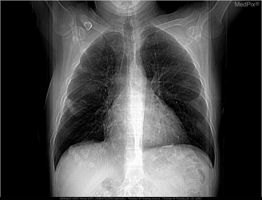
1. Chest X-ray interpretation in pneumoconioses
A posteroanterior (PA) chest X-ray is usually the first imaging test for suspected pneumoconiosis. Use a systematic reading method.
Stepwise approach
-
Check film quality
- Correct patient, date, PA view, inspiration, rotation, exposure/penetration.
- Compare with prior radiographs whenever possible.
-
Assess the general lungs
- Lung volumes: hyperinflation, volume loss
- Distribution: upper versus lower zones; central versus peripheral
- Type of opacity: small rounded, small irregular, or large opacity
- Extent/profusion of abnormalities
-
Assess pleura
- Pleural plaques, diffuse pleural thickening, calcification, costophrenic-angle blunting.
-
Assess hila and mediastinum
- Lymph-node enlargement or calcification, including eggshell calcification.
-
Look for complications
- Progressive massive fibrosis
- Tuberculosis or other infection
- Lung cancer
- Emphysema or chronic obstructive pulmonary disease
- Cor pulmonale in advanced disease
The International Labour Organization system is the standard method for recording radiographic abnormalities caused by inhaled dust. Its revised 2022 system uses digital reference images and is intended for PA chest radiographs. It supports surveillance and epidemiology but does not by itself diagnose disease severity or determine compensation. ILO classification guidance
ILO classification: what to report
| Feature | Description |
|---|---|
| Small-opacity shape | Rounded or irregular |
| Small-opacity size | Rounded: p, q, r. Irregular: s, t, u |
| Profusion | Number/concentration of small opacities, graded 0 to 3 using standard reference films |
| Zones affected | Upper, middle, lower lung zones; right and left |
| Large opacities | A, B, or C categories |
| Pleural abnormalities | Plaques, diffuse thickening, calcification, site and extent |
| Other findings | Hilar/mediastinal nodes, emphysema, tuberculosis, cardiac abnormalities |
Formal ILO classification requires appropriate image quality and comparison with standard radiographs, often by a trained reader. NIOSH technical guidance
Typical chest X-ray patterns
| Disease / exposure | Typical radiographic pattern | Important associated findings |
|---|---|---|
| Silicosis - crystalline silica | Multiple small rounded opacities, usually upper-zone predominant; may coalesce | Hilar/mediastinal lymph-node calcification, sometimes eggshell pattern; progressive massive fibrosis; increased tuberculosis risk |
| Coal workers' pneumoconiosis - coal mine dust | Small rounded upper-zone opacities, resembling silicosis | May progress to progressive massive fibrosis; emphysema and airflow obstruction may coexist |
| Asbestosis - asbestos fibers | Fine irregular/reticular opacities, generally peripheral and lower-zone predominant, with volume loss in advanced disease | Pleural plaques, diaphragmatic or parietal pleural calcification, diffuse pleural thickening, rounded atelectasis |
| Chronic beryllium disease | May resemble sarcoidosis with diffuse nodular/interstitial changes | Hilar adenopathy may occur; diagnosis requires exposure history and immunologic testing |
| Talcosis / mixed dust disease | Variable diffuse nodular or reticulonodular pattern | Depends on agent and co-exposures |
Progressive massive fibrosis
This is a severe complication of silica- and coal-dust disease. It appears as large, often bilateral upper-lung masses formed by coalescence of small opacities. It may cause marked volume loss, hilar distortion, traction emphysema, hypoxemia, and respiratory disability.
Key caution
The X-ray appearance does not reliably predict symptom burden or physiological impairment. Rounded-opacity pneumoconioses may look extensive with little functional loss, while irregular fibrosis, such as asbestosis, may produce significant restrictive impairment despite less striking radiographic disease. Harrison’s Principles of Internal Medicine, 22e, section on occupational lung disease.
2. Spirometry in occupational lung disease
Purpose
Spirometry measures airflow and is useful for:
- Baseline assessment before or early in exposure
- Periodic surveillance of exposed workers
- Detecting airway disease due to dust, fumes, sensitizers, or irritants
- Assessing disability and change over time
- Investigating suspected work-related asthma or chronic obstructive pulmonary disease
It is particularly helpful in silica-exposed workers and may identify early health effects from occupational dust exposure. Fishman’s Pulmonary Diseases and Disorders, section on lung functional abnormalities in silicosis.
Core variables
| Variable | Meaning |
|---|---|
| FEV1 | Forced expiratory volume exhaled in the first second |
| FVC | Forced vital capacity, the total forced exhaled volume |
| FEV1/FVC | Main indicator of airflow obstruction |
Interpret results using appropriate reference values, preferably expressed as z-scores and lower limit of normal rather than a fixed ratio alone.
Major spirometric patterns
| Pattern | Spirometry | Occupational examples |
|---|---|---|
| Obstructive | Reduced FEV1/FVC; reduced FEV1 | Occupational asthma, chronic bronchitis/COPD from dusts or fumes, coal-dust-associated airway disease |
| Restrictive pattern suspected | Reduced FVC with normal or increased FEV1/FVC | Asbestosis, advanced interstitial fibrosis, progressive massive fibrosis |
| Mixed pattern | Reduced FEV1/FVC and FVC | Combined fibrosis and airway disease, often with smoking-related COPD |
| Normal spirometry | Normal FEV1, FVC, and ratio | May occur in early pneumoconiosis, early silicosis, pleural plaques, or intermittent occupational asthma |
Important: Spirometry cannot confirm restriction. A low FVC suggests restriction, but total lung capacity must be measured to establish it. Diffusing capacity for carbon monoxide (DLCO) may be reduced in interstitial fibrosis, emphysema, and pulmonary vascular disease, sometimes before major spirometric abnormalities.
Application to specific occupational disorders
Silicosis and coal workers' pneumoconiosis
- Simple disease may have normal spirometry.
- Obstruction can occur due to dust-related small-airway disease, chronic bronchitis, emphysema, or smoking.
- Restriction, low DLCO, hypoxemia, and reduced exercise capacity are more likely in advanced fibrosis or progressive massive fibrosis.
- A normal test does not exclude radiographic pneumoconiosis.
Asbestosis
- Typical physiological pattern is restriction:
- Low FVC
- Preserved or elevated FEV1/FVC ratio
- Reduced TLC
- Often reduced DLCO
- Pleural plaques alone often cause little or no major spirometric abnormality.
Occupational asthma
- Baseline spirometry may be normal between episodes.
- Obstruction may improve after bronchodilator.
- Serial peak expiratory flow or serial FEV1 measurements at work and away from work are often more informative than a single clinic test.
Work-related COPD
- Persistent obstruction may be present.
- Consider additive effects of smoking plus occupational dust, fumes, biomass smoke, or chemical exposure.
Quality and interpretation principles
- Ensure patient effort is acceptable and repeatable.
- Record height, age, sex, and reference equation used.
- Compare with previous tests, not just predicted values.
- A consistent decline in FEV1 over time may signal harmful exposure even if individual values remain within the reference range.
- Interpret changes after considering smoking, weight change, respiratory infection, equipment variation, and test quality.
- Abnormal spirometry should trigger exposure review, clinical evaluation, and consideration of full pulmonary function testing, imaging, and occupational-medicine referral.
Examination correlation
In early simple pneumoconiosis, the respiratory examination may be normal. In advanced fibrotic disease, look for:
- Tachypnea and reduced exercise tolerance
- Reduced chest expansion
- Fine late inspiratory basal crackles, especially in asbestosis
- Wheeze or prolonged expiration when airflow obstruction is present
- Cyanosis, signs of pulmonary hypertension, peripheral edema, or raised JVP in advanced respiratory failure/cor pulmonale
Silicosis often has few signs unless complications develop, and inspiratory crackles may be absent. Fishman’s Pulmonary Diseases and Disorders, section on clinical picture of silicosis.
Recent-evidence note: the recent systematic-review search did not identify a directly relevant updated meta-analysis specifically validating spirometric interpretation in pneumoconiosis. Current practice still relies on quality-assured serial spirometry integrated with exposure history, imaging, lung volumes, and DLCO.
This is a shared conversation. Sign in to Orris to start your own chat.