
What is Trans Anal Minimal Invasive Surgery (TAMIS)? Give outlines of its indications, contraindications, advantages and limitations. How is TAMIS different from Trans-anal Endoscopic Microsurgery (TEM)
Reading File
Finding Sources
Finding Sources
Searching PubMed
TAMIS "transanal minimally invasive surgery" rectal
Reading File
Reading File
Finding Sources
Reading File
Reading File
Reading File
Reading File
Reading File
Finding Sources
Reading File
Finding Sources
Reading File
Reading File
Reading Article
PMID: 35237103
Reading File
I now have all the information needed to write a comprehensive answer. Let me compile it.
Transanal Minimally Invasive Surgery (TAMIS)
Overview and Historical Background
TAMIS was developed in 2009 (first performed on June 30, 2009) as a consequence of advances in single-port laparoscopic surgery. It was conceived specifically to give surgeons without access to the expensive rigid platforms (TEM/TEO) a means of performing advanced transanal local excision (LE) of rectal neoplasia. The original port used was the SILS Port (Covidien); the GelPOINT Path (Applied Medical), which has FDA approval specifically for transanal use, is now the most widely adopted platform in the United States.
TAMIS uses a flexible, disposable multichannel single-site laparoscopic port inserted into the anal canal, combined with standard laparoscopic instruments, a 5 mm 30°/45° angled camera, and CO₂ insufflation (pneumorectum at up to 18 mmHg). No proprietary or capital-intensive equipment beyond what a standard laparoscopic theatre already possesses is required.
- Fischer's Mastery of Surgery - 8th ed., p. 4362
Indications
The indications for TAMIS mirror those for all transanal LE platforms and include:
Benign lesions:
- Villous adenoma and tubular adenoma not amenable to simple endoscopic retrieval
- Submucosal lesions of unknown histology requiring excision for diagnosis
- Rectal neuroendocrine tumors ≤1 cm in diameter
Malignant lesions (highly selected):
- Clinical T1 rectal cancer with all of the following features:
- Histologically favorable (no lymphovascular invasion, no perineural invasion, no tumor budding, well-differentiated, no mucinous or signet ring features)
- Tumor occupying <1/3 of the rectal circumference
- Diameter <3 cm
- Situated in the distal or mid-third of the rectum (preferably ≤10 cm from the anal verge)
- Submucosa invasion level: SM1 or SM2 (not SM3)
- Selected T2 cancers after neoadjuvant chemoradiotherapy (watch-and-wait/near-complete response protocols)
Anatomic reach:
- TAMIS is especially valuable for mid-rectal lesions (beyond the reach of conventional Parks' TAE, i.e., above 5–6 cm) where TEM/TEO are not available.
Extended applications:
-
Transanal total mesorectal excision (TaTME / TAMIS-TME) for rectal cancer requiring radical surgery
-
Resection of rectal GISTs and other submucosal tumors
-
Fischer's Mastery of Surgery - 8th ed., pp. 4347–4349; Current Surgical Therapy 14e, p. 2807
Contraindications
Absolute:
- T2 or higher rectal cancer (without neoadjuvant therapy protocols) - requires formal segmental resection (SR) with TME
- Any rectal cancer with poor prognostic features (lymphovascular invasion, perineural invasion, tumor budding, poor differentiation, SM3 invasion) - standard recommendation is SR even after LE
- Tumors in the proximal third of the rectum (>10 cm from anal verge), particularly anterior tumors - peritoneal violation is unavoidable and oncologic principles are compromised
- Evidence of lymph node metastasis (N+) on staging MRI or ERUS
- Distant metastatic disease (M1) where cure is not the intent and LE does not change management
Relative:
-
Lesions occupying >1/3 of the rectal circumference (high risk of rectal stenosis and functional compromise)
-
Prior pelvic radiotherapy (increased risk of wound breakdown and fistulation)
-
Poor anal sphincter tone or significant anal stenosis (platform cannot be safely seated)
-
Very distal lesions just at the dentate line (the port sits above the anorectal ring and distal margins may be difficult to achieve; a hybrid conventional TAE/TAMIS approach can be used)
-
Immunocompromised patients or those with significant co-morbidity (relative)
-
Fischer's Mastery of Surgery - 8th ed., pp. 4349–4352, 4368–4369
Advantages of TAMIS
| Domain | Advantage |
|---|---|
| Cost & availability | Uses standard laparoscopic tower, instruments, and CO₂ insufflator - no capital investment in proprietary equipment |
| Disposable platform | Single-use port eliminates sterilization costs and scheduling constraints |
| Patient positioning | Patient stays in dorsal lithotomy regardless of lesion position in the rectal wall (anterior vs. posterior) - no repositioning needed |
| Instrument compatibility | Accepts standard 5 mm and 10 mm laparoscopic instruments including graspers, thermal energy devices, and needle drivers |
| Reach | Accesses mid and upper rectum, well beyond the 5–6 cm limit of conventional Parks' TAE |
| Oncologic quality | Allows full-thickness excision with mesorectal sampling, comparable specimen quality to TEM |
| Learning curve | Shorter and less steep than TEM, because surgeons already familiar with laparoscopic technique can adapt readily |
| Pneumodissection | CO₂ pneumorectum provides natural tissue plane separation, clarifying the dissection |
| Specimen retrieval | Simple - remove the port lid (GelPOINT) to retrieve the specimen without the need to remove the scope |
| Expandability | Compatible with robotic platforms (R-TAMIS) using DaVinci Xi or SP, further expanding capability |
| TME applications | The platform has enabled TaTME (TAMIS-TME) for radical rectal cancer surgery from below |
- Fischer's Mastery of Surgery - 8th ed., pp. 4362–4365; Current Surgical Therapy 14e, p. 2807
Limitations of TAMIS
| Domain | Limitation |
|---|---|
| Intraluminal suturing | Closing the rectal defect intraluminally is technically demanding due to the confined space and instrument triangulation within the port - the most consistently cited technical challenge |
| Optics | Standard 2D laparoscopic camera (vs. TEM's stereoscopic 3D view) - depth perception is inferior without robotic assistance |
| Instrument rigidity | Laparoscopic instruments are straight; lack of articulation limits maneuverability in the confined rectal lumen compared to robotic instruments |
| Pneumorectum stability | Standard insufflation can cause "bellowing" of the rectal wall during suctioning; AirSeal (valveless trocar with pressure regulation) helps but adds cost |
| Very distal lesions | The port seats just above the anorectal ring, making excision of lesions immediately above the dentate line difficult; the platform may obscure the distal margin |
| No tactile feedback | Standard laparoscopic instruments have limited haptic sense; this is exacerbated in R-TAMIS |
| Platform seat | The flexible port can migrate or lose its seal in some anatomic configurations (short anal canal, low sphincter tone) |
| Smoke evacuation | Standard CO₂ systems accumulate smoke during cautery; requires dedicated suction-irrigator management or AirSeal system |
| Complications | Subcutaneous emphysema (including scrotal), pneumoperitoneum, wound dehiscence, post-operative bleeding (1-10%), rectovaginal fistula (in females after anterior excision), rectal stenosis |
- Fischer's Mastery of Surgery - 8th ed., pp. 4364, 4368–4370
TAMIS vs. TEM - Key Differences
| Feature | TAMIS | TEM |
|---|---|---|
| Introduced | 2009 (Atallah & Albert) | 1983 (Gerhard Buess) |
| Platform | Flexible, disposable multichannel single-port (GelPOINT Path or SILS Port) | Rigid 4 cm diameter steel rectoscope (12 cm or 20 cm lengths), reusable, proprietary |
| Equipment cost | Minimal - uses standard laparoscopic tower already available | High capital investment - proprietary Karl Storz system required |
| Optics | 2D laparoscopic camera (5 mm, 30°/45°) | 3D stereoscopic magnified (6×) eyepiece; superior depth perception |
| Vision system | Standard HD 2D (unless R-TAMIS used) | Binocular stereoscopic = gold standard for precision excision |
| Patient positioning | Dorsal lithotomy always - no repositioning regardless of lesion location | Must position patient so the lesion is dependent (6 o'clock) - may require lateral decubitus or full prone position |
| Instrument articulation | Standard straight laparoscopic instruments | Specialized long-shaft TEM instruments with angled tips (right/left) for improved intra-lumen maneuverability |
| Stabilization | Port self-retains in anal canal | Martin Arm clamp fixes the scope rigidly to the table rail for a fixed orientation |
| Channels | 3 working channels | 4 working channels via airtight sealed rubber sleeves in faceplate |
| Insufflation | Standard CO₂ insufflator (laparoscopic); AirSeal recommended | Proprietary specialized insufflation system designed for small-volume pneumorectum |
| Learning curve | Shorter - adaptable by laparoscopic surgeons | Longer and steeper - unique instruments, scope, and 3D optics require dedicated training |
| Disposable vs. reusable | Disposable port (per-case cost) | Reusable scope (sterilization overhead, scheduling dependency) |
| Oncologic outcomes | Comparable to TEM for T1 rectal cancer (similar R0 rates, specimen quality) | Gold standard historically; 3D optics may confer marginal technical advantage |
| Robotic extension | R-TAMIS well established (DaVinci Xi / SP) | Robotic TEM less widely explored |
| TaTME use | Widely adopted as the platform for TaTME/TAMIS-TME | TEM used for conventional LE only; not suited for TME |
| Distal rectal lesions | Can be technically challenging (port sits above anorectal ring) | Rigid scope with beveled end can be more precisely oriented for mid-rectal lesions |
Summary: TEM remains the historical gold standard with superior 3D optics and a proven four-decade track record, but its high cost, steep learning curve, mandatory patient repositioning, and proprietary infrastructure requirements have driven widespread adoption of TAMIS. Oncologic outcomes (local recurrence, R0 resection, specimen quality) appear equivalent between the two platforms. TAMIS has additionally expanded its role to enable TaTME, an application TEM was not designed for.
- Fischer's Mastery of Surgery - 8th ed., pp. 4354–4365; Current Surgical Therapy 14e, p. 2807; Devane LA et al., TAMIS vs. rigid platforms, Clin Colon Rectal Surg 2022 (PMID: 35237103)
Robotic TAMIS (R-TAMIS) - Brief Note
R-TAMIS uses the DaVinci Xi or SP platform inserted through the GelPOINT Path and directly addresses TAMIS's two major technical limitations: the 2D optics and the lack of instrument articulation. High-definition 3D vision and endowristed instruments with 7 degrees of freedom make intraluminal suturing significantly easier and allow resection of near-circumferential lesions up to 13.5 cm. The DaVinci SP (25 mm port) additionally minimizes lateral sphincter distension. The main limitations of R-TAMIS are higher cost and longer operative time.
- Fischer's Mastery of Surgery - 8th ed., pp. 4365–4366
Discuss management of hyperhidrosis.
Reading File
Finding Sources
Finding Sources
Searching PubMed
hyperhidrosis treatment management
Reading File
Reading File
Reading File
Reading File
Reading File
Reading File
Reading File
Reading File
Reading File
Reading File
Reading File
Loading Image
I now have comprehensive information from all major textbooks. I can now write a detailed, well-organized answer.
Management of Hyperhidrosis
Classification - A Framework for Management
Hyperhidrosis is excessive sweating beyond what is physiologically required for thermoregulation. Treatment is guided by classification:
Primary (focal/idiopathic) hyperhidrosis
- Most common type; affects palms, soles, axillae, craniofacial region
- Occurs only during waking hours (diurnal); bilateral and symmetric
- Onset in childhood (palmoplantar) or adolescence (axillary)
- Up to 80% have a positive family history
- Not associated with any systemic disease
Secondary (generalized) hyperhidrosis
- Due to underlying systemic cause: endocrine disorders (hyperthyroidism, diabetes, pheochromocytoma, acromegaly, carcinoid, menopause), infections, malignancy (lymphoma), neurologic disorders (Parkinson's disease), or drugs (SSRIs, TCAs, opioids, anticholinesterases)
- Often nocturnal, generalized, or asymmetric
- Management is primarily directed at the underlying cause
Assessment Tools
- Minor's Starch-Iodine Test: Iodine solution is applied to the affected area and dried, then starch powder is dusted over it. Moisture from sweat glands turns the mixture dark blue-purple, mapping the extent and distribution of hyperhidrosis. This guides both diagnosis and treatment (e.g., targeting botulinum toxin injections).
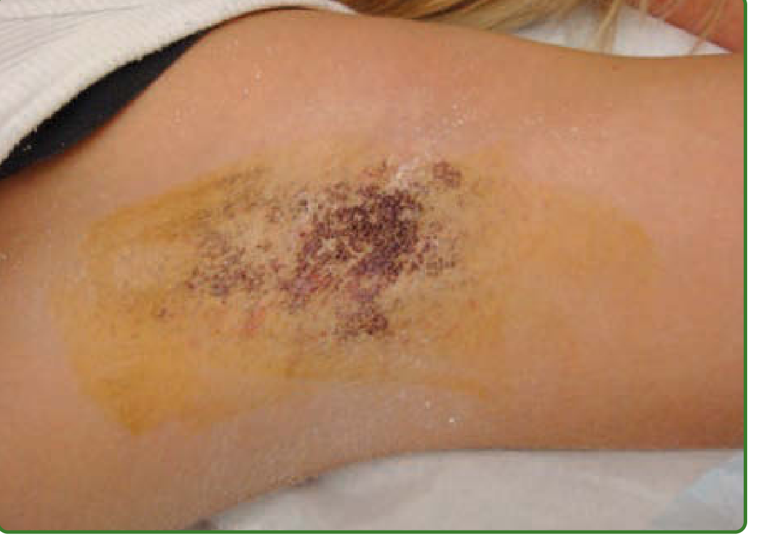
- Hyperhidrosis Disease Severity Scale (HDSS): A 4-point scale rating impact on daily activities (1 = never noticeable; 4 = intolerable and always interferes). Used to grade severity and monitor treatment response.
- Dermatology Life Quality Index (DLQI): Used to quantify quality-of-life impact.
- Gravimetric (weight-based) sweat measurement: accurate but mainly a research tool.
Management - Stepwise Approach
Treatment is selected based on body site, severity, and response to prior therapy. A stepwise escalation from least to most invasive is used.
Step 1 - Topical Therapy (First-Line)
Aluminum chloride hexahydrate (AlCl₃)
- Concentration: 10-35% for axillae; up to 50% (with occlusion under plastic gloves) for palms and soles
- Applied to completely dry skin at bedtime (blow-dry skin first); washed off after 6-8 hours
- Mechanism: precipitates within the eccrine duct lumen causing mechanical obstruction
- If irritation occurs, reduce frequency to every other night or apply over a hydrocortisone cream
- Once effective, frequency can be reduced to 1-2 times per week for maintenance
- Pitfall: Moisture on skin during application generates weak hydrochloric acid, causing burning and desquamation
Topical glycopyrrolate (0.5-2%)
-
An anticholinergic applied topically; especially useful for craniofacial hyperhidrosis
-
Can be compounded at specialty pharmacies
-
Reduces sweating with less systemic absorption than oral anticholinergics
-
Fitzpatrick's Dermatology, 9th ed., p. 1491; Andrews' Diseases of the Skin, p. 892
Step 2 - Physical Therapy Devices
Tap water iontophoresis
- A low-intensity direct electrical current (15-18 mA) is passed through tap water into the skin surface
- Mechanism not fully established; likely blocks eccrine duct function through pH changes or ion deposition
- Treatment sessions: 20-30 minutes daily or twice daily initially; maintenance once every 1-2 weeks
- Effective for palmar and plantar hyperhidrosis; less commonly used for axillae
- Adding glycopyrrolate 0.01%, botulinum toxin, or aluminum chloride 2% to the iontophoresis medium can accelerate response
- Main advantages: inexpensive, no systemic effects, can be self-administered at home (Drionic device or Fischer unit)
- Limitations: time-consuming, requires regular sessions to maintain effect; cannot be used over broken skin, in pregnancy, or with cardiac pacemakers
Microwave thermolysis (miraDry)
- Microwave energy selectively destroys eccrine and apocrine glands in the dermis-hypodermis interface via thermal coagulation
- Primarily used for axillary hyperhidrosis
- High reported patient satisfaction; long-lasting or near-permanent effect
- Side effects: transient edema, erythema, pain; longer-term effects include fibrous bands and possible muscle weakness
- Offers the advantage of also reducing axillary hair and odor
Radiofrequency microneedling and laser therapy
-
Fractional laser and microneedle radiofrequency can destroy sweat glands
-
Appear effective but require more robust study for response rates and adverse effects
-
Andrews' Diseases of the Skin, p. 892; Fitzpatrick's Dermatology, 9th ed., p. 1492
Step 3 - Botulinum Toxin A Injection (Second-Line, Most Evidence)
Botulinum toxin A (onabotA / Botox) is FDA-approved (2004) for axillary hyperhidrosis and has Level A evidence for this indication. It is also used off-label for palmar, plantar, and craniofacial hyperhidrosis.
Mechanism: Inhibits cholinergic synaptic transmission at eccrine sweat gland neuroeffector junctions by blocking acetylcholine release from presynaptic vesicles.
Axillary hyperhidrosis:
- Dose: 50-100 U of onabotulinumtoxinA (or up to 200 U of abobotulinumtoxinA / Dysport) per axilla
- Technique: Intradermal injections spaced 1-2 cm apart over the entire hyperhidrotic area (mapped by starch-iodine test); 10-20 injection sites
- Anesthesia: Usually not required (axillary skin relatively insensitive); EMLA cream may be applied
- Onset: Within 72 hours; peak at 5-7 days
- Duration: Anhidrosis lasts 6-12 months (up to 29 months with higher doses)
- Side effects: Minor bruising from needle trauma; no significant compensatory hyperhidrosis (axillary surface area is small compared to the area affected by sympathectomy)
- Repeated injections maintain efficacy and may extend duration over time
Palmar hyperhidrosis:
- More technically challenging than axillary treatment
- Dose: 100-200 U onabotulinumtoxinA per palm; 50-100 injection sites at 2-3 U each, covering the entire palmar surface including distal wrist crease, thenar and hypothenar eminences
- Anesthesia required: Regional wrist nerve blocks (median, ulnar, and radial nerves) using 1-2% lidocaine without epinephrine, OR ice applied to skin
- Duration: 3-4 months (shorter than axillary)
- Side effect: Temporary hand weakness (especially thumb-index pinch) lasting up to 2 weeks, due to toxin diffusion to intrinsic hand muscles - a predictable and important limitation
- Considered second-line after failure of iontophoresis and topical therapy
Plantar hyperhidrosis:
- Similar principles to palmar; requires regional nerve blocks
- Doses comparable to palmar treatment
- Response duration similar to palmar
Other sites (craniofacial, gustatory/Frey syndrome):
-
Botulinum toxin is highly effective for Frey syndrome (auriculotemporal nerve syndrome post-parotidectomy); remission for 1-10 years
-
Effective for craniofacial and compensatory hyperhidrosis post-sympathectomy
-
Andrews' Diseases of the Skin, p. 892; Fitzpatrick's Dermatology, 9th ed., p. 1491; Dermatology 2-Volume Set 5e, p. 3957
Step 4 - Systemic Pharmacotherapy
Used as monotherapy for generalized hyperhidrosis or as an adjunct when hyperhidrosis is multifocal or topical/injection therapy is impractical.
Anticholinergics (first choice):
| Drug | Notes |
|---|---|
| Glycopyrrolate (1-2 mg BD) | Quaternary amine - limited CNS/cardiac penetration; fewer central side effects; preferred |
| Oxybutynin (2.5-5 mg BD/TDS) | Tertiary amine - crosses blood-brain barrier; more CNS side effects; effective but causes more dry mouth |
| Propantheline bromide | Older agent; regulated by patient tolerance |
- Common anticholinergic side effects: xerostomia, xerophthalmia, constipation, mydriasis, blurred vision, urinary hesitancy/retention, tachycardia (high doses), bradycardia (low doses), mental confusion (especially in children and elderly)
- Contraindications: Myasthenia gravis, paralytic ileus, pyloric stenosis
- Caution: Closed-angle glaucoma, bladder outlet obstruction, GERD, cardiac insufficiency
- Sweating suppression generally lasts 4-6 hours per dose; many patients prefer to use intermittently for social events rather than continuously
Other systemic agents:
-
Clonidine (centrally acting α₂-adrenergic agonist): useful for stress-induced and menopausal hyperhidrosis
-
Propranolol (β-blocker): particularly useful in stress-induced or anxiety-related hyperhidrosis
-
Benzodiazepines: for anxiety-triggered episodes; not recommended long-term
-
Diltiazem: occasional reports of benefit in localized hyperhidrosis
-
Fitzpatrick's Dermatology, 9th ed., pp. 1491-1492; Andrews' Diseases of the Skin, p. 893
Step 5 - Surgical Treatment
Reserved for severe, refractory cases that have failed conservative measures.
A. Local Surgical Procedures for Axillary Hyperhidrosis:
-
Excision of axillary skin: Elliptical excision of the most actively sweating portion of axillary skin, followed by undercutting and subcutaneous resection of sweat glands for 1-2 cm on either side of the incision. Nearly always effective. Preoperative mapping with the starch-iodine test is essential as eccrine gland distribution does not always correlate with hair-bearing areas.
-
Liposuction / subcutaneous curettage (suction-curettage): Tumescent liposuction or surgical ultrasonic aspiration removes the subcutaneous fat containing the sweat glands. Less invasive than excision; avoids skin scarring. Can be combined with direct curettage for higher efficacy.
-
Laser treatments: Nd:YAG laser, diode laser, or fractional CO₂ laser can thermally destroy subdermal sweat glands. Effective for axillary disease with acceptable side effect profiles.
B. Endoscopic Thoracic Sympathectomy (ETS)
- The definitive, most effective surgical treatment for severe palmar and axillary hyperhidrosis refractory to all other modalities
- Procedure: Thoracoscopic interruption (clipping, cutting, or ablation) of the thoracic sympathetic chain at specific ganglia:
- T2: Craniofacial/upper limb hyperhidrosis
- T3-T4: Palmar hyperhidrosis (T3 preferred to reduce compensatory sweating)
- T4: Axillary hyperhidrosis
- Performed under general anesthesia; bilateral procedure done at the same sitting or in staged fashion
- Immediate and permanent cessation of sweating in the targeted areas
Complications of ETS - critical to counsel patients:
| Complication | Incidence | Notes |
|---|---|---|
| Compensatory (reflex) hyperhidrosis | 30-75% | Most important and often disabling complication; excessive sweating of trunk, thighs, and lower limbs to compensate for anhidrosis of treated areas. Severity correlates with the level and extent of sympathectomy |
| Horner's syndrome | ~1-2% | Ptosis, miosis, anhidrosis of the face - if T1 ganglia damaged |
| Pneumothorax | ~2-5% | Usually minor; managed with drainage |
| Bradycardia | Rare | If stellate ganglion affected |
| Gustatory sweating | Uncommon | Post-sympathectomy |
| Recurrence | Rare (nerve regeneration) | May occur months/years later |
-
ETS is considered irreversible; patient selection must be meticulous. Compensatory hyperhidrosis can be more debilitating than the original problem, and patients must be fully informed.
-
Sympathectomy clipping (rather than cutting/ablation) allows reversal if compensatory hyperhidrosis becomes intolerable, though reversal is not always successful.
-
For axillary hyperhidrosis, local surgical procedures are preferred over ETS due to the high rate of compensatory hyperhidrosis with sympathectomy.
-
Fitzpatrick's Dermatology, 9th ed., p. 1492; Andrews' Diseases of the Skin, p. 893; Bailey and Love's Surgery 28e, p. 664
Summary - Treatment Algorithm by Site
| Site | First-Line | Second-Line | Third-Line / Refractory |
|---|---|---|---|
| Axillae | Aluminum chloride 10-20% | Botulinum toxin A (50-100 U/axilla) | Microwave thermolysis; local excision/curettage; ETS (T4) |
| Palms | Aluminum chloride 20-50%; Iontophoresis | Botulinum toxin A (100-200 U/palm) with nerve block | ETS (T3) |
| Soles | Aluminum chloride; Iontophoresis | Botulinum toxin A (higher doses; painful) | ETS |
| Craniofacial | Topical glycopyrrolate; Systemic anticholinergics | Botulinum toxin A | Rarely ETS (T2) |
| Generalized | Treat underlying cause; Systemic anticholinergics | - | - |
| Frey syndrome | Botulinum toxin A | - | Tympanic neurectomy |
Key Points
-
The diagnosis of primary focal hyperhidrosis requires at least 6 months of visible focal sweating without apparent cause, plus at least two of: bilateral/symmetric distribution, onset <25 years, cessation during sleep, frequency ≥1 episode/week, positive family history, or impairment of daily activities.
-
Secondary hyperhidrosis should always be excluded before treating - it is often nocturnal, asymmetric, and generalized.
-
Botulinum toxin remains the most evidence-based non-surgical second-line therapy for focal hyperhidrosis and is the treatment of choice when topicals fail.
-
ETS is highly effective but compensatory hyperhidrosis (in up to 30-75% of patients) is a major limitation, and patients must be carefully counselled before surgery.
-
A newer FDA-approved oral anticholinergic, sofpironium bromide (a topical glycopyrrolate prodrug), and oxybutynin extended-release are emerging options reviewed in recent literature (Maazi et al., Drugs Context, 2025 - PMID: 40575073).
-
Fitzpatrick's Dermatology, 9th ed.; Andrews' Diseases of the Skin; Dermatology 2-Volume Set 5e; Devane et al., Clin Colon Rectal Surg 2022; Maazi M et al., Primary hyperhidrosis: an updated review, Drugs Context 2025 (PMID: 40575073)
Briefly discuss the etiology, evaluation and management of chronic umbilical discharge in a 30-year-old woman. [2+3+5]
Finding Sources
Finding Sources
Reading File
Reading File
Reading File
Reading File
I have sufficient information from multiple authoritative sources. I can now compile a focused, well-structured answer matching the [2+3+5] mark allocation.
Chronic Umbilical Discharge in a 30-Year-Old Woman
The marks [2+3+5] reflect: Etiology (2), Evaluation (3), Management (5).
Etiology [2 marks]
The causes of chronic umbilical discharge in an adult woman can be broadly grouped as:
1. Congenital Remnant Anomalies
-
Patent vitello-intestinal (omphalomesenteric) duct: Failure of obliteration of the duct connecting the fetal midgut to the yolk sac. Normally obliterates by the 7th week of gestation. Persistence results in a fistula between the ileum and the umbilicus, producing faecal/intestinal discharge. Prolapse of the distal mucosal end produces a raspberry tumour (enteroteratoma / umbilical adenoma) - a red, moist, bleeding nodule at the umbilicus.
-
Patent urachus: The urachus connects the fetal bladder to the allantois and normally obliterates to form the median umbilical ligament. Partial or complete failure gives rise to four variants:
- Patent urachus: Full connection between bladder and umbilicus - urinary discharge from the umbilicus
- Urachal sinus: Blind-ending tract opening at the umbilicus (no bladder connection) - mucoid/purulent discharge
- Urachal cyst: Isolated segment that does not drain - presents with infection/mass rather than discharge
- Urachal diverticulum: Opens only into the bladder - no umbilical discharge
2. Acquired Causes
-
Umbilical endometrioma: Deposition of ectopic endometrial tissue at the umbilicus. Highly relevant in a 30-year-old woman. Characteristically produces cyclical bleeding/discharge coinciding with menstruation (catamenial). The umbilicus appears as a blue-brown nodule that enlarges and bleeds during periods. Associated with pelvic endometriosis in many cases.
-
Umbilical sepsis/abscess: Poor umbilical hygiene leading to accumulation of desquamated epithelium, dirt, and bacteria. Umbilical concretion (calculus) - black/dark compacted debris - may be the nidus. Infection causes abscess formation and purulent discharge. Not the same as omphalitis (which is a neonatal condition).
-
Pilonidal sinus of the umbilicus: Rare; hair penetrates the umbilicus in hairy individuals, with sinus formation and discharge.
-
Intra-abdominal abscess/fistula: Any deep pelvic or abdominal abscess (e.g., Crohn's disease, appendiceal abscess, ovarian abscess, pelvic actinomycosis) may spontaneously drain through the umbilicus as it is the thinnest and weakest point of the anterior abdominal wall.
-
Secondary carcinomatous deposit (Sister Mary Joseph's nodule): Umbilical metastasis from primary gastric, colonic, ovarian, or breast cancer. In a 30-year-old woman, ovarian carcinoma is a relevant concern. Presents as a hard, indurated nodule that may ulcerate and discharge.
-
Primary umbilical carcinoma: Rare; involves both axillary and inguinal lymph nodes due to its unique lymphatic drainage.
-
S. Das - A Manual on Clinical Surgery, 13th ed., p. 521; Bailey and Love's Surgery 28e, p. 1517
Evaluation [3 marks]
History
- Character of discharge: faecal (vitello-intestinal fistula), urinary/clear (patent urachus), bloody/cyclical (endometrioma), purulent (sepsis/abscess), mucoid (urachal sinus)
- Timing: cyclical with menstruation strongly suggests endometrioma
- Duration, onset, aggravating factors
- Urinary symptoms (dysuria, frequency, haematuria) - suggest urachal anomaly
- History of abdominal/pelvic surgery, inflammatory bowel disease, pelvic pain (endometriosis)
- Family history of malignancy
Physical Examination
- Inspect umbilicus: note appearance of the discharge, colour, associated skin changes (erythema, induration), presence of a nodule or mass
- A red raspberry-like nodule = enteroteratoma/umbilical adenoma
- A blue-brown nodule bleeding cyclically = endometrioma
- A hard, indurated nodule with discharge = malignancy (Sister Mary Joseph's nodule)
- Palpate the abdomen for any deep mass, liver enlargement, ascites
- Pelvic examination: assess for uterine/adnexal pathology, pelvic endometriosis
Investigations
- Sinogram/fistulogram: Injection of contrast through the distal opening under fluoroscopy - delineates the tract and its connections (with bowel or bladder)
- Ultrasound (abdomen and pelvis): First-line imaging; can identify urachal cyst, pelvic endometriosis, adnexal/pelvic masses; Doppler to assess vascularity of any nodule
- MRI pelvis: Investigation of choice in adults for urachal anomalies (clearly demonstrates the tract and its relationship to the bladder); also excellent for pelvic endometriosis
- Micturating cystourethrogram (MCUG): Demonstrates a patent urachus (contrast fills the tract from the bladder to the umbilicus)
- Cystoscopy: If urachal fistula is suspected; can identify the urachal opening and guide catheter placement
- Biopsy/excision biopsy of any nodule: For tissue diagnosis - especially to exclude malignancy or confirm endometrioma
- Endoscopy (colonoscopy): If enteric fistula is suspected and to exclude IBD
- Tumour markers (CA-125, CEA): If malignancy suspected
- Routine bloods: FBC (infection/anaemia), CRP, serum CA-125 (if ovarian pathology suspected)
Management [5 marks]
Management is directed at the underlying cause. A stepwise approach from conservative to surgical is used.
1. Umbilical Endometrioma
- First-line in mild disease: GnRH analogues (leuprorelin) or combined oral contraceptive pill - suppresses menstruation and may shrink the deposit
- Definitive treatment: Wide local excision of the umbilical endometrioma, ensuring clear margins. The umbilicus may need to be sacrificed and reconstructed.
- Concurrent treatment of pelvic endometriosis (laparoscopic ablation/excision) is required if present, as umbilical deposits often recur if pelvic disease is left untreated.
2. Patent Vitello-Intestinal Duct / Fistula
- Surgical excision is the only treatment
- Laparotomy or laparoscopy: The patent duct is traced from the umbilicus down to the ileum; the fistulous tract is excised in continuity with the ileal segment if necessary, with bowel repair or limited resection and anastomosis
- A raspberry tumour (enteroteratoma) at the umbilicus is treated by local excision with obliteration of the underlying duct
3. Urachal Anomalies
- Infected urachal remnants: IV antibiotics first to control sepsis, followed by definitive surgical excision
- Complete surgical excision is recommended for all urachal anomalies - both symptomatic and asymptomatic - because of the risk of malignant transformation to adenocarcinoma (mucinous adenocarcinoma), which carries high mortality
- Technique: Open or laparoscopic/robotic approach; the urachus is excised with a wide bladder cuff (to include any bladder extension); the bladder is closed in two layers; cystoscopy and catheter placement into the urachal tract aids intraoperative identification; the umbilicus can be preserved for cosmesis by circumscribing it during excision
4. Umbilical Sepsis / Abscess / Concretion
- Conservative initially: Warm compresses, antiseptic soaks, topical antibiotics
- Incision and drainage if an abscess has formed
- Umbilical calculus removal: Under local anaesthesia; the concretion is extracted, the cavity curetted and dressed
- Antibiotics (oral or IV depending on severity): Cover gram-negatives and anaerobes
- Umbilicoplasty may be required if the umbilicus is permanently deformed
5. Intra-Abdominal Fistula (Crohn's, Pelvic Abscess)
- Treatment of the primary intra-abdominal pathology: resection of the affected bowel segment, drainage of abscess
- Umbilical fistula tract excision after eradication of the primary source
- Nutritional optimisation and immunosuppression adjustment (for Crohn's)
6. Malignancy (Sister Mary Joseph's Nodule)
- Biopsy to confirm the diagnosis and identify primary site
- Prognosis is generally poor (indicates advanced systemic disease)
- Palliative chemotherapy directed at the primary tumour; surgical excision of the umbilical nodule may be considered for local symptom control
- Multidisciplinary oncology input required
Summary Table
| Cause | Character of Discharge | Key Investigation | Treatment |
|---|---|---|---|
| Patent vitello-intestinal duct | Faecal/intestinal | Sinogram, contrast enema | Surgical excision of duct + bowel repair |
| Patent urachus | Clear/urinary | MCUG, MRI | Surgical excision with bladder cuff |
| Urachal sinus | Mucoid/purulent | MRI, ultrasound | Surgical excision |
| Umbilical endometrioma | Cyclical bloody | USS, MRI, biopsy | Hormonal suppression; wide local excision |
| Umbilical sepsis/abscess | Purulent | Clinical, USS | Antibiotics, I&D, calculus removal |
| Secondary carcinoma | Bloody/sero-sanguinous | Biopsy, CT staging | Palliative chemotherapy ± local excision |
- S. Das - A Manual on Clinical Surgery, 13th ed., p. 521; Bailey and Love's Short Practice of Surgery 28e, p. 1517; Yamada's Textbook of Gastroenterology 7e
Briefly discuss the use of tumor biomarkers in clinical practice.
Finding Sources
Finding Sources
Reading File
Reading File
Reading File
Reading File
Reading File
I now have comprehensive, authoritative content from multiple sources. Let me compile the final answer.
Tumor Biomarkers in Clinical Practice
Definition
A tumor (cancer) biomarker is any molecule - protein, glycoprotein, enzyme, hormone, oncofetal antigen, carbohydrate antigen, nucleic acid, or whole cell - that is produced by or in response to a neoplasm and can be detected and quantified in blood, urine, CSF, or other body fluids. Their concentrations correlate with the presence, activity, or extent of a tumor.
Classification of Tumor Biomarkers
By molecular nature:
| Category | Examples |
|---|---|
| Oncofetal proteins | CEA, AFP |
| Mucin-type carbohydrate antigens | CA-125, CA 19-9, CA 15-3, CA 72-4 |
| Hormones | hCG, calcitonin, ACTH |
| Enzymes | ALP (isoenzymes), LDH, NSE, PSA |
| Serum proteins | Monoclonal immunoglobulin (M-protein), β₂-microglobulin |
| Receptors | ER, PR, HER2/neu (tissue biomarkers) |
| DNA/genomic markers | ctDNA, microsatellite instability (MSI), KRAS, BRCA1/2 |
| RNA markers | mRNA, miRNA, lncRNA |
| Whole cells | Circulating tumor cells (CTCs) |
- Sabiston Textbook of Surgery, Box 60.2; Harper's Illustrated Biochemistry 32nd ed., p. 721
Ideal Properties of a Tumor Biomarker
- High sensitivity: positive in virtually all patients with the malignancy (few false negatives)
- High specificity: absent in benign conditions and healthy individuals (few false positives)
- Proportional to tumor burden: concentration rises with increasing disease and falls with successful treatment
- Lead time: detectable before clinical recurrence (typically 3-6 months)
- Easily and reproducibly measurable by simple, inexpensive assay
- Available in peripheral blood (non-invasive)
In reality, no existing marker perfectly satisfies all these criteria. Almost all currently used markers are also elevated in benign conditions, limiting their utility for primary diagnosis.
Clinical Uses
1. Screening
Screening uses are limited because most markers lack sufficient specificity - a positive test in a low-prevalence population yields many false positives (low positive predictive value). However, select markers are used in defined at-risk populations:
- PSA: Used for prostate cancer detection in men >50 years (especially African Americans with higher incidence); combined with digital rectal examination (DRE). Note: PSA is elevated in benign prostatic hyperplasia and prostatitis, making interpretation nuanced. PSA velocity and PSA density improve discrimination.
- AFP: Used for surveillance of hepatocellular carcinoma (HCC) in cirrhotic patients and high-incidence regions (e.g., China); combined with ultrasound every 6 months
- CA-125: Being evaluated for ovarian cancer screening, particularly in high-risk women (BRCA mutation carriers); not yet recommended for general population screening alone
- Calcitonin: Screening for medullary thyroid carcinoma in patients with thyroid nodules and in relatives with familial MTC (MEN2)
2. Diagnosis (Aid to)
Tumor markers alone are not diagnostic of cancer - they serve as adjuncts to imaging, biopsy, and clinical findings. They are most useful when:
- An elevated marker strongly correlates with a specific tumor in the right clinical context
- Multiple markers are used in combination to improve sensitivity and specificity
Key diagnostic applications:
| Marker | Cancer | Diagnostic Role |
|---|---|---|
| AFP + hCG | Testicular germ cell tumor | Elevated in ~85% of non-seminomatous GCTs; required for staging |
| AFP | Hepatocellular carcinoma | AFP >400 ng/mL is near-diagnostic in cirrhotic patient with liver mass |
| hCG | Choriocarcinoma / gestational trophoblastic disease | Markedly elevated; near-pathognomonic |
| Calcitonin | Medullary thyroid carcinoma | Highly specific; elevated in virtually all cases |
| Monoclonal Ig (M-protein) | Multiple myeloma | Essential for diagnosis and subtyping |
| PLAP | Pineal germ cell tumor | Elevation in CSF can confirm diagnosis and avoid surgery |
- Henry's Clinical Diagnosis, p. 2573; Harper's Illustrated Biochemistry, p. 721
3. Staging and Prognosis
- Preoperative marker levels reflect tumor burden and correlate with stage
- Pre-operative CEA is an independent predictor of survival in colorectal cancer - higher levels predict worse prognosis even after stratifying for stage and resectability
- AFP and hCG levels in testicular cancer are incorporated into the IGCCCG staging system (good/intermediate/poor prognosis groups) and directly influence treatment intensity
- CA 19-9 in pancreatic cancer: baseline elevation correlates with unresectability and shorter survival
- PSA in prostate cancer: PSA level guides risk stratification (low/intermediate/high risk), treatment decisions, and follow-up intensity
- β₂-microglobulin in myeloma: incorporated into the International Staging System (ISS)
4. Monitoring Treatment Response
This is the most valuable and universally accepted clinical application of tumor markers:
- A fall in marker level after surgery, chemotherapy, or radiotherapy indicates effective treatment
- Failure to normalize after surgery suggests residual or metastatic disease
- Important rule: Post-surgical marker levels should not be assessed before 2 weeks (preferably 1 month) to allow pre-existing circulating marker to clear, based on its half-life
Half-lives to remember:
- AFP: ~5 days
- hCG: ~24-36 hours
- CEA: ~2-8 days
- PSA: ~2-3 days
Specific examples:
- CEA in colorectal cancer: Post-resection CEA should normalize. Failure to do so indicates residual disease. CEA >5 ng/mL post-treatment has sensitivity 71%, specificity 88% for recurrence
- PSA after prostatectomy: Should become undetectable. Rising PSA (biochemical recurrence) precedes clinical recurrence by months
- AFP/hCG in testicular cancer: Return to normal = evidence of complete remission; re-elevation = relapse
- Henry's Clinical Diagnosis, p. 2580; Sabiston Textbook of Surgery, p. 1221
5. Detection of Recurrence
- Tumor markers detect recurrence 3-6 months before clinical or radiological evidence - an important lead time that can guide early salvage therapy
- Monitoring CEA 3-monthly for 2 years post-colorectal cancer resection is standard practice - rising levels prompt CT chest/abdomen/pelvis to identify resectable metastases
- Rising PSA post-prostatectomy/radiotherapy triggers bone scan, PSMA PET, or biopsy
6. Selecting and Guiding Targeted Therapy (Molecular / Tissue Biomarkers)
Modern oncology increasingly uses tissue-based biomarkers to guide targeted therapy:
| Biomarker | Cancer | Therapeutic Implication |
|---|---|---|
| HER2/neu (overexpression) | Breast, gastric cancer | Eligibility for trastuzumab (Herceptin) |
| ER/PR (hormone receptors) | Breast cancer | Eligibility for tamoxifen, aromatase inhibitors |
| KRAS/NRAS/BRAF wild-type | Colorectal cancer | Eligibility for anti-EGFR therapy (cetuximab, panitumumab) |
| BRCA1/2 mutation | Breast, ovarian cancer | Eligibility for PARP inhibitors (olaparib) |
| MSI-H / dMMR | Colorectal, endometrial cancer | Eligibility for immune checkpoint inhibitors (pembrolizumab) |
| ALK/ROS1 rearrangement | Non-small cell lung cancer | Eligibility for crizotinib, alectinib |
| PD-L1 expression | Multiple cancers | Guides immunotherapy eligibility |
7. Emerging: Liquid Biopsy and Circulating Tumor DNA (ctDNA)
- ctDNA refers to cell-free DNA shed from tumor cells into blood, carrying tumor-specific mutations
- Liquid biopsy using ctDNA allows:
- Early cancer detection (pre-symptomatic)
- Identifying actionable mutations without tissue biopsy
- Monitoring minimal residual disease (MRD) after treatment
- Tracking clonal evolution and emergence of resistance mutations
- This is a rapidly evolving field with potential to transform early diagnosis and personalized therapy
- Harper's Illustrated Biochemistry 32nd ed., p. 721
Common Tumor Markers - Summary Table
| Marker | Primary Associated Tumor | Other Causes of Elevation | Main Clinical Use |
|---|---|---|---|
| CEA | Colorectal, lung, breast, pancreas | UC, cirrhosis, smoking, COPD | Monitoring CRC recurrence; prognosis |
| AFP | HCC, non-seminomatous GCT | Cirrhosis, hepatitis, pregnancy | Diagnosis (HCC); staging/monitoring GCT |
| CA-125 | Ovarian cancer | Endometriosis, PID, pregnancy, liver disease | Monitoring treatment response; recurrence |
| CA 19-9 | Pancreatic, biliary cancer | Pancreatitis, cholangitis, cirrhosis | Baseline prognosis; monitoring pancreatic Ca |
| PSA | Prostate cancer | BPH, prostatitis, UTI | Screening (selected); staging; monitoring; recurrence |
| hCG | Trophoblastic disease, GCT | Pregnancy | Diagnosis + monitoring choriocarcinoma/GCT |
| Calcitonin | Medullary thyroid carcinoma | - | Diagnosis; monitoring; family screening |
| LDH | Lymphoma, germ cell tumors, melanoma | Tissue injury (non-specific) | Prognosis (staging); monitoring |
| CA 15-3 / CA 27-29 | Breast cancer | Liver disease, other malignancies | Monitoring treatment response |
| Beta-HCG + AFP | Testicular GCT | - | Staging (IGCCCG); treatment monitoring |
| Monoclonal Ig | Multiple myeloma, Waldenström's | - | Diagnosis; monitoring treatment response |
| β₂-microglobulin | Myeloma, lymphoma | Renal failure | Staging (ISS); prognosis |
| NSE | Small cell lung cancer, neuroblastoma | Haemolysis | Monitoring treatment |
Limitations of Tumor Markers
- Low specificity: Most markers are elevated in benign conditions; cannot diagnose cancer alone
- Low sensitivity for early disease: CEA elevated in only 5-40% of localized colorectal cancer
- Inter-laboratory variability: Different assay platforms give different absolute values; serial monitoring must be done with the same assay at the same laboratory
- Heterogeneous tumors: Not all tumors of the same type produce the same marker
- Post-treatment delay: Must wait 2-4 weeks post-surgery before interpreting marker levels
Key Principle
"Their main uses are in following the effectiveness of treatments and in detecting early recurrences - not in primary diagnosis or population screening."
- Harper's Illustrated Biochemistry, 32nd ed., p. 721; Henry's Clinical Diagnosis and Management by Laboratory Methods
- Harper's Illustrated Biochemistry 32nd ed., p. 720-721; Henry's Clinical Diagnosis and Management by Laboratory Methods; Sabiston Textbook of Surgery, Box 60.2, p. 1221; Bailey and Love's Surgery 28e
This is a shared conversation. Sign in to Orris to start your own chat.