
Role of PBS examination for parasitic infection diagnosis microbiology apurba shastry
peripheral blood smear Plasmodium malaria thick thin film Giemsa stain
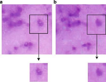
This Comparison Chart displays two panels of Giemsa-stained thick blood film microscopy, labeled 'a' (left) and 'b' (right), demonstrating the effects of color consistency correction on Extended Depth of Field (EDoF) composite images. The images depict dark purple foreground objects, representing cells or malarial parasites (Plasmodium species), against a light pink/lavender background. Panel 'a' serves as the pre-correction baseline, exhibiting visible shading aberrations and color inconsistencies where pixels from different focal planes have been merged. Black bounding boxes with magnified insets highlight these artifacts, showing uneven distribution of color within the cellular structures. Panel 'b' shows the results after applying a 5x5 filtering mask for color correction. The magnified inset in panel 'b' demonstrates improved color uniformity, reduced shading artifacts, and sharper edge definition of the foreground objects. This educational visual illustrates critical digital pathology preprocessing steps necessary for automated malaria diagnosis and pattern recognition, where consistent color and contrast are essential for accurate object identification.
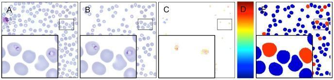
This composite educational graphic illustrates an automated diagnostic workflow for detecting malaria (Plasmodium falciparum) in human blood smears. The sequence transitions from raw clinical imaging to final automated classification. Panel A shows a Giemsa-stained thin blood film with erythrocytes and potential parasites. Panel B demonstrates the candidate segmentation phase, where individual red blood cells are isolated from the background. Panel C presents a diagnostic heat map generated by a sliding-window support vector machine (SVM) classifier, where colored pixels represent the probability of infection. Panel D provides the heat map scale, ranging from blue (low probability) to red (high probability). Panel E shows the final binary classification result, with infected erythrocytes color-coded in red and uninfected cells in blue. Each panel includes a magnified inset to highlight morphological details, such as trophozoite ring stages. This image demonstrates the application of computer-aided detection (CAD) in parasitology for calculating parasitemia levels in hematological samples.
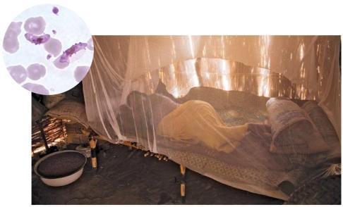
This composite educational image illustrates public health interventions and the pathophysiology of malaria. The main clinical photograph depicts a person in a rural setting in eastern Chad sleeping under a white, semi-transparent insecticide-treated bed net (ITN), a primary preventative measure against vector-borne diseases. The environment reflects a high-risk tropical region with simple housing structures. An inset diagnostic micrograph shows a Giemsa-stained thin blood smear at high magnification, highlighting Plasmodium falciparum. Visible are pale purple erythrocytes, several of which contain characteristic banana-shaped or crescentic gametocytes, diagnostic of P. falciparum infection. The visual contrast between the bedside preventative measure and the microscopic evidence of infection serves as a teaching tool for infectious disease management, epidemiology, and the importance of vector control in endemic regions. This material is suitable for medical students and public health professionals studying parasitology and global health strategies.
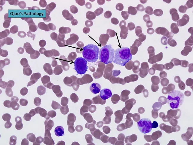
Peripheral blood smear prepared from a patient with suspected plasma cell leukemia (PCL) was examined by bright‑field light microscopy after Wright‑Giemsa staining. The smear shows several circulating plasma‑cell–like elements, including four conspicuous proplasmacytes with dispersed nuclear chromatin and prominent nucleoli, as indicated by arrows in the original figure. In addition, background erythrocytes exhibit rouleaux formation, a common feature in paraproteinemic states. The cells display eccentrically placed nuclei with pronounced basophilic cytoplasm and a coarse chromatin pattern; occasional cells show a perinuclear clearing (Golgi rim) suggestive of plasmacytic differentiation. Immunoglobulin light‑chain restriction is not determined on this smear alone but flow cytometry or immunohistochemistry would typically confirm clonal plasma cells. Relative proportions indicate that neoplastic plasma cells constitute a substantial fraction of leukocytes, consistent with plasma cell leukemia rather than reactive plasmacytosis. PCL is an aggressive myelomatous process often presenting with cytopenias, organomegaly, and sometimes lymphadenopathy; prognosis is poor, and treatment responsiveness is limited. This image illustrates characteristic morphologic features used for differential diagnosis against multiple myeloma, Waldenström macroglobulinemia, and other causes of plasmacytosis. Clinically relevant keywords include plasma cells, rouleaux, Wright‑Giemsa stain, 100X oil, peripheral blood, leukemia, myeloma, and hematologic malignancy.
Role of Peripheral Blood Smear (PBS) Examination in Parasitic Infection Diagnosis
Overview
Types of Blood Smear Preparations
1. Direct (Wet) Mount
- One drop of blood on a glass slide, covered with a cover slip, examined immediately
- Best for trypanosomiasis and filariasis - the motile trypomastigotes and microfilariae are seen actively moving
- Not for species identification; stained smears are required for that
2. Thin Blood Smear
- Made exactly like a hematological smear
- Fixed in anhydrous methanol, then stained with Giemsa stain (pH 7.2 preferred)
- Examined under oil immersion at x1000 magnification
- Gold standard for species identification of Plasmodium spp., Babesia spp., Trypanosoma spp., and microfilariae
- Parasitemia expressed as number of parasitized erythrocytes per 1000 RBCs
- First scan at low power (for microfilariae, which are 100-200 µm long and found at lateral edges), then high dry for trypanosomes, then oil immersion for Plasmodium and Babesia
3. Thick Blood Smear
- One drop of blood spread to cover 1 cm² on a glass slide
- Dried but NOT fixed before staining with Giemsa (allows dehemoglobinization)
- Concentrates parasites 40-100 fold compared to a thin film - greatly increases diagnostic sensitivity
- Useful for detecting all blood parasites, especially at low-level parasitemia
- Parasites counted per 200 WBCs under oil immersion; converted to parasites/µL
- A minimum of 100-200 fields should be examined before calling the smear negative
- Limitation: requires experience; artifacts are common; cannot speciate as well as thin films
4. Buffy Coat Film
- Concentrates white blood cells
- Particularly useful for detecting microfilariae (which concentrate with the buffy coat)
- Also helpful for Trypanosoma detection
Parasites Detected and Specific Features
A. Malaria (Plasmodium spp.)
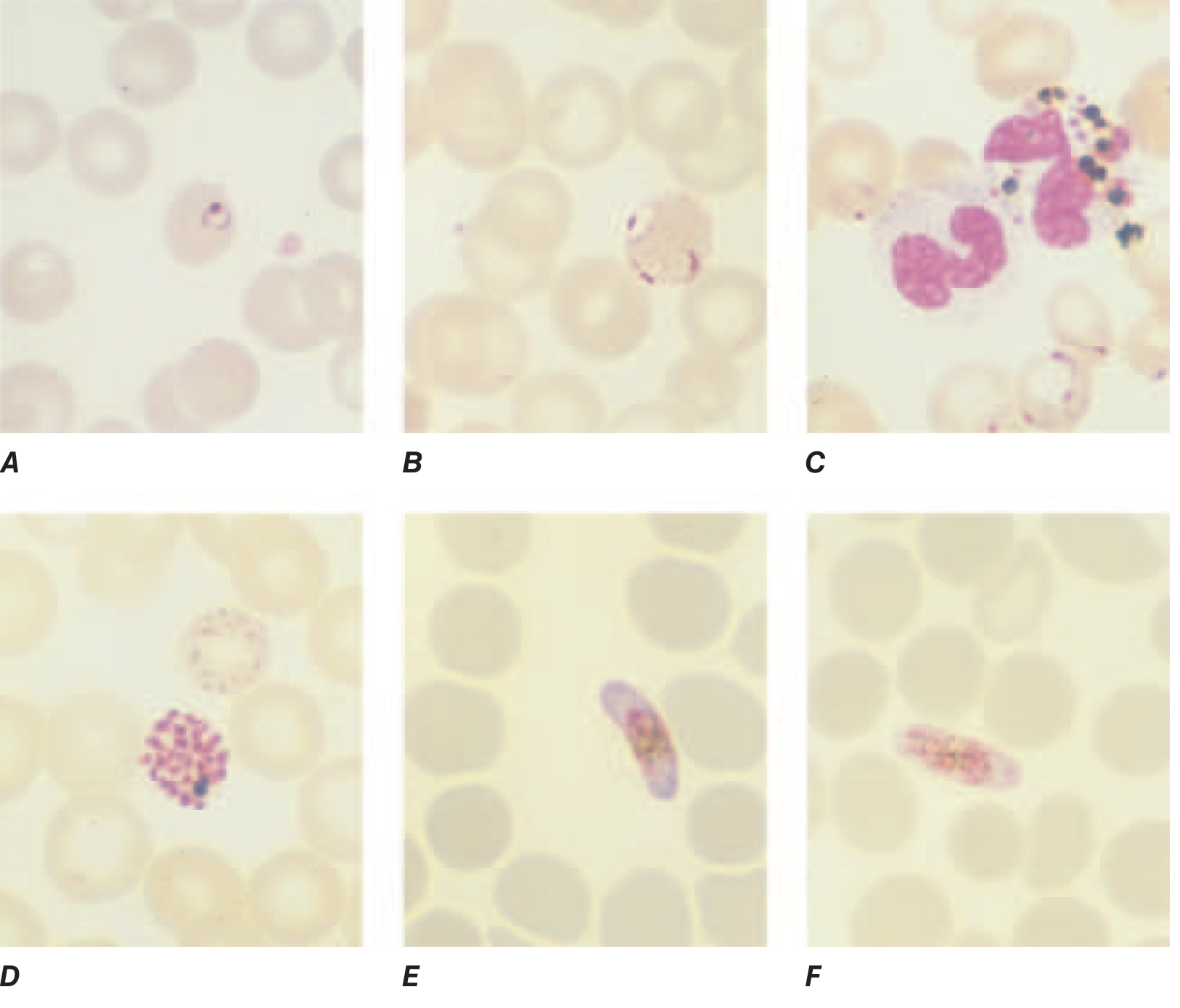
| Feature | Significance |
|---|---|
| >20% parasites with visible pigment | Poor prognosis in severe malaria |
| Malarial pigment in >5% neutrophils | Indicator of recent schizogony |
| Gametocyte count peaks 1 week after asexual peak | Not a sign of drug resistance |
| >10⁵ parasites/µL | Increased risk of death |
B. Filariasis (Wuchereria bancrofti, Brugia malayi, Loa loa, Mansonella spp.)
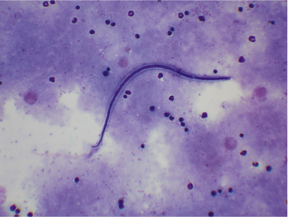
- Microfilariae exist extracellularly in the blood (unlike malaria which is intraerythrocytic)
- Demonstrable in Giemsa-stained blood films
- Periodicity matters for timing of blood collection:
- W. bancrofti: Nocturnal periodicity - maximum microfilariae at night; blood should be drawn between 10 PM and 2 AM
- Subperiodic forms: microfilariae present throughout the day, peaking in afternoon
- B. malayi sheath stains bright pink
- W. bancrofti and L. loa sheaths do not stain
- Further ID based on head and tail nuclear structures
C. Trypanosomiasis (Trypanosoma spp.)
- Trypomastigotes seen in blood during acute phase
- Direct wet mount excellent for initial detection (motile forms visible)
- Thin smear with Giemsa for definitive identification
- For T. brucei (sleeping sickness): also demonstrated in CSF during late-stage (CNS) disease
- For T. cruzi (Chagas disease): parasites seen on blood smear or via PCR during initial (acute) phase
D. Babesiosis (Babesia spp.)
- Intraerythrocytic ring forms on Giemsa-stained thin smear
- Pathognomonic feature: "Maltese cross" or tetrad form (2 pairs of joined ring forms in a single RBC) - distinguishes it from P. falciparum
- No malarial pigment or hemozoin
- Can infect >1 RBC at a time (multiple ring forms per cell)
Practical Technique Summary
| Step | Thin Smear | Thick Smear |
|---|---|---|
| Preparation | Air-dry, fix in methanol | Air-dry, NO fixation |
| Stain | Giemsa pH 7.2 | Giemsa (dehemoglobinization occurs) |
| Magnification | Oil immersion x1000 | Oil immersion x1000 |
| Sensitivity | Lower (standard) | 40-100x higher |
| Main use | Species ID / morphology | Screening / low parasitemia |
| Minimum fields | 200 WBCs counted | 100-200 fields before calling negative |
Limitations of PBS and Adjunct Methods
| Limitation | Adjunct Method |
|---|---|
| Cannot detect below threshold parasitemia | PCR (most sensitive; used in reference labs) |
| Species ID may be difficult | RDTs for P. falciparum (HRP2, LDH, aldolase antigens) |
| Requires experienced microscopist | Acridine orange fluorescence for rapid screening |
| Low-density microfilaremia missed | Membrane filtration / Nucleopore filter |
| Cryptic (amicrofilaremic) filariasis | Circulating antigen detection (ELISA, ICT card test for W. bancrofti) |
| Chronic trypanosomiasis may have undetectable parasitemia | Serology, NAAT |
Key Points for Exams (Apurba Shastry-style)
- Giemsa stain at pH 7.2 is the stain of choice for PBS examination in parasitology
- Thick film - screening, higher sensitivity; Thin film - species identification
- Thick film stained without fixation to allow dehemoglobinization (lysis of RBCs)
- Microfilariae are scanned at low power first (lateral edges of smear); identified under oil immersion
- Blood for nocturnal-periodic filariasis collected at night (10 PM - 2 AM)
- B. malayi sheath - pink with Giemsa; W. bancrofti sheath - does not stain
- PBS alone is sufficient for malaria diagnosis in most settings; PCR is reserved for reference labs
- In severe falciparum malaria, pigment in >5% neutrophils = poor prognosis
- Buffy coat films - useful for microfilariae and trypanosomes
- A PBS is only reported as negative after examining 100-200 fields (thick) or 200 WBCs (thick)