
Ear anatomy ent
Reading File
Finding Sources
Finding Sources
Searching PubMed
ear anatomy
Searching the Web
ear anatomy external middle inner ear NIDCD
Searching Images
labeled diagram anatomy of external middle and inner ear
Reading File
Loading Image
Ear anatomy
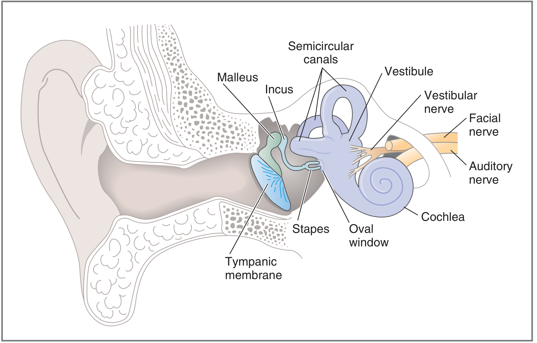
The ear has three parts: external, middle, and inner ear.
1. External ear
- Auricle (pinna): Visible outer part that collects sound.
- External auditory canal (external acoustic meatus): Conducts sound to the eardrum.
- Tympanic membrane (eardrum): Boundary between external and middle ear; vibrates in response to sound.
2. Middle ear
An air-filled space within the temporal bone containing three auditory ossicles:
- Malleus (hammer): Attached to the tympanic membrane.
- Incus (anvil): Links malleus and stapes.
- Stapes (stirrup): Its footplate rests on the oval window.
Other structures:
- Oval window: Transfers ossicular vibration into the fluid of the inner ear.
- Round window: Permits pressure release within the cochlea.
- Pharyngotympanic (Eustachian) tube: Connects the middle ear to the nasopharynx and equalizes pressure across the eardrum.
- Mastoid air cells: Communicate posteriorly with the middle ear.
3. Inner ear
A fluid-filled system consisting of a bony labyrinth enclosing a membranous labyrinth.
For hearing
- Cochlea: Spiral structure that converts sound-induced vibrations into neural signals.
- Organ of Corti: Sensory receptor organ within the cochlea; contains hair cells.
- Cochlear division of CN VIII: Carries hearing signals to the brain.
For balance
- Vestibule: Contains the utricle and saccule, which detect linear acceleration and head position.
- Semicircular canals: Superior, posterior, and lateral canals detect rotational head movements.
- Vestibular division of CN VIII: Carries balance information to the brain.
Sound pathway
Pinna → auditory canal → tympanic membrane → malleus → incus → stapes → oval window → cochlear fluid → organ of Corti hair cells → cochlear nerve (CN VIII) → brain.
The external ear directs sound inward, the middle ear transmits and amplifies vibration across the air-fluid boundary, and the inner ear performs hearing and balance functions. This organization is described in Costanzo Physiology, p. 98. The NIDCD ear illustration provides a useful clinical overview.
This is a shared conversation. Sign in to Orris to start your own chat.