
Muscles of thigh and leg
Reading File
Now let me search the medical library for muscles of the thigh and leg.
Finding Sources
Finding Sources
Good results from the THIEME Atlas and other sources. Let me read the key sections in parallel.
Reading File
Reading File
Reading File
Now let me get the detailed muscle tables from the THIEME Atlas - the anterior thigh muscles section and leg muscles sections with full detail.
Reading File
Reading File
Reading File
Now let me get the adductor/medial thigh muscles and leg muscles detailed sections.
Finding Sources
Reading File
Now let me get the adductor muscles and the anterior/posterior compartment of the leg detailed muscles.
Finding Sources
Finding Sources
Finding Sources
Finding Sources
Let me read the THIEME Atlas section on medial thigh and leg compartments directly, and also fetch the key anatomy images.
Reading File
Loading Image
Loading Image
Excellent! Now I have comprehensive data. Let me read the leg anterior/lateral compartment details and posterior compartment details, and fetch a leg muscle image.
Reading File
Loading Image
Loading Image
Now let me get the adductor/medial thigh muscle details and the posterior leg compartment details.
Reading File
Reading File
Let me now fetch the posterior leg muscles image and get the superficial posterior compartment (gastrocnemius, soleus) details.
Reading File
Loading Image
Now let me get the triceps surae/gastrocnemius/soleus detail from the THIEME Atlas.
Finding Sources
Reading File
I need to find where gastrocnemius/soleus details are. Let me search paragraphs for that info.
Finding Sources
Reading File
I now have all the information needed for a comprehensive response. Let me compile it.
Muscles of the Thigh and Leg
Sources: THIEME Atlas of Anatomy - General Anatomy and Musculoskeletal System; Imaging Anatomy Vol. 3; Miller's Review of Orthopaedics 9th Ed.
THIGH MUSCLES
The thigh is divided into three compartments: anterior (extensors), medial (adductors), and posterior (flexors/hamstrings).
1. Anterior Compartment - Extensor Group
Innervation: Femoral nerve (L1-L4)
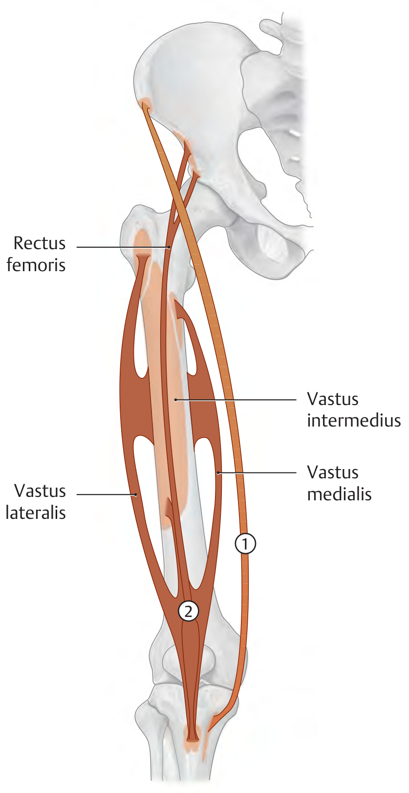
Here is the anatomy of the quadriceps femoris group:
Sartorius
| Origin | Anterior superior iliac spine (ASIS) |
| Insertion | Medial tibial surface (pes anserinus, with gracilis & semitendinosus) |
| Actions | Hip: flexion, abduction, external rotation; Knee: flexion, internal rotation |
| Nerve | Femoral nerve (L1-L4) |
| Note | Longest muscle in the body; crosses two joints; migrates posteriorly at knee level to become a knee flexor |
Quadriceps Femoris (4 heads + articularis genus)
| Head | Origin | Insertion | Special Actions |
|---|---|---|---|
| Rectus femoris | AIIS (straight head); acetabular roof (reflected head) | Tibial tuberosity via patellar ligament | Hip flexion + knee extension |
| Vastus medialis | Medial lip of linea aspera, distal intertrochanteric line | Tibial tuberosity; medial patellar retinaculum | Knee extension; stabilizes patella medially |
| Vastus lateralis | Lateral lip of linea aspera, lateral greater trochanter | Tibial tuberosity; lateral patellar retinaculum | Knee extension; stabilizes patella laterally |
| Vastus intermedius | Anterior femoral shaft | Tibial tuberosity via patellar ligament | Knee extension |
| Articularis genus | Distal femoral shaft (deep to vastus intermedius) | Suprapatellar recess of knee capsule | Prevents capsule entrapment during extension |
Innervation of all heads: Femoral nerve (L1-L4) Key clinical point: Paralysis of quadriceps destabilizes the knee in stance phase. Walking is possible only if the patient hyperextends the knee to lock it, using the line of gravity in front of the knee axis.
2. Medial Compartment - Adductor Group
Primary innervation: Obturator nerve (L2-L4); adductor magnus also by tibial part of sciatic nerve (L4-S1)
From anterior to posterior: pectineus → adductor longus → adductor brevis → adductor magnus; gracilis lies medially.
| Muscle | Origin | Insertion | Actions | Nerve |
|---|---|---|---|---|
| Pectineus | Pectineal line of pubis | Pectineal line of femur | Adduction, flexion, medial rotation of hip | Femoral + obturator (L2-L4) |
| Adductor longus | Body of pubis | Medial lip of linea aspera (middle third) | Adduction, flexion, medial rotation | Obturator (L2-L4) |
| Adductor brevis | Body and inferior ramus of pubis | Medial lip of linea aspera (upper third) | Adduction, flexion, medial rotation | Obturator (L2-L4) |
| Adductor magnus | Inferior pubic ramus to ischial tuberosity | Linea aspera + adductor tubercle of femur | Adduction (ant. part), extension (post. part) | Obturator + tibial nerve (L2-S1) |
| Gracilis | Body and inferior ramus of pubis | Medial tibial surface (pes anserinus) | Hip adduction; knee flexion & internal rotation | Obturator (L2-L3) |
Key facts: Adductor magnus is the largest adductor (27% of thigh muscle mass). It has a dual nerve supply. The adductor hiatus in its tendon transmits the femoral artery into the popliteal fossa. Most common injured adductor in athletes: adductor longus.
3. Posterior Compartment - Hamstrings (Flexor Group)
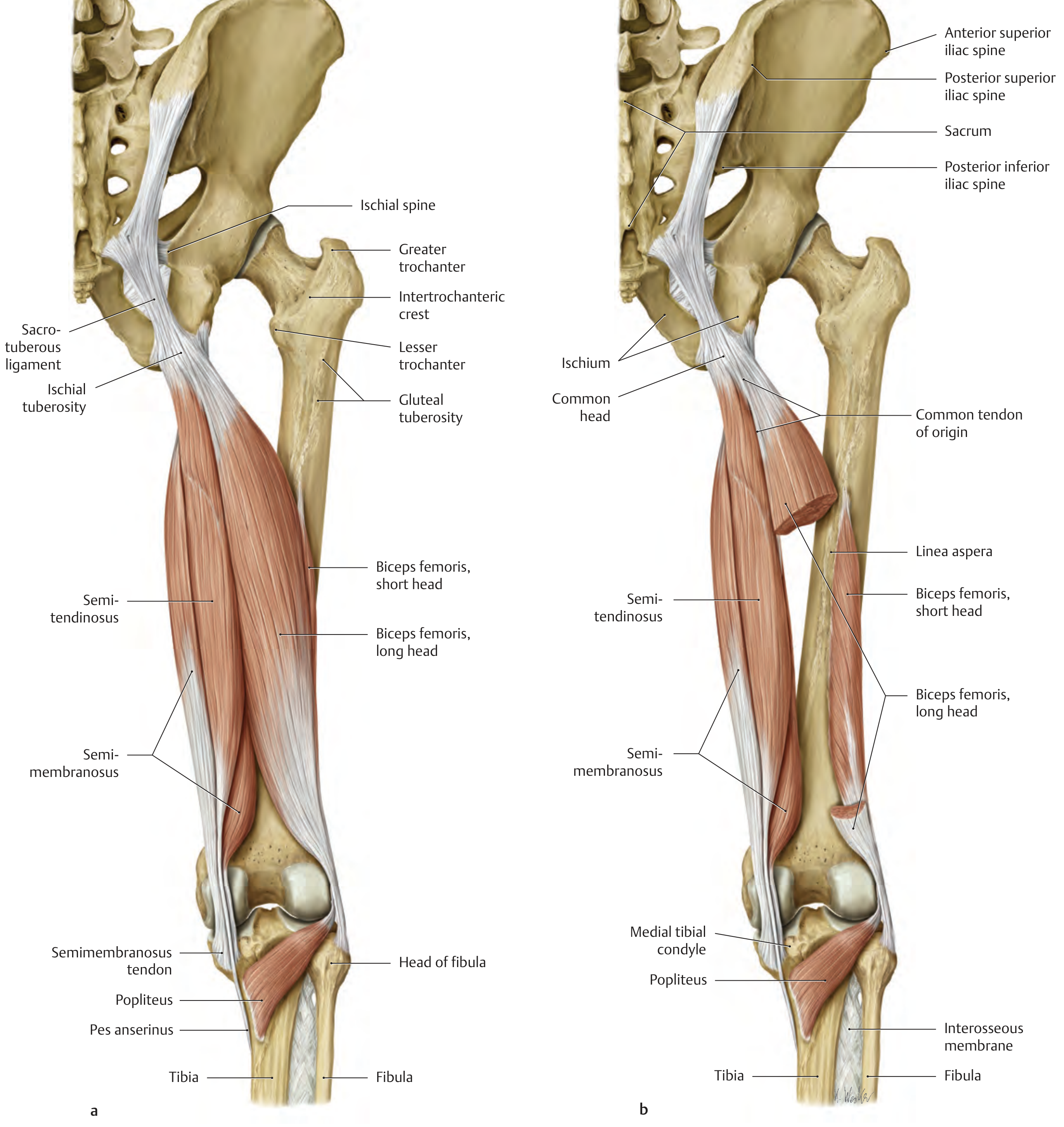
The hamstrings = biceps femoris + semimembranosus + semitendinosus. All originate from the ischial tuberosity (except the short head of biceps femoris) and cross both hip and knee joints.
| Muscle | Origin | Insertion | Actions | Nerve |
|---|---|---|---|---|
| Biceps femoris (long head) | Ischial tuberosity, sacrotuberous ligament (common head with semitendinosus) | Head of fibula | Hip: extension, adduction; Knee: flexion, external rotation | Tibial nerve, L5-S2 |
| Biceps femoris (short head) | Lateral lip of linea aspera (middle third of femur) | Head of fibula | Knee: flexion, external rotation | Common fibular nerve, L5-S2 |
| Semimembranosus | Ischial tuberosity | Medial tibial condyle, oblique popliteal ligament, popliteus fascia | Hip: extension, adduction; Knee: flexion, internal rotation | Tibial nerve (L5-S2) |
| Semitendinosus | Ischial tuberosity (common head with biceps long head) | Medial tibial tuberosity - pes anserinus (with sartorius & gracilis) | Hip: extension, adduction; Knee: flexion, internal rotation | Tibial nerve (L5-S2) |
Pes anserinus ("goose's foot"): The combined tendon insertion of sartorius + gracilis + semitendinosus at the medial tibial surface. A frequent site of bursitis.Hamstring insufficiency: With the knee extended, the hamstrings are too short to allow full hip flexion (passive insufficiency). With the hip extended, they cannot shorten enough to allow full knee flexion (active insufficiency).
LEG MUSCLES
The leg has four compartments. Anterior and lateral compartments = supplied by common fibular nerve; Posterior compartments = supplied by tibial nerve.
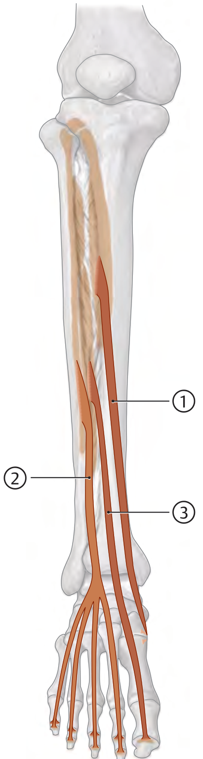
1. Anterior Compartment (Extensors / Dorsiflexors)
Innervation: Deep fibular nerve (L4, L5)
| Muscle | Origin | Insertion | Actions |
|---|---|---|---|
| Tibialis anterior | Upper 2/3 of lateral tibial surface + crural interosseous membrane | Medial cuneiform + base of 1st metatarsal (medial & plantar) | Dorsiflexion; inversion (supination) of subtalar joint |
| Extensor digitorum longus | Lateral tibial condyle, head & medial surface of fibula, interosseous membrane | Dorsal aponeuroses of toes 2-5, bases of distal phalanges 2-5 | Dorsiflexion; eversion; extends toes 2-5 |
| Extensor hallucis longus | Middle 1/3 of medial fibula surface + interosseous membrane | Dorsal aponeurosis of great toe + base of distal phalanx | Dorsiflexion; extends great toe; active in both inversion and eversion |
| Fibularis (peroneus) tertius | Anterior border of distal fibula (split of EDL) | Base of 5th metatarsal | Dorsiflexion; eversion |
2. Lateral Compartment (Evertors)
Innervation: Superficial fibular nerve (L5, S1)
| Muscle | Origin | Insertion | Actions |
|---|---|---|---|
| Fibularis (peroneus) longus | Head of fibula + proximal 2/3 of lateral fibular surface | Plantar surface of medial cuneiform + base of 1st metatarsal | Plantarflexion; eversion; supports transverse arch of foot |
| Fibularis (peroneus) brevis | Distal 1/2 of lateral fibular surface + intermuscular septa | Tuberosity at base of 5th metatarsal | Plantarflexion; eversion; tenses plantar aponeurosis |
3. Posterior Compartment - Superficial (Plantarflexors)
Innervation: Tibial nerve (S1, S2)
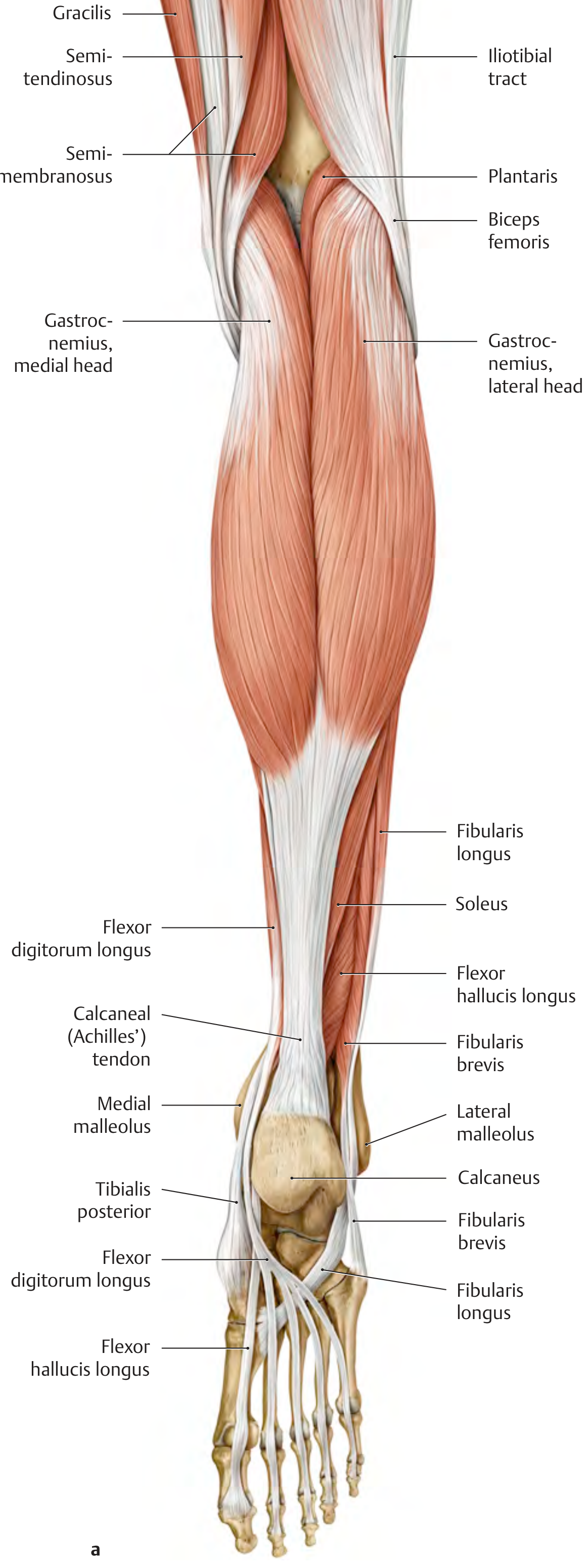
Triceps Surae (= Gastrocnemius + Soleus)
| Head | Origin | Insertion | Actions |
|---|---|---|---|
| Gastrocnemius (medial head) | Medial epicondyle of femur | Calcaneal tuberosity via Achilles tendon | Plantarflexion (talocrural); knee flexion |
| Gastrocnemius (lateral head) | Lateral epicondyle of femur | Calcaneal tuberosity via Achilles tendon | Plantarflexion; knee flexion |
| Soleus | Posterior fibular head/neck + soleal line of tibia | Calcaneal tuberosity via Achilles tendon | Plantarflexion only (single-joint muscle) |
Plantaris: Origin at lateral femoral epicondyle (just proximal to lateral gastrocnemius head); inserts via Achilles tendon; small cross-section - functionally negligible; may prevent compression of posterior musculature during knee flexion. Nerve: Tibial (S1, S2).
Achilles tendon: Average length 20-25 cm; breaking strength ~60-100 N/mm² (can bear nearly 1 ton). Weakest blood supply (and most common rupture site) = 2-6 cm proximal to calcaneal insertion.
4. Posterior Compartment - Deep (Plantarflexors + Invertors)
Innervation: Tibial nerve
| Muscle | Origin | Insertion | Actions | Nerve |
|---|---|---|---|---|
| Tibialis posterior | Crural interosseous membrane + adjacent tibia and fibula borders | Navicular tuberosity + cuneiforms + 2nd-4th metatarsal bases (fan-shaped) | Plantarflexion; inversion (supination); supports longitudinal & transverse arches | Tibial (L4, L5) |
| Flexor digitorum longus | Middle 1/3 of posterior tibial surface | Bases of distal phalanges 2-5 | Plantarflexion; inversion; flexes toes 2-5 at MTP & IP joints | Tibial (L5-S2) |
| Flexor hallucis longus | Distal 2/3 of posterior fibular surface + interosseous membrane | Base of distal phalanx of great toe | Plantarflexion; inversion; flexes great toe; supports medial longitudinal arch | Tibial (L5-S2) |
| Popliteus | Lateral femoral condyle + posterior horn of lateral meniscus | Posterior tibial surface above soleus origin | Flexes knee; unlocks knee by internally rotating femur on fixed tibia 5° ("key to the knee") | Tibial (L4-S1) |
"Tom, Dick, ANd Harry" mnemonic for the order of structures passing posterior to the medial malleolus: Tibialis posterior → Digitorum flexor longus → (posterior tibial) Artery & nerve → Hallucis flexor longus.
Summary Table: Compartments and Innervation
| Region | Compartment | Key Muscles | Nerve |
|---|---|---|---|
| Thigh | Anterior | Sartorius, Quadriceps femoris | Femoral (L1-L4) |
| Medial | Pectineus, Add. longus/brevis/magnus, Gracilis | Obturator (L2-L4) | |
| Posterior | Biceps femoris, Semimembranosus, Semitendinosus | Tibial + Common fibular (L5-S2) | |
| Leg | Anterior | Tibialis anterior, EDL, EHL, Fibularis tertius | Deep fibular (L4-L5) |
| Lateral | Fibularis longus & brevis | Superficial fibular (L5-S1) | |
| Post. (superficial) | Triceps surae, Plantaris | Tibial (S1-S2) | |
| Post. (deep) | Tibialis posterior, FDL, FHL, Popliteus | Tibial (L4-S2) |
- THIEME Atlas of Anatomy - General Anatomy and Musculoskeletal System, pp. 488-515
- Imaging Anatomy Vol. 3, pp. 268-276
- Miller's Review of Orthopaedics 9th Ed., p. 18
Hip bone it’s landmarks and attachments in the landmarks
Finding Sources
Finding Sources
Finding Sources
Finding Sources
Finding Sources
Finding Sources
Excellent - found rich sources. Let me read the key sections in parallel.
Reading File
Reading File
Reading File
Now let me get the detailed iliac bone landmarks and attachments, and the THIEME Atlas hip bone section.
Reading File
Reading File
Now let me fetch the key images and read more detail on the ischium and pubis sections.
Loading Image
Loading Image
Reading File
Excellent! Now let me read the ischium and pubis sections, and get the THIEME Atlas hip bone attachment diagrams.
Reading File
Loading Image
Loading Image
I now have comprehensive images and detailed text on all hip bone landmarks and their attachments. Let me compile the full response.
The Hip Bone (Os Coxae) - Landmarks and Attachments
The hip bone (os coxae) is formed by the fusion of three bones - the ilium (superior), ischium (posteroinferior), and pubis (anteroinferior) - which meet in the acetabulum at the triradiate cartilage (fuses between ages 14-16 years). The ilium and ischium each contribute 2/5 of the acetabulum; the pubis contributes 1/5.
Here are the right hip bone from lateral and medial views with all major landmarks labeled:
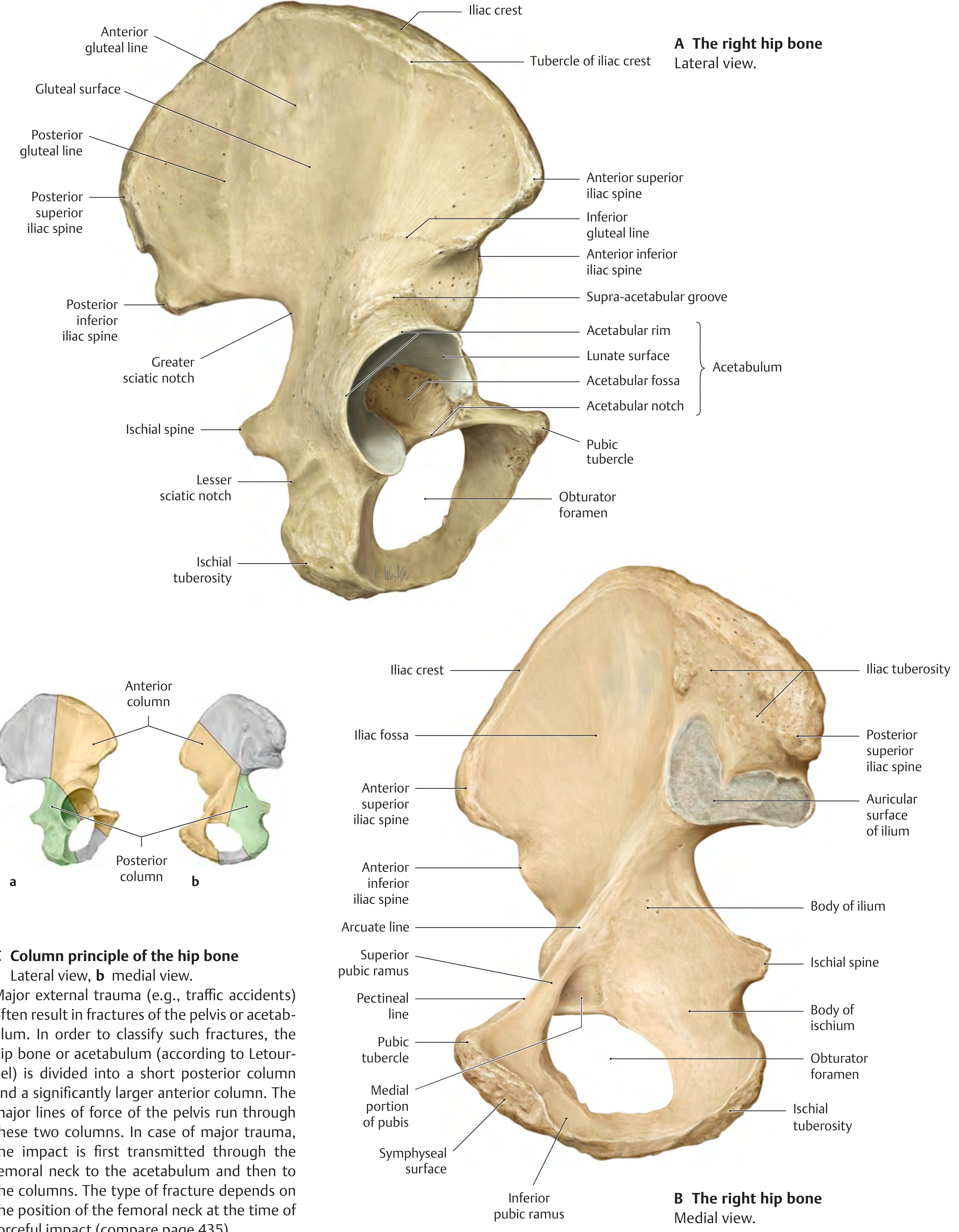
PART I: THE ILIUM
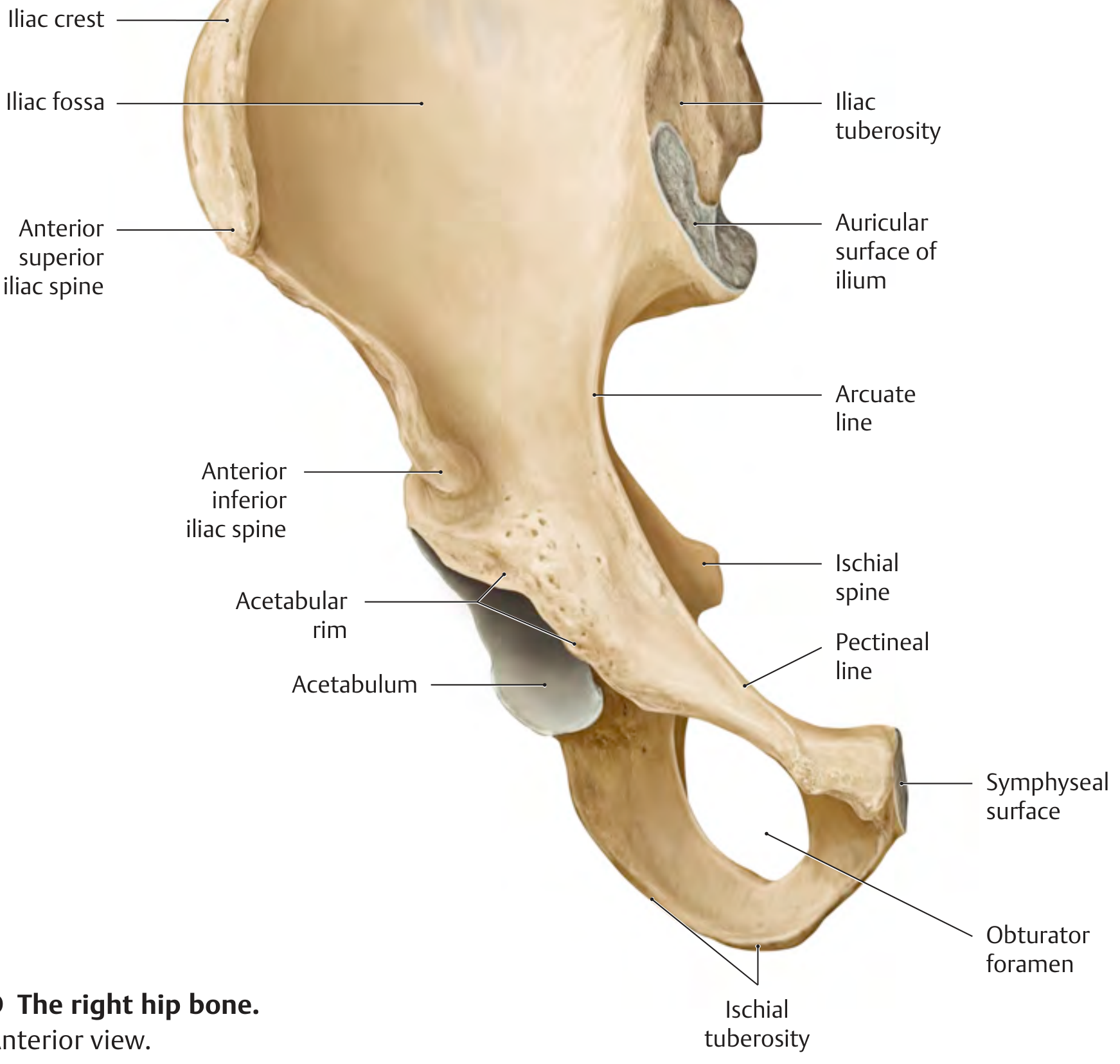
The ilium has an ala (wing) superiorly and a body inferiorly. The ala has two surfaces: the iliac fossa (anterior/medial, concave) and the gluteal surface (posterior/lateral, with three gluteal lines).
1. Iliac Crest
The bony ridge running from ASIS to PSIS - the highest landmark of the ilium. The crest has an outer lip, intermediate zone, and inner lip.
| Zone | Attachments |
|---|---|
| Outer lip | External oblique abdominis, tensor fasciae latae, latissimus dorsi |
| Intermediate zone | Internal oblique abdominis |
| Inner lip | Transversus abdominis, iliacus, quadratus lumborum, iliocostalis (sacrospinalis) |
| Tubercle of crest (~5 cm behind ASIS) | Tensor fasciae latae, iliotibial tract |
Clinical note: Avulsion of the iliac crest can occur from forceful contraction of the abdominal muscles in young athletes.
2. Anterior Superior Iliac Spine (ASIS)
- Sartorius (origin)
- Tensor fasciae latae (origin)
- Inguinal ligament (lateral attachment - runs from ASIS to pubic tubercle)
3. Anterior Inferior Iliac Spine (AIIS)
- Rectus femoris - straight head (origin)
- Iliofemoral ligament (upper limb - the strongest ligament of the hip joint; extends to the anterior hip joint/intertrochanteric line)
4. Posterior Superior Iliac Spine (PSIS)
- Sacrotuberous ligament (partial attachment)
- Multifidus (origin)
- Iliolumbar ligament (posterior iliac wing to L5 transverse process)
- Surface landmark: corresponds to the dimple of Venus; lies at level of S2 vertebra
5. Posterior Inferior Iliac Spine (PIIS)
- Forms the superior boundary of the greater sciatic notch
- Attachment for the posterior capsule of the sacroiliac joint
6. Iliac Fossa (Inner/Medial Surface)
- Iliacus muscle - large, broad attachment covering most of the iliac fossa
- Iliac fascia (inner lip of crest)
7. Gluteal Surface (Outer/Lateral Surface) - Three Gluteal Lines
| Line | Location | Between Lines: Muscle Origin |
|---|---|---|
| Posterior gluteal line | Most posterior vertical line | Posterior to it: Gluteus maximus |
| Anterior gluteal line | Curved line from iliac crest to greater sciatic notch | Between posterior & anterior lines: Gluteus medius |
| Inferior gluteal line | Below anterior, arches over acetabulum | Between anterior & inferior lines: Gluteus minimus |
- Piriformis - small attachment at inferior/posterior aspect of gluteal surface (near greater sciatic notch)
8. Auricular Surface
- Articulates with the sacrum forming the sacroiliac joint (hyaline inferiorly, fibrous superiorly)
9. Iliac Tuberosity (posterior to auricular surface)
- Posterior sacroiliac ligaments and interosseous ligaments
10. Arcuate Line (Iliopectineal Line)
- Separates the greater pelvis (above) from the lesser pelvis (below)
- Forms part of the pelvic brim (inlet)
- Continuous with the pectineal line of pubis
PART II: THE ISCHIUM
The ischium has a body (fused to ilium above and pubis anteroinferiorly) and a ramus (ischiopubic ramus).
11. Ischial Tuberosity
The most prominent roughened projection - the "sitting bone." Carries the entire body weight in the seated position.
| Muscle/Structure | Attachment Type |
|---|---|
| Biceps femoris (long head) | Origin (common head with semitendinosus) |
| Semitendinosus | Origin (common head with biceps long head) |
| Semimembranosus | Origin (from superolateral impression) |
| Adductor magnus (hamstring/posterior part) | Origin (inferior ischial tuberosity) |
| Quadratus femoris | Origin (lateral border of ischial tuberosity) |
| Inferior gemellus | Origin (upper part of ischial tuberosity) |
| Sacrotuberous ligament | Attachment (ischial tuberosity to sacrum) |
| Ischial bursa | Overlies it |
Avulsion fracture: Forceful contraction of the hamstrings can avulse the ischial tuberosity apophysis - classic in sprinters and football players.
12. Ischial Spine
A pointed projection separating the greater sciatic notch (above) from the lesser sciatic notch (below).
| Structure | Attachment |
|---|---|
| Superior gemellus | Origin (lateral surface) |
| Coccygeus muscle | Origin (medial surface, from ischial spine to coccyx) |
| Levator ani | Origin (medial surface - ischial spine to pubic body via tendinous arch) |
| Sacrospinous ligament | Runs from ischial spine to lateral sacrum/coccyx |
Clinical: The sacrospinous ligament, with the ischial spine, separates the greater from lesser sciatic foramina.
13. Greater Sciatic Notch (above ischial spine)
Converted into the greater sciatic foramen by the sacrospinous ligament. Transmits:
- Piriformis muscle (exits the pelvis through it)
- Sciatic nerve
- Superior and inferior gluteal vessels/nerves
- Pudendal nerve and internal pudendal vessels (exit here, re-enter via lesser foramen)
- Nerve to obturator internus, nerve to quadratus femoris
14. Lesser Sciatic Notch (below ischial spine)
Converted into the lesser sciatic foramen by sacrospinous + sacrotuberous ligaments. Transmits:
- Obturator internus tendon (exits to reach greater trochanter)
- Internal pudendal vessels
- Pudendal nerve
15. Ischiopubic Ramus (Body/Ramus of Ischium meeting inferior pubic ramus)
| Muscle | Attachment |
|---|---|
| Adductor magnus (adductor/anterior part) | Origin (inferior pubic ramus and ischial ramus) |
| Adductor brevis | Origin |
| Gracilis | Origin |
| Obturator externus | Origin (outer surface of obturator membrane & surrounding bone) |
| Obturator internus | Origin (inner surface of obturator membrane & surrounding bone) |
| Ischiocavernosus | Origin (crus of penis/clitoris) |
| Transverse perineal | Attachment |
PART III: THE PUBIS
The pubis consists of a body, superior ramus, and inferior ramus.
16. Pubic Tubercle
- Inguinal ligament (medial attachment)
- Rectus abdominis (lateral slip)
- Pyramidalis (origin)
- Adductor longus - some fibers attach near the body/crest of pubis
17. Pubic Crest (superior border of pubic body)
- Rectus abdominis (insertion onto crest)
- Pyramidalis (origin from crest)
- Inguinal ligament medial end
18. Pectineal Line (Pecten Pubis) - superior ramus
- Part of the pelvic brim
- Pectineus muscle (origin)
- Pectineal ligament (Cooper's ligament)
19. Symphyseal Surface
- Articulates with the opposite pubis at the pubic symphysis (secondary cartilaginous joint / fibrocartilage)
20. Iliopubic Eminence (Iliopectineal Eminence)
A ridge where the ilium meets the superior pubic ramus.
- Iliopsoas passes over this ridge (potential site of iliopsoas bursitis from friction)
PART IV: THE ACETABULUM
The cup-shaped socket for the femoral head, formed at the junction of all three bones.
| Feature | Description | Attachment |
|---|---|---|
| Acetabular rim | Bony lip around socket | Acetabular labrum attached here |
| Lunate surface | Articular horseshoe-shaped surface (lined with hyaline cartilage) | Covered with articular cartilage |
| Acetabular fossa | Non-articular central depression | Fat pad (Haversian fat pad); ligamentum teres attaches here |
| Acetabular notch | Inferior gap in the rim | Transverse acetabular ligament bridges it; transmits blood supply to femoral head via ligamentum teres (obturator artery) |
PART V: OBTURATOR FORAMEN
Large oval foramen formed by the pubic and ischial rami. Mostly closed by the obturator membrane.
| Structure | Attachment |
|---|---|
| Obturator externus | Origin from outer membrane surface and bony margin |
| Obturator internus | Origin from inner membrane surface and bony margin |
| Obturator canal | Superior gap transmits obturator nerve and vessels into the medial thigh |
Summary Attachment Map
Here is the complete muscle attachment diagram of the pelvis and femur (anterior and posterior views):
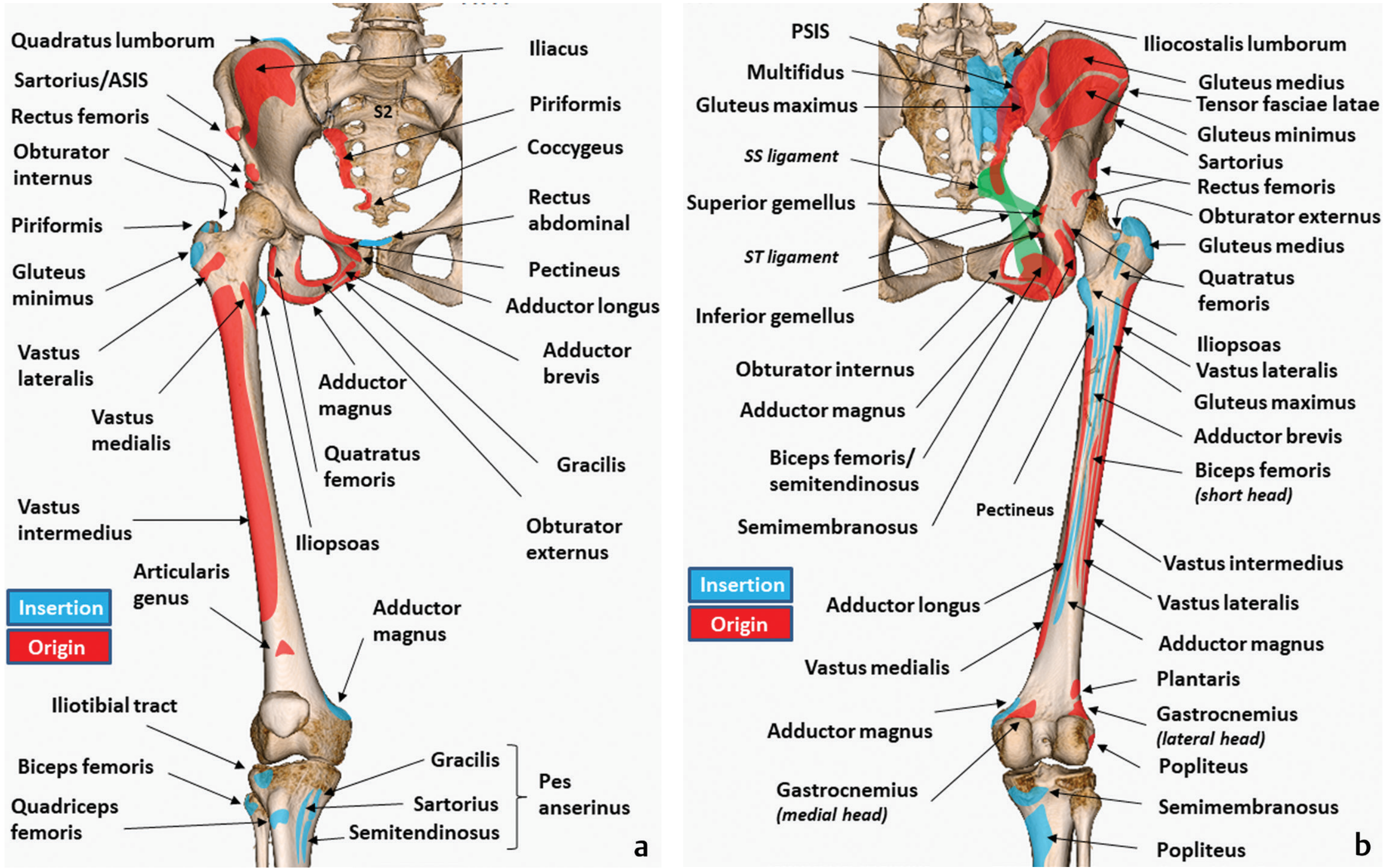
And the detailed 3D CT reconstruction showing iliac crest and pelvic ring attachments from anterior, medial, and posterior views:
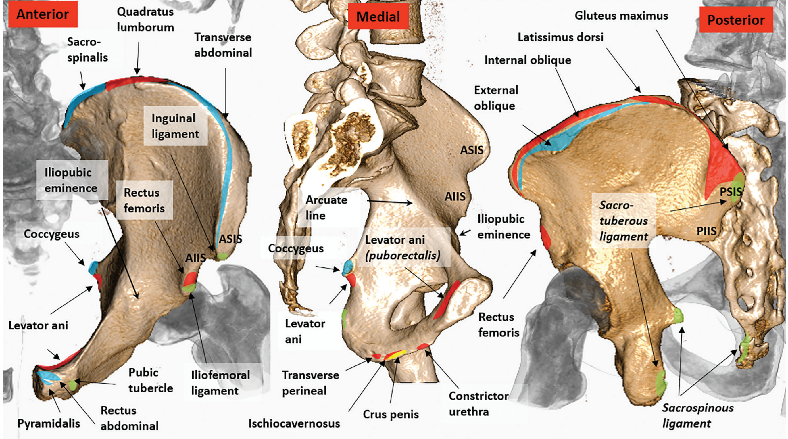
Quick Reference: Landmark → Key Attachment
| Landmark | Key Attachment(s) |
|---|---|
| ASIS | Sartorius, TFL, inguinal ligament |
| AIIS | Rectus femoris, iliofemoral ligament |
| PSIS | Sacrotuberous lig., multifidus, iliolumbar lig. |
| Iliac crest (outer lip) | External oblique, TFL, latissimus dorsi |
| Iliac crest (inner lip) | Internal oblique, transversus abdominis, iliacus, quadratus lumborum |
| Iliac fossa | Iliacus |
| Gluteal surface (post. to post. line) | Gluteus maximus |
| Gluteal surface (post.-ant. lines) | Gluteus medius |
| Gluteal surface (ant.-inf. lines) | Gluteus minimus |
| AIIS | Rectus femoris (straight head) |
| Ischial tuberosity | Biceps femoris (long head), semimembranosus, semitendinosus, adductor magnus (post. part), quadratus femoris, inferior gemellus |
| Ischial spine | Superior gemellus, coccygeus, levator ani, sacrospinous lig. |
| Ischiopubic ramus | Adductor magnus (ant.), adductor brevis, gracilis, obturator externus/internus |
| Pubic crest & tubercle | Rectus abdominis, pyramidalis, inguinal ligament |
| Pectineal line | Pectineus, Cooper's ligament |
| Obturator foramen/membrane | Obturator externus (outer), obturator internus (inner) |
| Acetabular fossa/notch | Ligamentum teres |
- Imaging Anatomy Vol. 3 - Bones, Joints, Vessels and Nerves, pp. 227-234
- THIEME Atlas of Anatomy - General Anatomy and Musculoskeletal System, pp. 433-434
- Gray's Anatomy for Students, p. 638
Tibia its landmarks and attachments
Finding Sources
Finding Sources
Finding Sources
Finding Sources
Reading File
Reading File
Finding Sources
Reading File
Loading Image
Excellent! Now let me read the distal tibia section and get more on the shaft attachments (soleal line, etc.), and search for an image of the full tibia with all attachment markings.
Reading File
Finding Sources
Finding Sources
Let me get the THIEME Atlas section on tibia and its attachment diagram, plus the distal tibia/medial malleolus section.
Finding Sources
Finding Sources
Reading File
Finding Sources
Now let me fetch the remaining key tibia images - the full anterior/posterior tibia with attachment diagram.
Loading Image
Loading Image
Now let me get the THIEME tibia attachment diagram and the leg bone origin/insertion image.
Loading Image
Loading Image
I now have everything needed. Let me compile the comprehensive response.
The Tibia - Landmarks and Attachments
The tibia is the medial and larger bone of the leg. It is the only leg bone that articulates with the femur at the knee joint, and it is the primary weight-bearing bone of the leg. It articulates with the femur and patella (proximally), the fibula (proximally and distally), and the talus (distally).
Shape of shaft in cross-section: Triangular, with 3 surfaces (medial, lateral/anterior, posterior) and 3 borders (anterior, medial, interosseous).
PROXIMAL TIBIA
Here is the tibial plateau with all ligament/meniscus attachments labeled, plus anterior and posterior views:
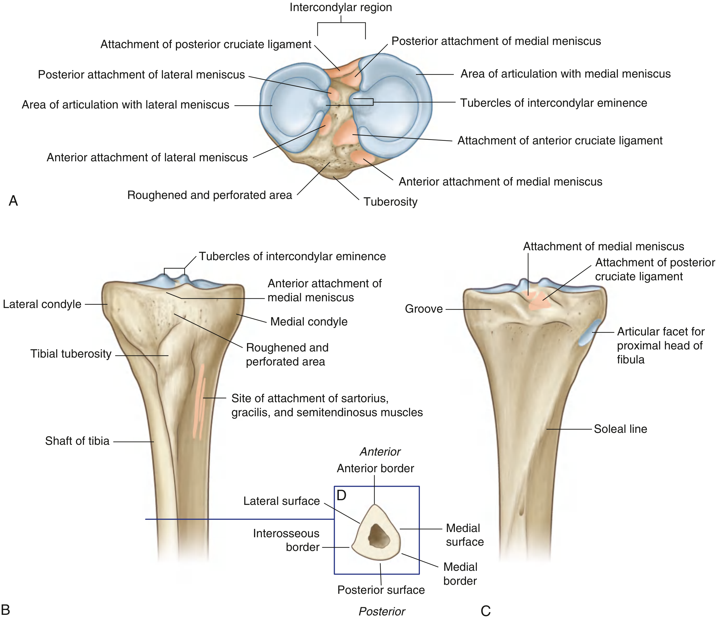
1. Medial Condyle (Tibial Plateau)
- Larger than lateral condyle; oval articular surface
- Articulates with the medial condyle of the femur and the medial meniscus
- Posterior surface bears a distinct horizontal groove for attachment of semimembranosus (direct head insertion)
2. Lateral Condyle (Tibial Plateau)
- Circular articular surface; articulates with the lateral condyle of the femur and lateral meniscus
- Undersurface bears a circular facet for articulation with the proximal head of the fibula (superior tibiofibular joint)
- Proximal anterior margin: Iliotibial band (ITB) inserts here (Gerdy's tubercle - anterolateral aspect of the lateral condyle)
3. Intercondylar Eminence (Area)
The non-articular region between the two condylar surfaces, raised centrally to form two intercondylar tubercles (medial and lateral). The area has 6 attachment facets:
Anterior intercondylar area (front to back):
| Structure | Attachment |
|---|---|
| Anterior horn of medial meniscus | Most anterior |
| Anterior cruciate ligament (ACL) | Just posterior to medial meniscus horn |
| Anterior horn of lateral meniscus | Just lateral to ACL |
Posterior intercondylar area (front to back):
| Structure | Attachment |
|---|---|
| Posterior horn of lateral meniscus | Most anterior |
| Posterior horn of medial meniscus | Posteromedial |
| Posterior cruciate ligament (PCL) | Most posterior (largest facet) |
Memory aid for the intercondylar area (front to back): M-A-L / L-M-P (medial meniscus anterior, ACL, lateral meniscus anterior / lateral meniscus posterior, medial meniscus posterior, PCL)
4. Tibial Tuberosity
A palpable inverted triangular roughened elevation on the anterior surface of the proximal tibia, just inferior to the condylar junction.
| Structure | Attachment |
|---|---|
| Patellar ligament | Insertion (continuation of quadriceps tendon below patella) |
| Deep infrapatellar bursa | Lies between patellar ligament and upper tibia |
| Superficial infrapatellar bursa | Lies in front of tibial tuberosity |
Osgood-Schlatter disease: Apophysitis/traction injury of the tibial tuberosity in adolescent athletes due to repetitive stress from the patellar ligament.
SHAFT OF TIBIA
The shaft is triangular in cross-section. Three borders and three surfaces:
5. Anterior Border ("Shin")
A sharp subcutaneous ridge running from the tibial tuberosity to the anterior aspect of the medial malleolus.
- No direct muscle attachments (subcutaneous throughout)
- Most vulnerable site for direct blows ("shin splints" occur in the surrounding periosteum and musculature)
6. Medial Surface
Smooth and entirely subcutaneous - palpable along its full length.
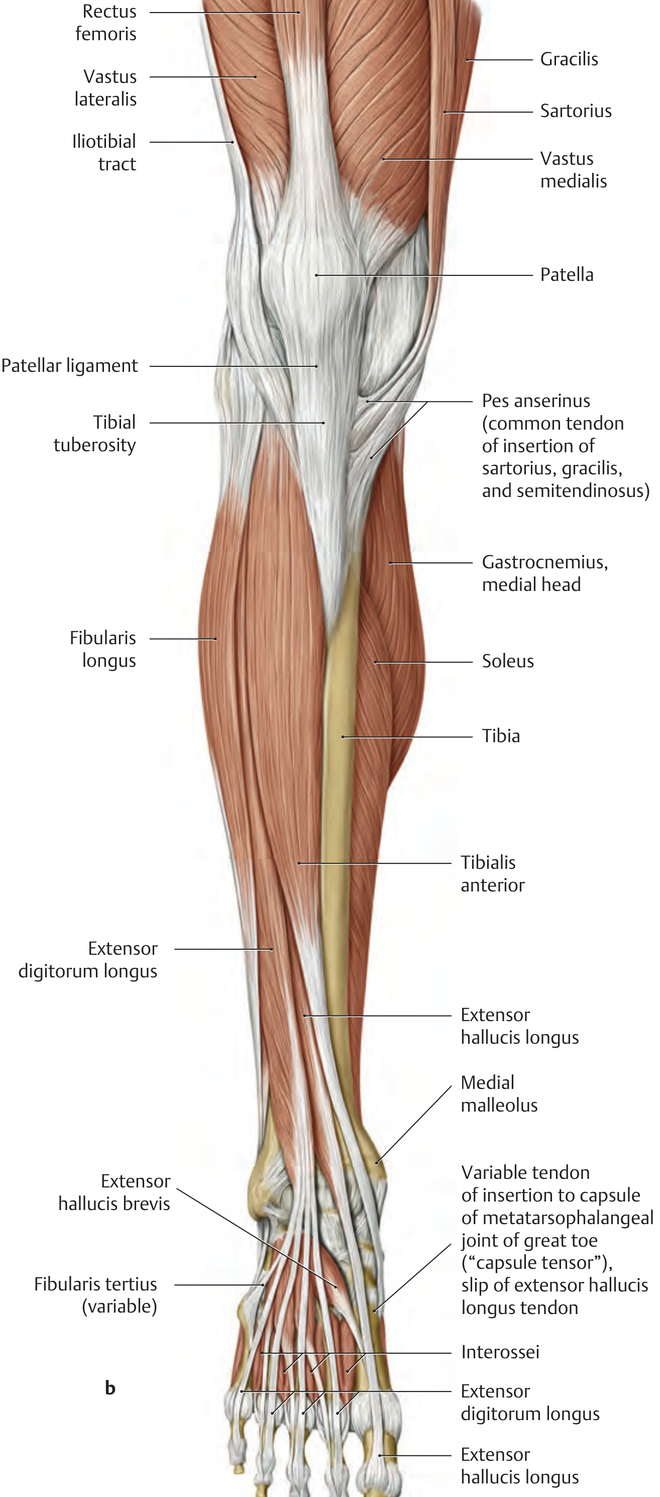
- Medial and slightly inferior to the tibial tuberosity: the pes anserinus insertion
| Muscle | Attachment (all insert via pes anserinus) |
|---|---|
| Sartorius | Most superficial/anterior slip |
| Gracilis | Middle slip |
| Semitendinosus | Deepest/posterior slip |
Pes anserinus bursitis occurs here between the pes anserinus tendons and the medial collateral ligament.
7. Lateral Surface
- Between the anterior and interosseous borders
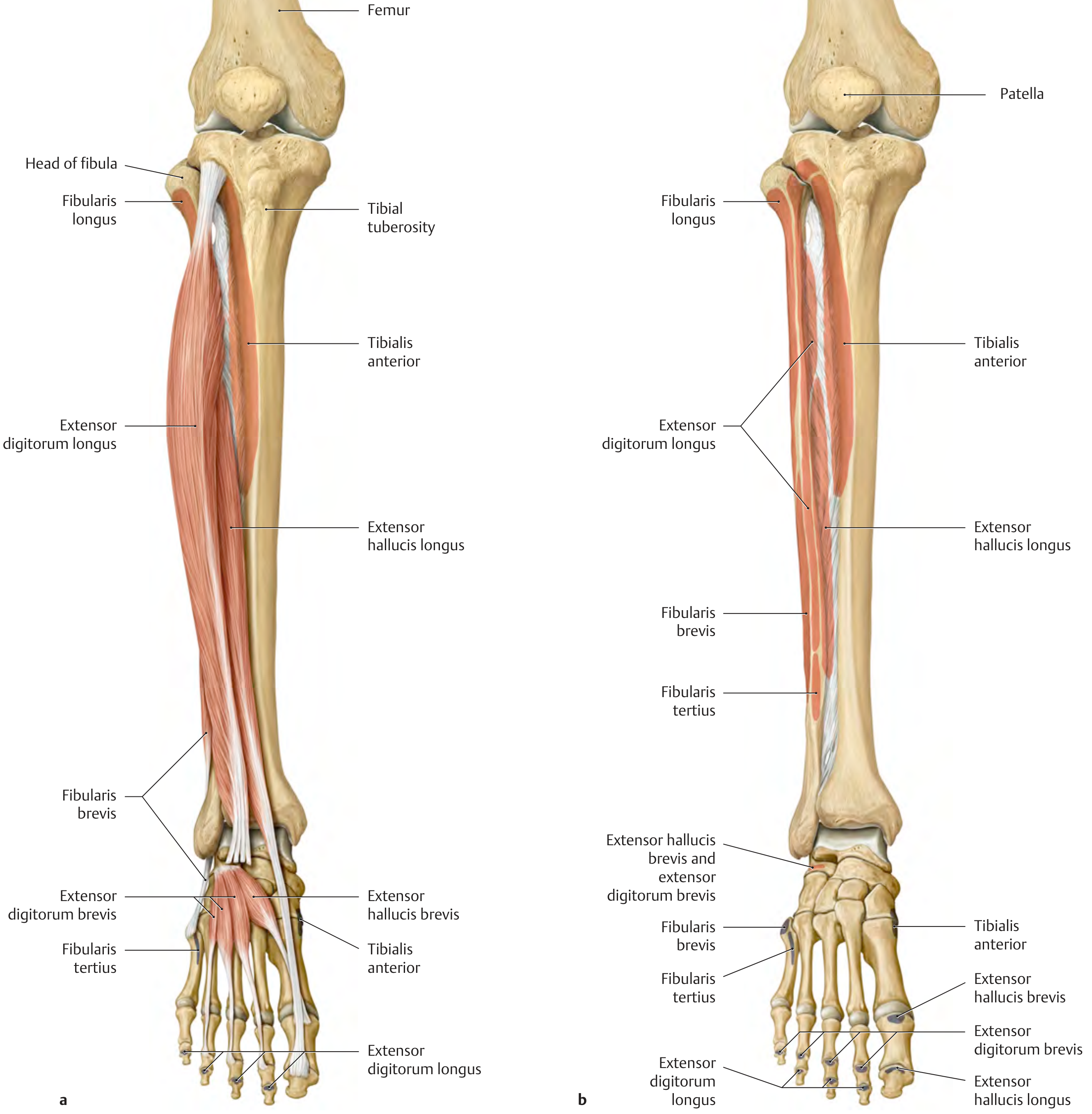
- Origin of tibialis anterior (upper 2/3 of lateral tibial surface)
8. Posterior Surface
- Upper part crossed by the soleal line (roughened oblique line running inferomedially from the fibular facet region)
| Structure | Attachment at Soleal Line |
|---|---|
| Soleus | Origin (medial part - from soleal line of tibia; lateral from posterior fibular head/neck) |
| Popliteus | Inserts onto the posterior surface above the soleal line |
| Flexor digitorum longus | Origin from the middle third of the posterior tibial surface (below soleal line) |
| Tibialis posterior | Origin from the posterior tibia (adjacent to interosseous membrane) |
9. Interosseous Border
A vertical ridge along the lateral shaft - attachment for the crural interosseous membrane that binds the tibia and fibula along their shafts.
- Also gives origin to tibialis anterior (its posterior aspect)
- Tibialis posterior and FHL also originate from the interosseous membrane
10. Medial Border
Indistinct superiorly (begins from the semimembranosus groove on the posterior medial condyle), sharp in midshaft. No major muscle attachments.
11. Nutrient Foramen
Located on the posterior surface proximal to midshaft.
- Opens away from the knee ("flees the knee") - a useful anatomical rule for determining the nutrient foramen direction
Here is the anterior leg showing muscles arising from the lateral surface and interosseous membrane of the tibia:
And the medial view of the leg showing pes anserinus, patellar ligament, tibial tuberosity, and tibialis anterior:
DISTAL TIBIA
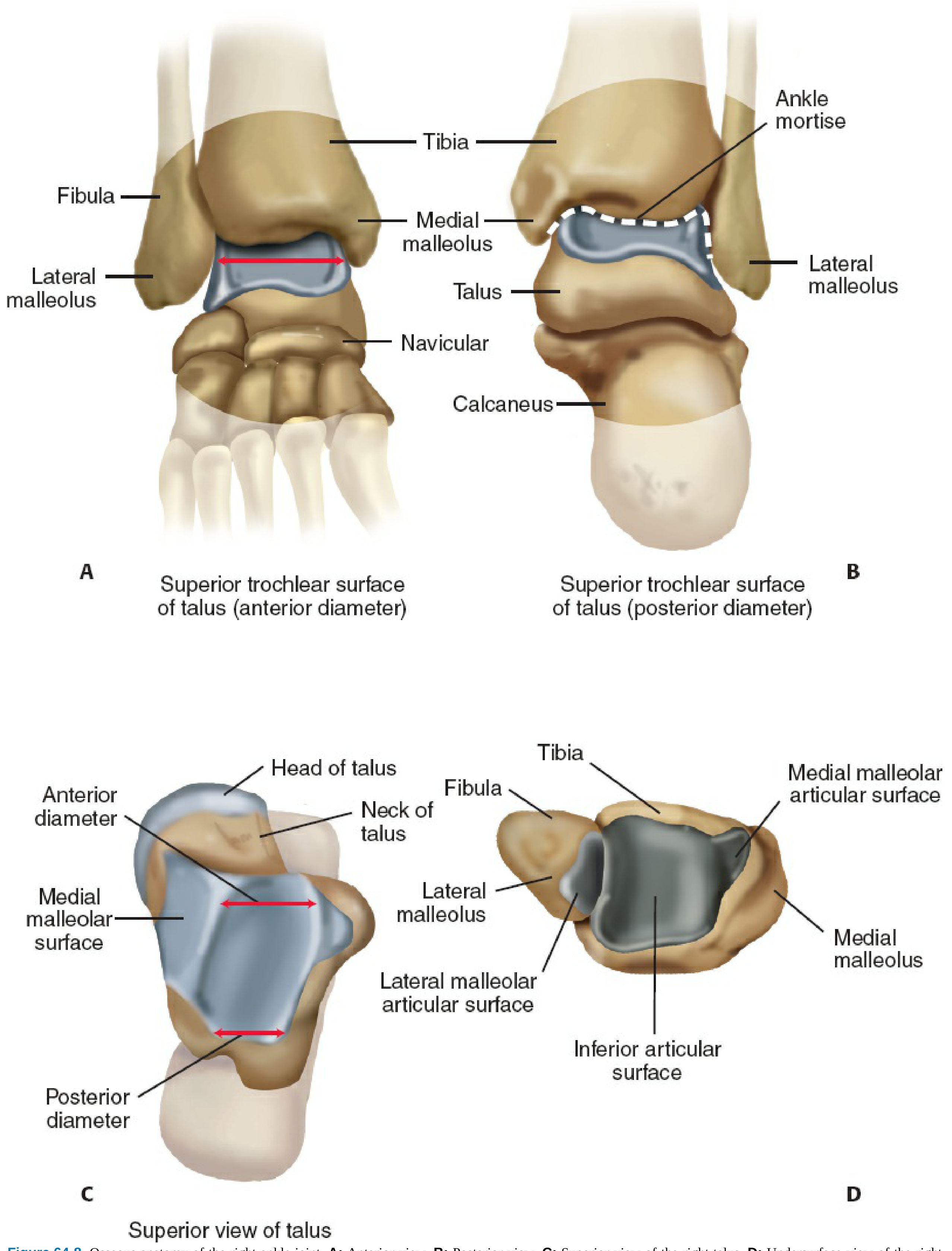
12. Medial Malleolus
The medial bony projection of the distal tibia, forming the medial wall of the ankle mortise. Has two bony projections (colliculi):
- Anterior colliculus - larger, more prominent
- Posterior colliculus - smaller, separated by the intercollicular groove
| Structure | Attachment |
|---|---|
| Deltoid ligament (medial collateral ligament of ankle) | Origin from the malleolus (superficial and deep parts) |
| Anterior tibiotalar ligament (deep deltoid) | From anterior colliculus |
| Posterior tibiotalar ligament (deep deltoid) | From posterior colliculus and intercollicular groove |
| Tibionavicular, tibiocalcaneal, tibiospring ligaments | Superficial deltoid layer |
| Posterior tibialis tendon groove | Runs posterior to the malleolus in a groove |
The deltoid ligament is the strongest ligament of the ankle.
13. Inferior Articular Surface (Tibial Plafond)
- The horizontal weight-bearing surface that articulates with the superior dome of the talus
- Together with the medial malleolus and lateral malleolus (fibula), forms the ankle mortise
14. Fibular Notch (Distal Lateral Tibia)
A roughened concave area on the lateral side of the distal tibia.
- Articulates with the distal fibula
- Distal tibiofibular syndesmosis formed by:
- Anterior tibiofibular ligament (from Tubercle of Chaput on anterolateral distal tibia)
- Posterior tibiofibular ligament (from Posterior tibial tubercle)
- Interosseous tibiofibular ligament
- Inferior transverse ligament
15. Posterior Malleolus
- A dorsal/posterior projection of the distal tibia
- Deepens the ankle mortise posteriorly
- Attachment of posterior tibiofibular ligament
Here is the ankle mortise showing the distal tibia, medial malleolus, lateral malleolus, and the talus:
Complete Landmark-to-Attachment Reference Table
| Landmark | Key Attachments |
|---|---|
| Medial condyle (articular surface) | Medial meniscus (articulates); semimembranosus (posterior groove) |
| Lateral condyle (articular surface) | Lateral meniscus; proximal fibula (inferior facet); Gerdy's tubercle = iliotibial band |
| Anterior intercondylar area | ACL; anterior horn of medial meniscus; anterior horn of lateral meniscus |
| Posterior intercondylar area | PCL; posterior horn of medial meniscus; posterior horn of lateral meniscus |
| Tibial tuberosity | Patellar ligament (= quadriceps femoris tendon via patella) |
| Medial surface (pes anserinus) | Sartorius + Gracilis + Semitendinosus |
| Lateral surface | Tibialis anterior (upper 2/3) |
| Posterior surface above soleal line | Popliteus (insertion) |
| Soleal line | Soleus (medial origin) |
| Posterior surface below soleal line | Flexor digitorum longus (middle 1/3) |
| Interosseous border/membrane | Tibialis anterior, tibialis posterior, FHL, extensor digitorum longus, extensor hallucis longus (shared with fibula) |
| Medial malleolus | Deltoid ligament (all layers) |
| Tibial plafond | Ankle joint articular surface (with talus) |
| Tubercle of Chaput (anterolateral distal tibia) | Anterior tibiofibular ligament |
| Posterior tibial tubercle | Posterior tibiofibular ligament |
Quick Clinically Important Points
- The medial surface is entirely subcutaneous - commonly fractured by direct blows (most tibial shaft fractures are open due to lack of overlying muscle)
- Compartment syndrome most commonly involves the anterior compartment of the leg where tibialis anterior and extensors arise from the lateral tibial surface and interosseous membrane
- Osgood-Schlatter = tibial tuberosity apophysitis (adolescents, patellar ligament traction)
- Tibial stress fractures most commonly occur in the anterior cortex of the midshaft (the "tension side") in runners
- Pes anserinus bursitis - pain over the medial tibial surface 3-4 cm below the joint line, between pes tendons and the MCL of the knee
Sources: Gray's Anatomy for Students, pp. 689-691; Forensic Anthropology - A Comprehensive Introduction, pp. 4567-4581; Rockwood & Green's Fractures in Adults 10th Ed., p. 3249; THIEME Atlas of Anatomy, pp. 513-515
Femur landmarks and its attachments
Finding Sources
Finding Sources
Finding Sources
Finding Sources
Finding Sources
Finding Sources
Reading File
Reading File
Reading File
Finding Sources
Reading File
Finding Sources
Reading File
Loading Image
Loading Image
Excellent! Now let me get the THIEME Atlas femur attachment diagram and the greater trochanter facets detail.
Finding Sources
Loading Image
Now let me fetch one more key image - the THIEME femur neck angle diagram and the distal femur with condyle/epicondyle attachments.
Loading Image
Loading Image
I now have comprehensive text and images for a complete femur landmarks and attachments answer. Let me compile the full response.
The Femur - Landmarks and Attachments
The femur is the longest, heaviest, and strongest bone in the human body. It extends from the hip to the knee and is divided into three regions: proximal femur, shaft (diaphysis), and distal femur.
Here are the anterior and posterior views of the femur with all major landmarks labeled, alongside the pelvis:
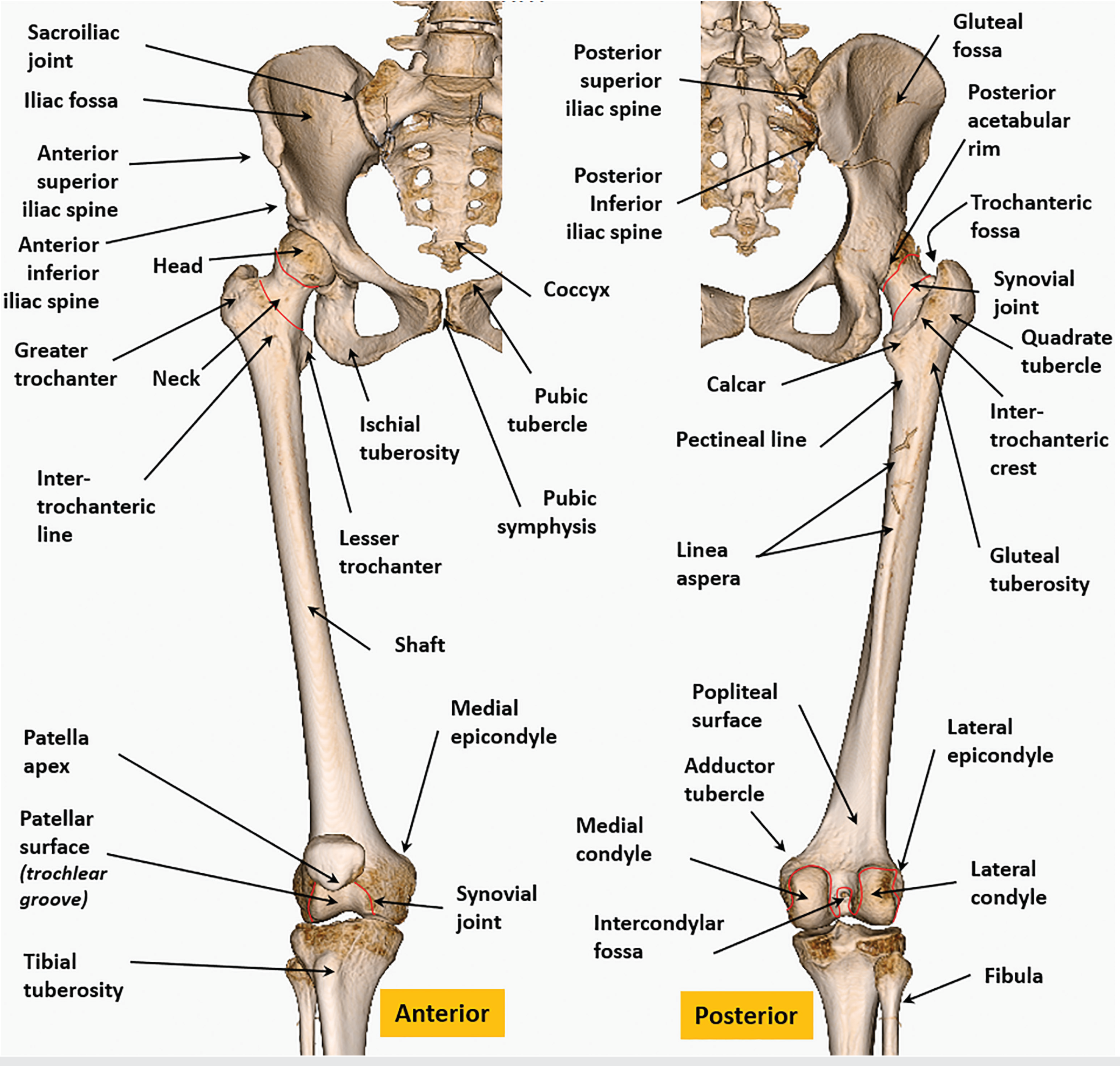
PART I: PROXIMAL FEMUR
1. Femoral Head
- Almost perfectly spherical (2/3 of a sphere)
- Covered entirely with hyaline cartilage except at the fovea centralis (a small pit on the medial surface)
- Articulates with the acetabulum to form the hip joint
| Landmark | Attachment |
|---|---|
| Fovea centralis (fovea capitis) | Ligamentum teres (round ligament) - carries the obturator artery branch to the femoral head in children |
2. Femoral Neck
- ~5 cm long; connects the head to the shaft
- Projects superomedially at a neck-shaft angle of ~122-135° (decreases with age; coxa valga = increased angle; coxa vara = decreased)
- Also has anteversion of 10-15° (the neck is twisted anteriorly relative to the shaft)
- The anterior surface of the neck is entirely intracapsular (hip joint capsule attaches to the intertrochanteric line anteriorly)
- Posterior neck: only the medial part is intracapsular (capsule attaches halfway along the posterior neck)
Clinical importance: Femoral neck fractures disrupt the retinacular vessels (branches of the medial circumflex femoral artery) running along the posterior neck, causing avascular necrosis of the femoral head. Intracapsular neck fractures are especially at risk.
3. Greater Trochanter
The large bony projection at the lateral neck-shaft junction. Has four facets:
| Facet | Muscle Attachment |
|---|---|
| Superoposterior (superior) facet | Piriformis |
| Posterior facet | Obturator internus (with superior and inferior gemelli) |
| Lateral facet | Gluteus medius (main insertion - posterior and lateral parts) |
| Anterior facet | Gluteus minimus |
Additional attachments at the greater trochanter region:
- Tensor fasciae latae - inserts via the iliotibial band (to lateral condyle of tibia, not GT directly)
- Gluteus medius - main abductor of the hip (posterior fibers to posterior-superior GT)
- Trochanteric bursa overlies the greater trochanter deep to the iliotibial band
Trochanteric fossa: A depression on the medial surface of the greater trochanter, posterior aspect.
- Obturator externus tendon inserts here (trochanteric fossa, not the main GT surface)
- Obturator internus also inserts in this region
4. Lesser Trochanter
A conical bony projection on the posteromedial aspect of the neck-shaft junction.
| Muscle | Attachment |
|---|---|
| Iliopsoas (iliacus + psoas major) | Insertion - primary hip flexor |
5. Intertrochanteric Line (Anterior)
A roughened bony ridge running obliquely from the base of the greater trochanter to the lesser trochanter, on the anterior surface.
| Structure | Attachment |
|---|---|
| Iliofemoral ligament (Y-ligament of Bigelow) | Insertion - strongest ligament in the body; upper band to greater trochanter tubercle, lower band to intertrochanteric line |
| Hip joint capsule | Attaches anteriorly along the entire intertrochanteric line |
| Vastus lateralis | Origin from lower part of intertrochanteric line |
| Vastus medialis | Origin from lower part of medial intertrochanteric line |
6. Intertrochanteric Crest (Posterior)
A prominent rounded ridge on the posterior surface connecting the two trochanters.
| Structure | Attachment |
|---|---|
| Quadrate tubercle | Located on the upper half of the crest; Quadratus femoris inserts here |
| Ischiofemoral ligament | Posterior hip capsule attaches near the crest |
Calcar femorale: A dense vertical plate of bone within the medial femoral neck, continuous with the posteromedial cortex - important in distributing compressive forces.
PART II: FEMORAL SHAFT (DIAPHYSIS)
The shaft is slightly bowed anteriorly, and angles medially as it descends (giving the normal valgus alignment of the knee).
Here is the linea aspera and all posterior shaft attachments with axial CT cross-sections:
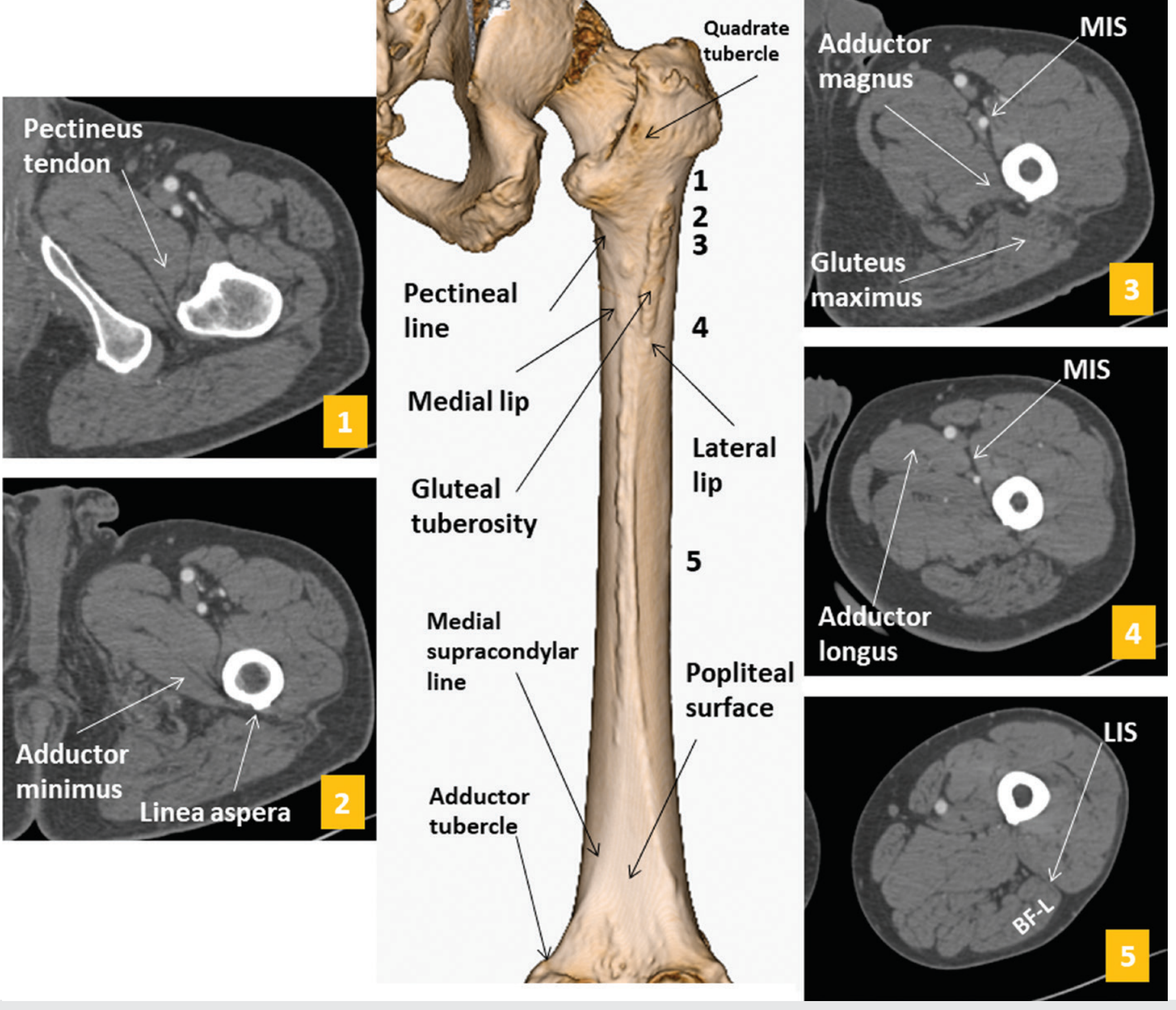
7. Gluteal Tuberosity (Posterolateral Shaft)
A roughened area on the upper posterior-lateral femoral shaft, lateral to the linea aspera - the extension of the lateral lip of the linea aspera.
| Muscle | Attachment |
|---|---|
| Gluteus maximus | Insertion (lower half of the gluteal tuberosity; upper half inserts into the iliotibial band) |
8. Pectineal Line (Spiral Line)
A roughened line on the posterior upper femoral shaft running from the lesser trochanter to the medial lip of the linea aspera.
| Muscle | Attachment |
|---|---|
| Pectineus | Insertion (flexion, adduction, lateral rotation of thigh) |
| Adductor brevis | Insertion (upper part, with pectineal line and upper medial linea aspera) |
9. Linea Aspera (Posterior Shaft)
The most important landmark of the femoral shaft. A prominent longitudinal double-lipped ridge running along the middle third of the posterior shaft. It has a medial lip and a lateral lip.
| Lip/Region | Muscle | Action |
|---|---|---|
| Lateral lip | Vastus lateralis (origin) | Knee extension |
| Lateral lip | Short head of biceps femoris (origin) | Knee flexion/ext rotation |
| Lateral lip | Gluteus maximus (lower insertion fibers) | Hip extension |
| Lateral lip | Lateral intermuscular septum | Divides anterior from posterior compartment |
| Medial lip | Vastus medialis (origin) | Knee extension |
| Medial lip | Adductor longus (insertion - middle third) | Hip adduction/flexion |
| Medial lip | Adductor brevis (insertion - upper medial lip) | Hip adduction/flexion |
| Medial lip | Adductor magnus (insertion - medial lip, throughout) | Hip adduction |
| Medial lip | Medial intermuscular septum | Divides anterior from medial compartment |
| Between both lips | Adductor minimus (upper part of adductor magnus) | Hip adduction |
"BADGE" mnemonic for linea aspera attachments (medial to lateral): Biceps femoris (short head), Adductor magnus/longus/brevis, Vastus medialis/lateralis, Gluteus maximus, and the two intermuscular septa.
10. Anterior Surface of Shaft
- Vastus intermedius originates from the anterior surface of the femoral shaft (upper 2/3)
- Articularis genus (deepest fibers of vastus intermedius) - origin from anterior distal shaft at level of suprapatellar recess; inserts into the suprapatellar recess of the knee capsule to prevent it from being pinched during extension
PART III: DISTAL FEMUR
The distal femur broadens to form two condyles separated posteriorly by the intercondylar fossa.
Here is the comprehensive anterior and posterior view of all thigh muscle attachments to the femur:
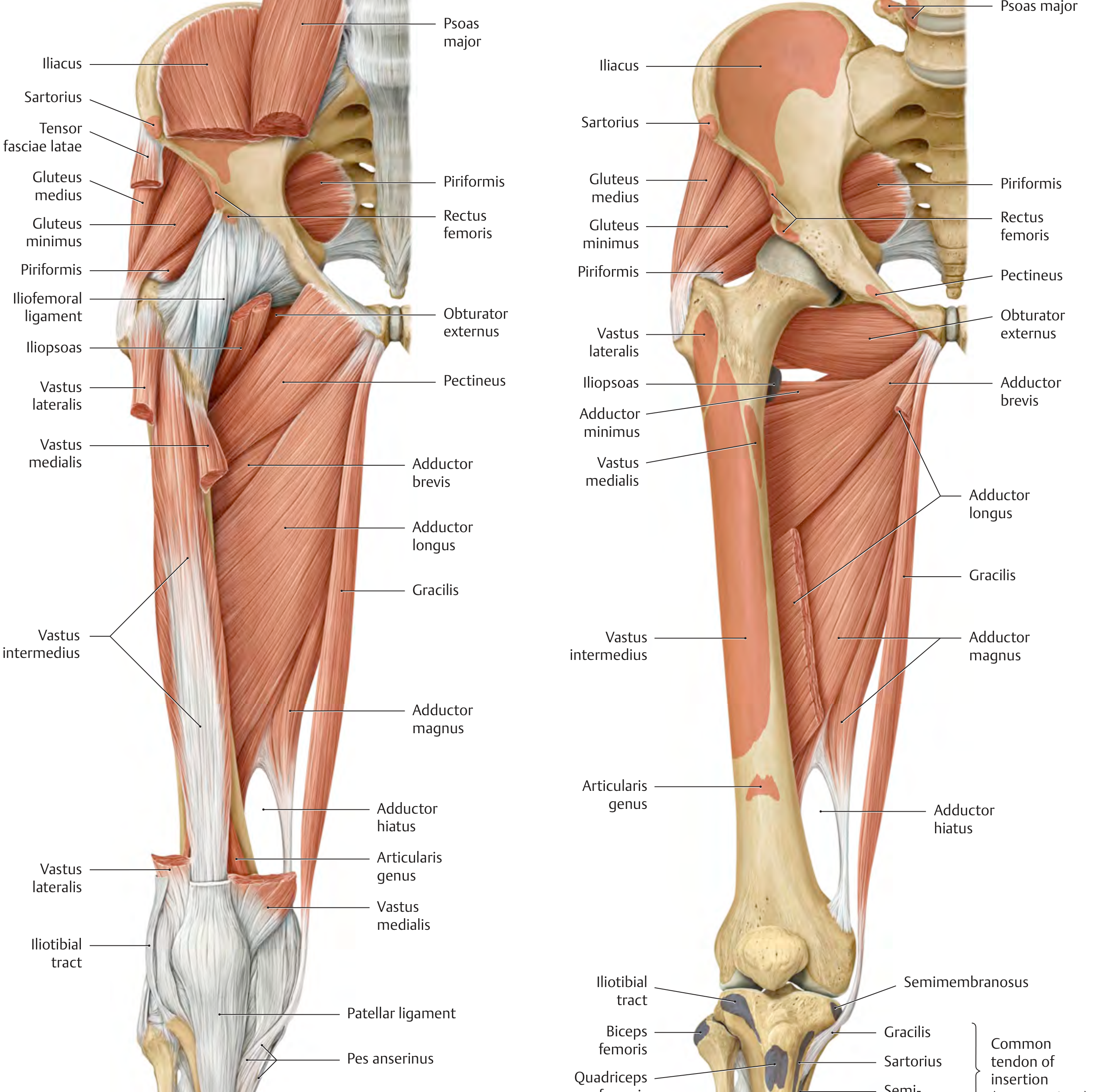
And with deep layers removed showing quadratus femoris, obturator externus, adductor magnus, and articularis genus:
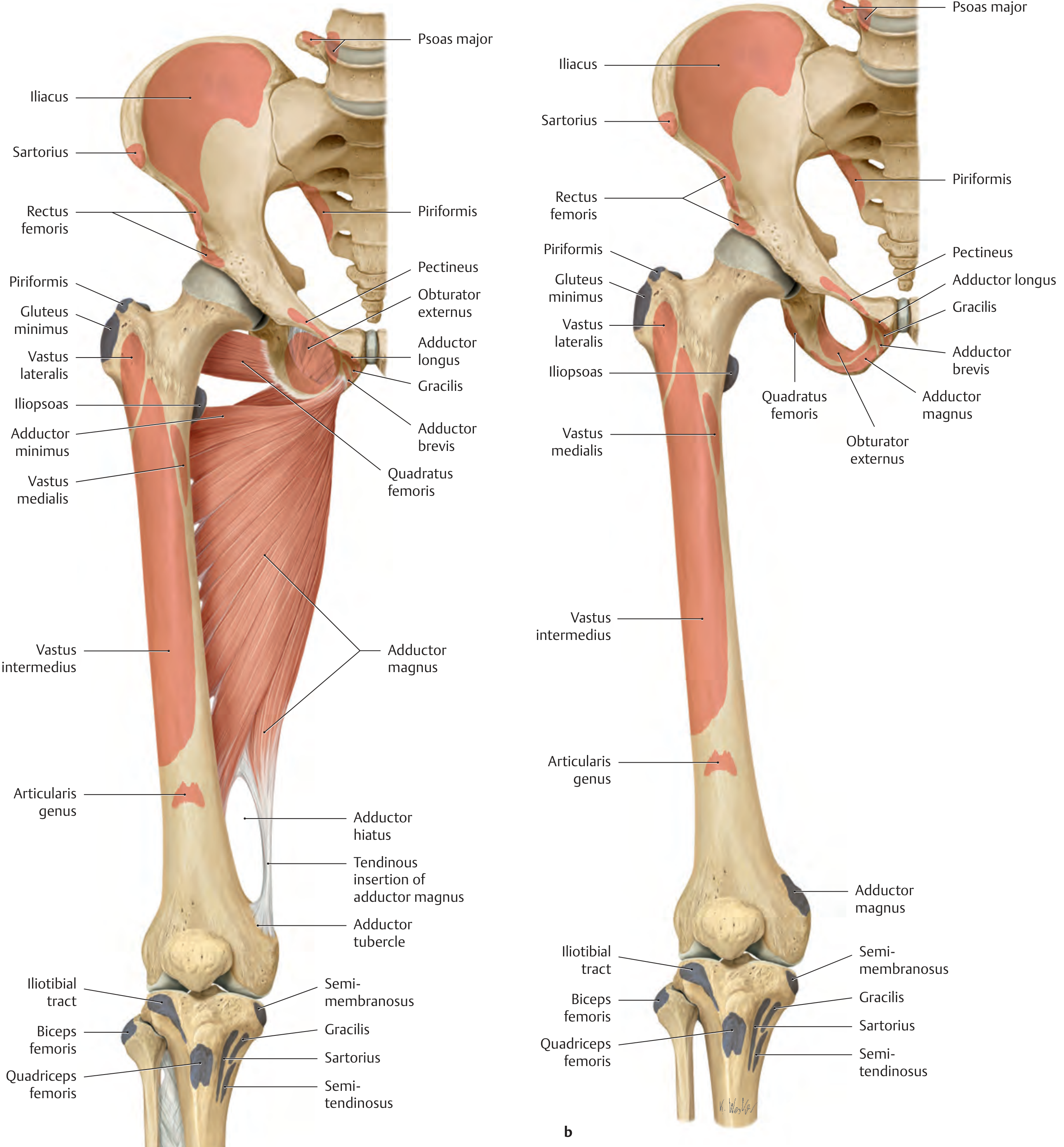
11. Medial Supracondylar Line
Continuation of the medial lip of the linea aspera distally toward the medial epicondyle; ends at the adductor tubercle.
- Vastus medialis continues to arise from this line distally
- Medial head of gastrocnemius - origin from the posterior aspect of the medial supracondylar region / medial epicondyle
12. Lateral Supracondylar Line
Continuation of the lateral lip of the linea aspera distally toward the lateral epicondyle.
- Vastus lateralis continues to arise from this line distally
- Lateral head of gastrocnemius - origin from posterior lateral supracondylar region / lateral epicondyle
- Plantaris - origin from the lower part of the lateral supracondylar line (just proximal to the lateral gastrocnemius head)
13. Popliteal Surface
A smooth flat triangular area on the posterior shaft between the diverging medial and lateral supracondylar lines. Forms the floor of the popliteal fossa.
- No direct muscle origins (smooth surface)
- Popliteal artery lies on it
14. Medial Epicondyle
A bony prominence on the medial non-articular surface of the medial condyle.
| Structure | Attachment |
|---|---|
| Tibial collateral ligament (MCL) | Origin (from medial epicondyle to medial tibia) |
| Medial head of gastrocnemius | Origin (just posterior to epicondyle, from the medial supracondylar area) |
| Posterior capsule of knee joint | Attachment |
15. Adductor Tubercle
A small sharp projection just superior to the medial epicondyle, at the end of the medial supracondylar line.
| Structure | Attachment |
|---|---|
| Adductor magnus | Tendinous insertion (hamstring/posterior part) - the most important insertion |
| Medial collateral ligament (superficial fibers) | Proximal attachment area |
Adductor hiatus: The gap in the distal tendon of adductor magnus between its linea aspera insertion and adductor tubercle insertion - transmits the femoral artery and vein into the popliteal fossa.
16. Lateral Epicondyle
A bony prominence on the lateral non-articular surface of the lateral condyle.
| Structure | Attachment |
|---|---|
| Fibular collateral ligament (LCL) | Origin (to head of fibula) |
| Popliteus tendon | Origin from a pit on the anterior/inferior aspect of the lateral epicondyle |
| Lateral head of gastrocnemius | Origin (just posterior to lateral epicondyle) |
| Plantaris | Origin (distal to lateral gastrocnemius head) |
17. Medial Femoral Condyle
- Larger and more prominent than the lateral condyle
- Extends further distally
- Articular surface covered with hyaline cartilage for articulation with the medial tibial condyle
- Posterior cruciate ligament (PCL): attachment on the medial wall of the intercondylar fossa (lateral surface of medial condyle)
18. Lateral Femoral Condyle
- Smaller, more anterior
- Articular surface for lateral tibial condyle
- Anterior cruciate ligament (ACL): attachment on the lateral wall of the intercondylar fossa (medial surface of lateral condyle - the "resident's ridge")
- Popliteus tendon: has a groove on the articular margin of the lateral condyle (popliteal sulcus)
- Iliotibial band (ITB): passes over the lateral condyle (does not attach here directly; attaches to Gerdy's tubercle on the tibia)
19. Intercondylar Fossa (Notch)
A deep posterior groove between the two condyles.
| Structure | Attachment |
|---|---|
| ACL | Lateral wall (posterosuperior aspect of medial surface of lateral condyle) |
| PCL | Medial wall (anterolateral surface of medial condyle) |
| Intercondyloid line | Ridge across the roof of the fossa |
20. Patellar Surface (Trochlea)
An anterior groove between the two condyles on the distal anterior femur - the trochlear groove.
- Articulates with the patella (forming the patellofemoral joint)
- Lateral trochlear ridge is higher (more prominent) than the medial - prevents lateral patellar subluxation
- Articularis genus inserts into the suprapatellar recess above this surface
Summary Attachment Table
| Landmark | Key Muscle/Ligament Attachment |
|---|---|
| Fovea capitis | Ligamentum teres |
| Greater trochanter (superoposterior) | Piriformis |
| Greater trochanter (posterior) | Obturator internus + gemelli |
| Greater trochanter (lateral) | Gluteus medius |
| Greater trochanter (anterior) | Gluteus minimus |
| Trochanteric fossa (medial GT) | Obturator externus |
| Lesser trochanter | Iliopsoas (iliacus + psoas major) |
| Intertrochanteric line (anterior) | Iliofemoral ligament; hip capsule; vastus medialis/lateralis (origin) |
| Intertrochanteric crest (posterior) | Hip capsule posterior; quadratus femoris (at quadrate tubercle) |
| Gluteal tuberosity | Gluteus maximus (lower portion) |
| Pectineal line | Pectineus; adductor brevis |
| Linea aspera (lateral lip) | Vastus lateralis; biceps femoris short head; gluteus maximus |
| Linea aspera (medial lip) | Vastus medialis; adductor longus; adductor brevis; adductor magnus |
| Anterior shaft | Vastus intermedius; articularis genus (distal) |
| Medial supracondylar line | Vastus medialis; medial gastrocnemius |
| Lateral supracondylar line | Vastus lateralis; lateral gastrocnemius; plantaris |
| Adductor tubercle | Adductor magnus (tendinous insertion) |
| Medial epicondyle | MCL (tibial collateral ligament); medial gastrocnemius |
| Lateral epicondyle | LCL (fibular collateral ligament); popliteus; lateral gastrocnemius; plantaris |
| Intercondylar fossa - lateral wall | ACL origin |
| Intercondylar fossa - medial wall | PCL origin |
| Patellar surface | Patella articulates here (patellofemoral joint) |
Key Clinical Correlations
- Femoral neck fractures: Disrupt the retinacular vessels → avascular necrosis of femoral head. Garden classification (I-IV). Intracapsular = higher risk of AVN.
- Intertrochanteric fractures: Extracapsular, preserve blood supply - treated with dynamic hip screw (DHS). Evans-Jensen classification.
- Adductor hiatus is the entry point of the femoral artery into the popliteal fossa - a potential compression site.
- Coxa valga (neck-shaft angle >135°) and coxa vara (<115°) affect hip biomechanics and gait.
- Greater trochanteric pain syndrome: Bursitis and/or gluteus medius/minimus tendinopathy at the GT facets.
Sources: Imaging Anatomy Vol. 3 - Bones, Joints, Vessels and Nerves, pp. 237-240; THIEME Atlas of Anatomy - General Anatomy and Musculoskeletal System, pp. 488-502; Imaging Anatomy Vol. 3, pp. 728-732
This is a shared conversation. Sign in to Orris to start your own chat.