
Hii
Hey! How can I help you today?
I'm a 1st year physiotherapy student can you help me with with anatomy
Absolutely! Anatomy is a big subject, so let me know what you'd like to focus on.
Asking for Preferences
What anatomy topic do you need help with?
Neuroanatomy is a fascinating area - especially relevant for physiotherapy! Let me narrow it down a bit more.
Asking for Preferences
Which area of neuroanatomy would you like to explore?
Explain midbrain
The midbrain (mesencephalon) is the uppermost part of the brainstem, connecting the pons below to the diencephalon above. It contains critical relay nuclei, cranial nerve nuclei (CN III & IV), and major motor/sensory tracts passing through it.
Searching sources...
Reading File
Finding Sources
Reading File
Reading File
Reading File
Finding Sources
Loading Image
Loading Image
Finding Sources
Reading File
Here is a thorough breakdown of the midbrain, tailored for a 1st year physiotherapy student:
The Midbrain (Mesencephalon)
Overview
The midbrain is the shortest segment of the brainstem - about 2 cm long. It sits between:
- Above: Diencephalon (thalamus/hypothalamus)
- Below: Pons
- Behind: Cerebellum
It contains ascending sensory tracts, descending motor tracts, cranial nerve nuclei, and several important reflex centers.
External Features
Anterior Surface
- A deep midline depression called the interpeduncular fossa
- On each side of this fossa: the crus cerebri (also called cerebral peduncles) - large bundles of descending motor fibers
- CN III (Oculomotor nerve) exits from the medial side of each crus cerebri
Posterior Surface (Tectum)
- Four rounded bumps called the corpora quadrigemina:
- 2 Superior colliculi (rostral) - visual reflexes
- 2 Inferior colliculi (caudal) - auditory relay
- CN IV (Trochlear nerve) emerges between the inferior colliculi, crosses the midline, and wraps around the midbrain - the only cranial nerve to exit from the posterior surface
Internal Structure: 3 Divisions (Anterior to Posterior)
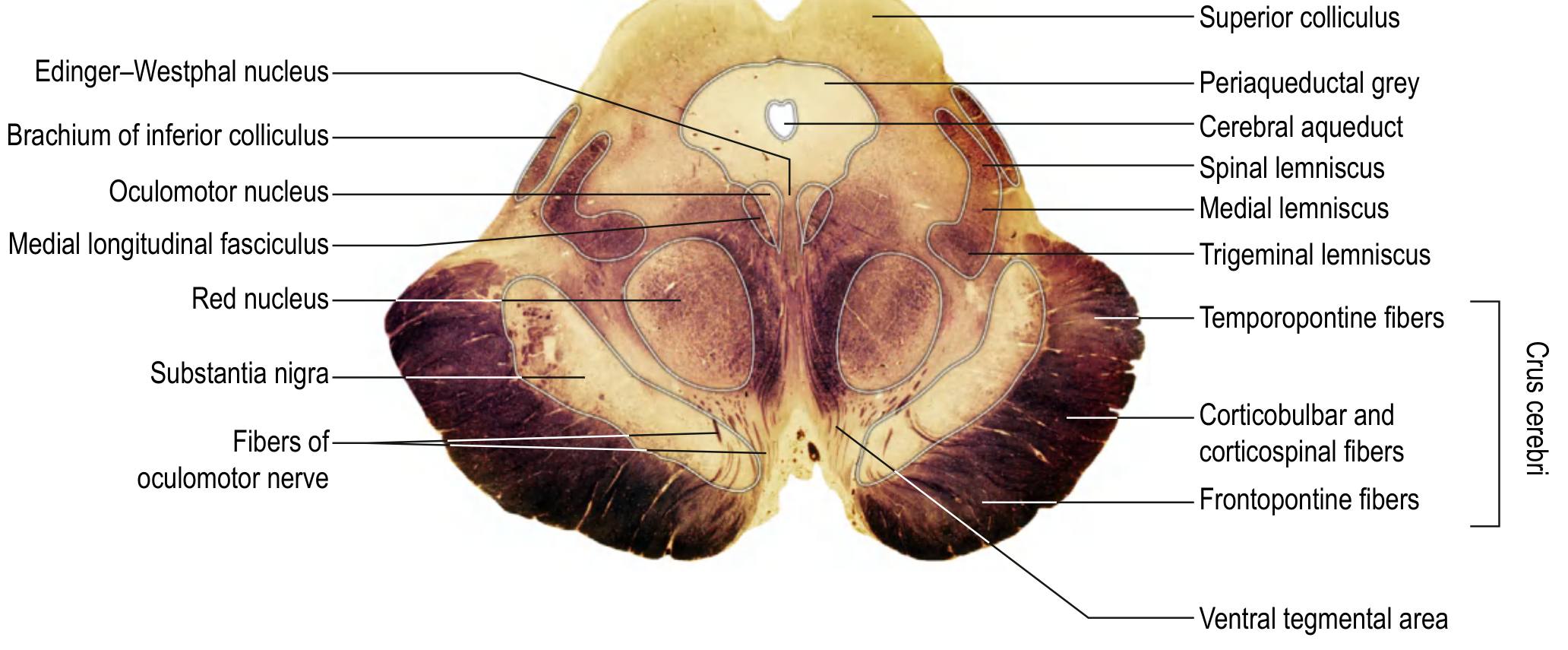
Here is a cross-section of the midbrain showing all the key structures:
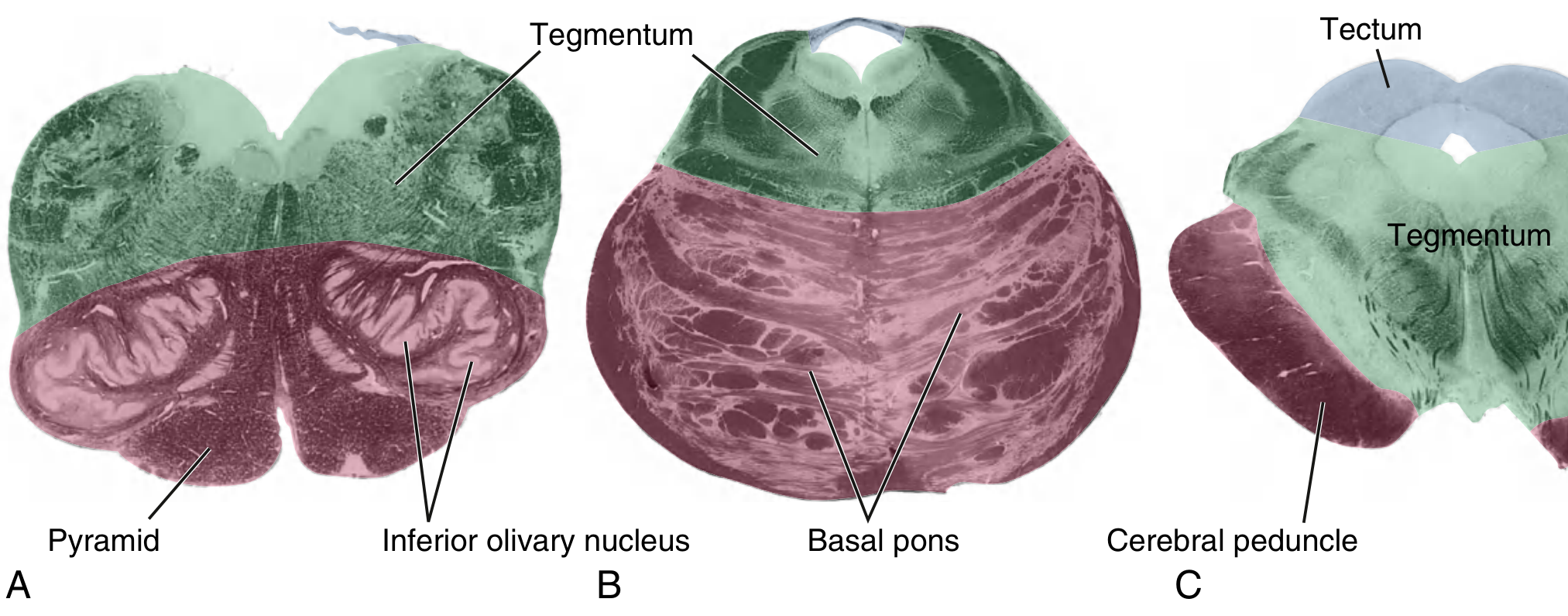
And here is how the midbrain compares to the medulla and pons in cross-section (midbrain = panel C on right):
The midbrain is divided into three parts:
1. Tectum ("Roof") - Posterior
- Only found in the midbrain (not pons or medulla)
- Contains the superior and inferior colliculi
- Superior colliculi: Relay center for visual tracking and pupillary light reflexes
- Inferior colliculi: Relay center for auditory pathways
2. Tegmentum ("Floor/Middle") - Central
The largest region, containing:
| Structure | Location / Function |
|---|---|
| Cerebral aqueduct | Channel carrying CSF from 3rd to 4th ventricle |
| Periaqueductal gray (PAG) | Surrounds the aqueduct; involved in pain modulation and defensive behavior |
| Red nucleus | Motor relay; receives input from cerebellum and cortex; gives rise to rubrospinal tract |
| Substantia nigra | Pigmented (melanin); divided into zona compacta (dopaminergic neurons) and zona reticulata; key for motor control - degeneration causes Parkinson's disease |
| CN III nucleus (Oculomotor) | Rostral midbrain; controls eye movement and pupil constriction |
| Edinger-Westphal nucleus | Near CN III nucleus; parasympathetic - controls pupil constriction and lens accommodation |
| CN IV nucleus (Trochlear) | Caudal midbrain; controls superior oblique muscle (downward/inward eye movement) |
| Medial longitudinal fasciculus (MLF) | Coordinates eye and head movements |
| Medial lemniscus | Ascending fine touch/proprioception (from opposite side) |
| Spinothalamic tract | Ascending pain/temperature |
| Reticular formation | Consciousness and arousal |
3. Cerebral Peduncles (Crus Cerebri) - Anterior
Large descending motor tracts arranged somatotopically:
- Medial 1/5: Frontopontine fibers (from frontal cortex to pons)
- Middle 3/5: Corticospinal + corticobulbar fibers (main motor pathway to spinal cord and brainstem)
- Lateral 1/5: Temporopontine fibers
Levels of the Midbrain
| Level | Key Structures |
|---|---|
| Rostral (superior colliculus level) | CN III nucleus, Edinger-Westphal nucleus, Red nucleus, Substantia nigra |
| Caudal (inferior colliculus level) | CN IV nucleus, Substantia nigra, Mesencephalic nucleus of CN V |
Blood Supply
The midbrain gets blood from branches of:
- Posterior cerebral artery (PCA)
- Superior cerebellar artery (SCA)
- Basilar artery (paramedian branches)
- Posterior communicating artery
Key: Peduncular arteries supply the oculomotor nucleus, red nucleus, and substantia nigra.
Why It Matters for Physiotherapy
| Structure | Clinical Relevance |
|---|---|
| Substantia nigra | Parkinson's disease - degeneration of dopaminergic neurons causes rigidity, bradykinesia, tremor |
| Red nucleus | Lesions contribute to movement disorders (ataxia, tremor) |
| CN III | Midbrain stroke can cause ipsilateral CN III palsy (drooping eyelid, dilated pupil) |
| Corticospinal tracts | Damage causes contralateral hemiplegia |
| PAG | Involved in pain perception - relevant to pain management in PT |
Sources: Gray's Anatomy for Students, p. 1316-1318 | Localization in Clinical Neurology, 8e, p. 943-944
Would you like me to go deeper on any specific part - for example, the blood supply and stroke syndromes, Parkinson's disease connections, or the cranial nerves of the midbrain?
This is a shared conversation. Sign in to Orris to start your own chat.