
Brain anatomy
Reading File
Brain anatomy is a broad topic - this is a Visual/Broad query. Let me search the library for dedicated sections and images simultaneously.
Finding Sources
Finding Sources
Reading File
Finding Sources
This is the table of contents - the actual content is in the atlas format. Let me get the substantive brain anatomy paragraphs from both the neuroscience textbook and check for images.
Finding Sources
Finding Sources
I found images and text. Let me now read the key anatomy content and fetch the brain images in parallel.
Reading File
Reading File
Loading Image
Excellent! I have a beautiful brain anatomy diagram. Now let me get more content on deep structures (brainstem, cerebellum, diencephalon, basal ganglia, ventricular system) and fetch another diagram.
Reading File
Reading File
Loading Image
I have rich content from multiple textbooks and excellent diagrams. Let me now get the brainstem/diencephalon/basal ganglia details and one more image.
Reading File
Finding Sources
Reading File
Reading File
I now have comprehensive content from multiple authoritative textbooks. Let me fetch one more key image - the brainstem diagram.
Loading Image
Loading Image
I now have everything needed to write a comprehensive brain anatomy overview. Here is the full answer:
Brain Anatomy
The human brain is the most complex structure in the known universe - roughly 1,400 g of tissue that coordinates every thought, movement, sensation, and bodily function. It divides into three broad regions: the cerebrum (forebrain), the cerebellum, and the brainstem.
Overview: Major Divisions
| Division | Embryonic Origin | Key Structures |
|---|---|---|
| Telencephalon | Forebrain | Cerebral cortex, basal ganglia, white matter |
| Diencephalon | Forebrain | Thalamus, hypothalamus, subthalamus, epithalamus |
| Mesencephalon | Midbrain | Midbrain (tectum, tegmentum, cerebral peduncles) |
| Metencephalon | Hindbrain | Pons, cerebellum |
| Myelencephalon | Hindbrain | Medulla oblongata |
1. Cerebral Hemispheres (Telencephalon)
The cerebral cortex is not smooth - it has numerous infoldings called sulci (grooves) and elevated ridges called gyri, which dramatically increase surface area. The outer surface is gray matter (six layers of cell bodies); beneath it is white matter (myelinated axons).
Each hemisphere is divided into four major lobes:
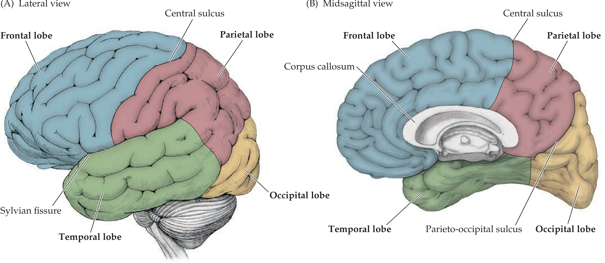
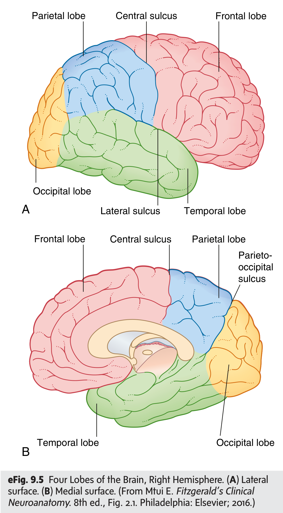
Frontal Lobe
- Bounded posteriorly by the central sulcus (of Rolando) and inferolaterally by the Sylvian (lateral) fissure
- Contains the precentral gyrus - the primary motor cortex (voluntary movement)
- The inferior frontal gyrus includes Broca's area (speech production, dominant hemisphere)
- Prefrontal cortex: executive function, personality, decision-making
- Key gyri: superior, middle, inferior frontal gyri
Parietal Lobe
- Bounded anteriorly by the central sulcus
- Postcentral gyrus = primary somatosensory cortex
- The intraparietal sulcus separates the superior parietal lobule from the inferior parietal lobule
- The inferior parietal lobule contains the supramarginal gyrus and angular gyrus (language processing)
- No sharp lateral demarcation from the occipital or temporal lobes
Temporal Lobe
- Lies below the Sylvian fissure
- Contains the primary auditory cortex (Heschl's gyri on the superior temporal plane)
- Wernicke's area (speech comprehension) in the posterior superior temporal gyrus of the dominant hemisphere
- Medial temporal lobe: hippocampus and amygdala (memory and emotion)
Occipital Lobe
- Most posterior lobe, separated from parietal lobe medially by the parieto-occipital sulcus
- Contains the primary visual cortex (V1) along the calcarine fissure
- The cuneus (above calcarine fissure) and lingula (below) are key medial landmarks
Insula (5th lobe)
- Buried within the depths of the Sylvian fissure, covered by the frontal, parietal, and temporal opercula
- Involved in interoception, taste, pain, and autonomic regulation
Key White Matter Structures
- Corpus callosum: the large C-shaped commissure connecting homologous areas of both hemispheres; divided into rostrum, genu, body, and splenium
- Internal capsule: V-shaped white matter tract containing corticospinal and corticobulbar fibers; divided into anterior limb, genu, and posterior limb
- Corona radiata: the fan-shaped expansion of the internal capsule radiating to the cortex
2. Diencephalon
Thalamus
- Paired egg-shaped gray matter masses flanking the third ventricle
- Acts as the relay station for almost all sensory input to the cortex (except olfaction)
- Key nuclei: VPL (somatosensory relay), LGN (visual), MGN (auditory), VL/VA (motor)
Hypothalamus
- Below the thalamus, around the third ventricle
- Controls the autonomic nervous system, pituitary gland, hunger, thirst, temperature regulation, circadian rhythms, and reproductive behavior
- Connected to the pituitary via the infundibular stalk
Other Diencephalon structures
- Subthalamus: contains the subthalamic nucleus (STN), part of the basal ganglia circuit
- Epithalamus: contains the pineal gland (melatonin synthesis) and habenular nuclei
3. Basal Ganglia
Deep gray matter nuclei embedded in the white matter of the cerebral hemispheres:
- Caudate nucleus and putamen together = striatum (main input structure)
- Globus pallidus (internal and external segments)
- Substantia nigra (in the midbrain) and subthalamic nucleus are functionally part of this circuit
- Role: regulation of voluntary movement, procedural learning, habit formation
4. Brainstem
The brainstem = midbrain + pons + medulla. It connects the forebrain to the spinal cord and houses most cranial nerve nuclei. All ascending and descending tracts pass through it.
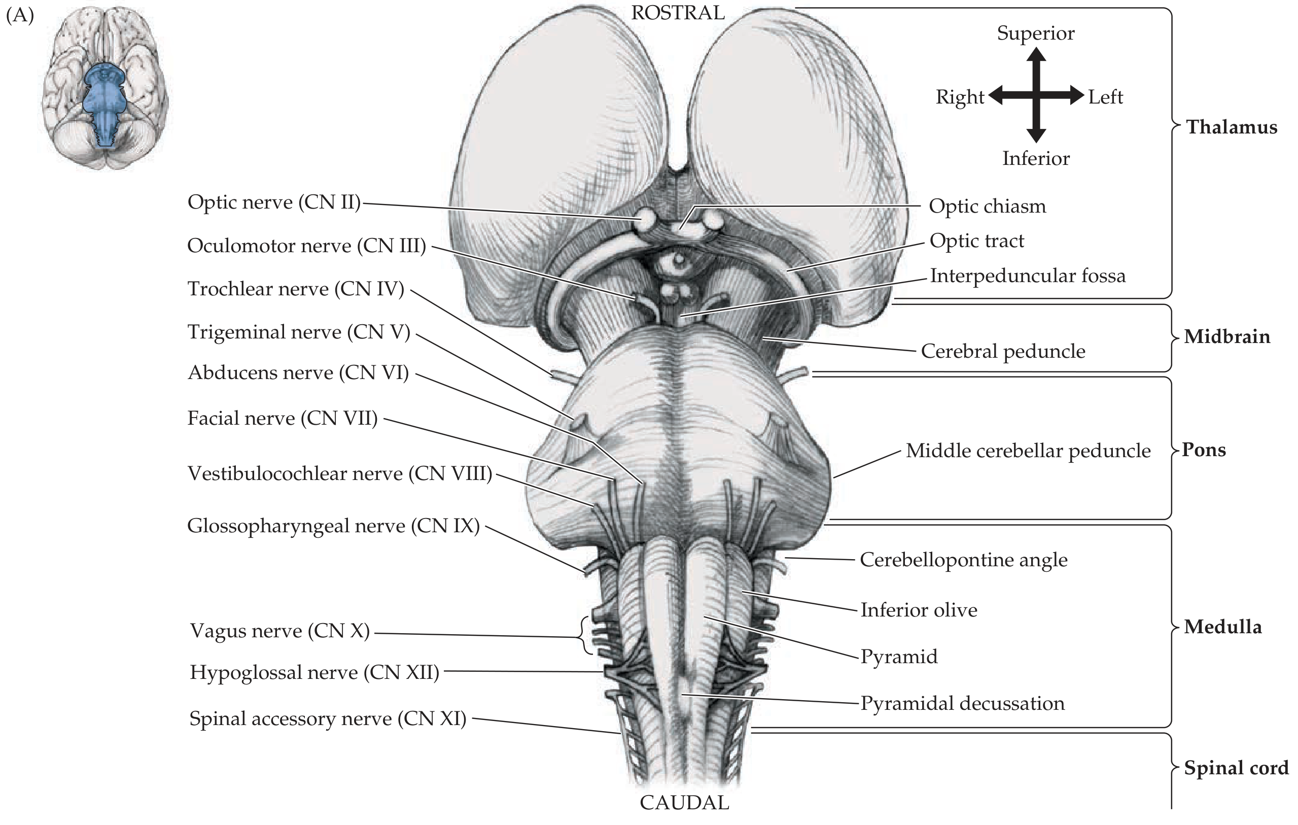
Midbrain (Mesencephalon)
- Most rostral brainstem segment, connects to the diencephalon
- Tectum: dorsal plate containing the superior colliculi (visual reflexes) and inferior colliculi (auditory reflexes)
- Tegmentum: contains cranial nerve nuclei (CN III, IV), red nucleus, substantia nigra, periaqueductal gray (pain modulation)
- Cerebral peduncles: large ventral columns carrying corticospinal fibers
- Cranial nerves: CN III (oculomotor) exits ventrally; CN IV (trochlear) exits dorsally - the only CN to exit dorsally
Pons
- Bulging ventral structure; connects to the cerebellum via the middle cerebellar peduncles
- Contains nuclei for CN V (trigeminal), VI (abducens), VII (facial), VIII (vestibulocochlear)
- The pontine reticular formation plays a key role in arousal and consciousness
- The locus coeruleus (norepinephrine) and raphe nuclei (serotonin) are located here
Medulla Oblongata
- Most caudal brainstem segment, continuous with the spinal cord at the foramen magnum
- Houses cardiovascular, respiratory, and vomiting centers - vital for survival
- Contains CN IX (glossopharyngeal), X (vagus), XI (spinal accessory), XII (hypoglossal)
- Key surface landmarks: pyramids (corticospinal tracts), inferior olives (cerebellar coordination), pyramidal decussation (where corticospinal fibers cross)
- The reticular formation extends throughout the brainstem, regulating consciousness, pain, and autonomic function
5. Cerebellum
- Located in the posterior cranial fossa, behind the pons and medulla
- Connected to the brainstem by three pairs of cerebellar peduncles (superior, middle, inferior)
- Divided into two hemispheres and a midline vermis
- Cortex has a distinctive folded appearance (folia)
- Function: coordination of movement, balance, fine motor control, motor learning
- Does NOT initiate movement but refines and coordinates it
6. Ventricular System and CSF
The ventricular system is a series of interconnected cavities filled with cerebrospinal fluid (CSF):
| Ventricle | Location | Connection |
|---|---|---|
| Lateral ventricles (x2) | Within cerebral hemispheres | Via foramen of Monro → 3rd ventricle |
| 3rd ventricle | Between the thalami | Via cerebral aqueduct → 4th ventricle |
| 4th ventricle | Between pons/medulla and cerebellum | Via foramina of Luschka (x2) and Magendie → subarachnoid space |
- Choroid plexus (modified ependymal cells) produces ~0.5 L of CSF per day
- CSF circulates around the brain and spinal cord in the subarachnoid space, then is reabsorbed into the venous system (mainly via arachnoid granulations)
7. Meninges (Protective Coverings)
From deep to superficial (mnemonic: PAD):
- Pia mater - delicate, adheres directly to brain surface
- Arachnoid mater - spiderweb-like; CSF flows in the subarachnoid space between pia and arachnoid
- Dura mater - tough outer layer; forms folds like the falx cerebri and tentorium cerebelli
8. Cortical Functional Areas (Brodmann Map Summary)
| Area | Location | Function |
|---|---|---|
| Primary motor cortex (M1) | Precentral gyrus (BA 4) | Voluntary movement |
| Primary somatosensory (S1) | Postcentral gyrus (BA 3, 1, 2) | Touch, proprioception |
| Primary visual (V1) | Calcarine fissure (BA 17) | Vision |
| Primary auditory | Heschl's gyri (BA 41, 42) | Hearing |
| Broca's area | Inferior frontal gyrus (BA 44, 45) | Speech production |
| Wernicke's area | Posterior superior temporal (BA 22) | Speech comprehension |
| Prefrontal cortex | Anterior frontal (BA 9, 10, 46) | Executive function |
Sources: Neuroanatomy through Clinical Cases, 3rd Edition (Blumenfeld) | Neuroscience: Exploring the Brain, 5th Edition (Bear et al.) | Gray's Anatomy for Students | Grainger & Allison's Diagnostic Radiology
This is a shared conversation. Sign in to Orris to start your own chat.