Tell me every crazy or funny or weird fact about cns tumors make me understand them and refer Robbins textbook I must know every conceptual and important thing about them

Reading File
Finding Sources
Finding Sources
Finding Sources
Finding Sources
Finding Sources
Reading File
Reading File
Reading File
Reading File
Reading File
Reading File
Searching Images

glioblastoma brain tumor butterfly necrosis histology

Reading File
Searching Images

oligodendroglioma fried egg cells chicken wire capillaries

Searching Images

meningioma psammoma bodies whorled histology
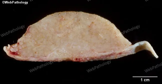
Cross-section gross pathology photograph of a dural-attached intracranial meningioma. The specimen shows a broad-based, lobulated mass arising from arachnoid cap cells and adherent to the dura mater. Cut surfaces are coarse and tan‑white with a gritty texture due to abundant psammoma bodies and dystrophic calcifications forming concentric lamellations. The tumor is relatively well circumscribed but intimately apposed to dura; no obvious necrosis or hemorrhage is evident in this section. Psammoma bodies produce the characteristic gritty feel and calcified foci on gross examination. Meningiomas account for roughly 20–30% of intracranial neoplasms and show a female preponderance (Female:Male ≈ 1.7:1). High‑grade variants occur more frequently in males and may demonstrate increased cellularity, mitotic activity, and necrosis. Overall, meningiomas are slow‑growing, dural‑based tumors derived from meningothelial (arachnoid cap) cells and are often calcified. Radiographically they appear as extra‑axial, sometimes hyperostotic lesions; histology commonly reveals whorled sheets of meningothelial cells with psammoma bodies. Clinically, presentation depends on tumor location and mass effect, including headaches, seizures, or focal neurologic deficits. Management typically involves complete surgical excision; adjuvant radiotherapy is considered for residual, recurrent, or anaplastic disease. This image provides a classic educational example of gross meningioma morphology for teaching and differential diagnosis with other dural‑based lesions such as fibrous meningioma, metastasis, or hemangiopericytoma.
Cross-section gross pathology photograph of a dural-attached intracranial meningioma. The specimen shows a broad-based, lobulated mass arising from arachnoid cap cells and adherent to the dura mater. Cut surfaces are coarse and tan‑white with a gritty texture due to abundant psammoma bodies and dystrophic calcifications forming concentric lamellations. The tumor is relatively well circumscribed but intimately apposed to dura; no obvious necrosis or hemorrhage is evident in this section. Psammoma bodies produce the characteristic gritty feel and calcified foci on gross examination. Meningiomas account for roughly 20–30% of intracranial neoplasms and show a female preponderance (Female:Male ≈ 1.7:1). High‑grade variants occur more frequently in males and may demonstrate increased cellularity, mitotic activity, and necrosis. Overall, meningiomas are slow‑growing, dural‑based tumors derived from meningothelial (arachnoid cap) cells and are often calcified. Radiographically they appear as extra‑axial, sometimes hyperostotic lesions; histology commonly reveals whorled sheets of meningothelial cells with psammoma bodies. Clinically, presentation depends on tumor location and mass effect, including headaches, seizures, or focal neurologic deficits. Management typically involves complete surgical excision; adjuvant radiotherapy is considered for residual, recurrent, or anaplastic disease. This image provides a classic educational example of gross meningioma morphology for teaching and differential diagnosis with other dural‑based lesions such as fibrous meningioma, metastasis, or hemangiopericytoma.

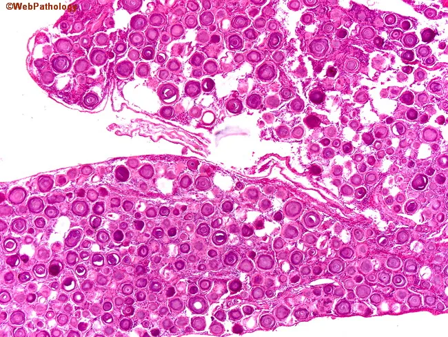
Light microscopy of a spinal meningioma specimen. Imaging modality: histology with Hematoxylin and Eosin staining. The tissue shows classic whorled sheets of uniform meningothelial cells with eosinophilic cytoplasm and oval to vesicular nuclei. Interspersed throughout the tumor are numerous psammoma bodies, concentric laminated calcifications that range from small to large; these calcifications can be basophilic or eosinophilic depending on staining and maturation. The psammomatous variant demonstrates dense sandstone-like lamination within the tumor matrix and occasional focal ossification in advanced foci. The background stroma is fibrous, with low cellular atypia and low mitotic activity, consistent with a benign WHO grade I meningioma. The overall architecture is organized into interrupted lobules and tight cellular whorls arising from meningothelial cells, with abundant extracellular collagen. The presence of multiple psammoma bodies is a diagnostic hallmark distinguishing psammomatous meningioma from other spinal neoplasms. Diagnostic significance: histopathology confirms meningothelial differentiation with psammomatous calcifications; supports a benign prognosis but may cause compressive myelopathy depending on size and location. Clinical correlation: spinal canal mass in older women presenting with progressive myelopathy or radiculopathy; treatment is surgical resection; prognosis is favorable with complete excision; radiologic correlation (MRI) typically shows dural-based enhancement.
Light microscopy of a spinal meningioma specimen. Imaging modality: histology with Hematoxylin and Eosin staining. The tissue shows classic whorled sheets of uniform meningothelial cells with eosinophilic cytoplasm and oval to vesicular nuclei. Interspersed throughout the tumor are numerous psammoma bodies, concentric laminated calcifications that range from small to large; these calcifications can be basophilic or eosinophilic depending on staining and maturation. The psammomatous variant demonstrates dense sandstone-like lamination within the tumor matrix and occasional focal ossification in advanced foci. The background stroma is fibrous, with low cellular atypia and low mitotic activity, consistent with a benign WHO grade I meningioma. The overall architecture is organized into interrupted lobules and tight cellular whorls arising from meningothelial cells, with abundant extracellular collagen. The presence of multiple psammoma bodies is a diagnostic hallmark distinguishing psammomatous meningioma from other spinal neoplasms. Diagnostic significance: histopathology confirms meningothelial differentiation with psammomatous calcifications; supports a benign prognosis but may cause compressive myelopathy depending on size and location. Clinical correlation: spinal canal mass in older women presenting with progressive myelopathy or radiculopathy; treatment is surgical resection; prognosis is favorable with complete excision; radiologic correlation (MRI) typically shows dural-based enhancement.

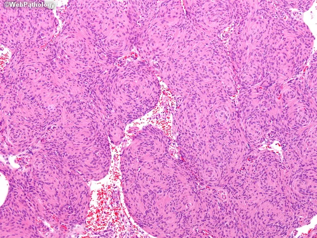
Imaging modality and technique: Light microscopy of Hematoxylin and Eosin (H&E) stained paraffin section from intracranial meningeal tumor tissue. Anatomical context: dura-based CNS neoplasm consistent with meningothelial meningioma. Specimen type: surgical biopsy/resection specimen of a brain/meningeal neoplasm. Primary subject: monomorphic meningothelial cells arranged in cohesive, syncytial sheets and whorls. Visual features: uniform oval nuclei with vesicular chromatin, indistinct cell borders, and scant cytoplasm; overall cellular architecture shows prominent whorled growth with peripherally arranged cells and occasional radial polarity. Background stroma is fibrous with minimal inflammatory infiltrate; psammoma bodies are not conspicuous in this field. Cytologic features are bland, with essentially no mitotic activity evident in this high-power view. Diagnostic interpretation: consistent with a World Health Organization (WHO) grade I meningothelial meningioma, reflecting low proliferative index and benign histology. Differential considerations: transitional or fibrous meningioma variants; metastatic carcinoma is unlikely given cohesive cell groups and meningioma-like whorls. Clinical significance: histopathologic confirmation aids prognosis and guides management after surgical resection. Clinical relevance: relevant for clinical decision-making, imaging-pathology correlation, and research into meningioma subtypes and outcomes. Note that there is no evidence of anaplasia or high grade histologic features such as increased mitotic activity, necrosis, or brain invasion visible in this field, supporting a favorable long term prognosis.

Imaging modality and technique: Light microscopy of Hematoxylin and Eosin (H&E) stained paraffin section from intracranial meningeal tumor tissue. Anatomical context: dura-based CNS neoplasm consistent with meningothelial meningioma. Specimen type: surgical biopsy/resection specimen of a brain/meningeal neoplasm. Primary subject: monomorphic meningothelial cells arranged in cohesive, syncytial sheets and whorls. Visual features: uniform oval nuclei with vesicular chromatin, indistinct cell borders, and scant cytoplasm; overall cellular architecture shows prominent whorled growth with peripherally arranged cells and occasional radial polarity. Background stroma is fibrous with minimal inflammatory infiltrate; psammoma bodies are not conspicuous in this field. Cytologic features are bland, with essentially no mitotic activity evident in this high-power view. Diagnostic interpretation: consistent with a World Health Organization (WHO) grade I meningothelial meningioma, reflecting low proliferative index and benign histology. Differential considerations: transitional or fibrous meningioma variants; metastatic carcinoma is unlikely given cohesive cell groups and meningioma-like whorls. Clinical significance: histopathologic confirmation aids prognosis and guides management after surgical resection. Clinical relevance: relevant for clinical decision-making, imaging-pathology correlation, and research into meningioma subtypes and outcomes. Note that there is no evidence of anaplasia or high grade histologic features such as increased mitotic activity, necrosis, or brain invasion visible in this field, supporting a favorable long term prognosis.

Loading Image
Loading Image
Loading Image
I now have all the content I need from both Robbins textbooks plus excellent histology images. Let me write the comprehensive answer.

CNS Tumors - Every Crazy, Weird & Conceptual Fact (Robbins Edition)


The First Big Mind-Bender: "Benign" Can Still Kill You

The CNS breaks all the usual rules of oncology. Elsewhere in the body, "benign" means safe. In the brain, a benign posterior fossa meningioma can cause cardiorespiratory arrest simply by compressing the medulla. Location is everything. A WHO grade 1 tumor sitting on a vital brainstem center is deadlier than a grade 3 tumor sitting in a silent frontal lobe. - Robbins & Cotran Pathologic Basis of Disease, p. 1193

The Grading Paradox: Grades Without the Usual Meaning

Most brain tumors are graded 1-4 (CNS WHO 2021 classification), but grade does NOT cleanly map to "benign vs. malignant" the way it does in other tissues. The grade reflects biologic behavior, not just histology. Malignant gliomas rarely metastasize outside the CNS - they kill not by spreading to the liver or lungs but by eating the brain from inside. Some pediatric tumors (medulloblastoma) do spread through the CSF, showering seeds down the spinal cord. - Robbins & Cotran, p. 1193

PART 1: GLIOMAS - The Infiltrators

The IDH Story: The Single Most Important Concept in Modern CNS Tumor Biology

In 2008, researchers discovered mutations in IDH1 and IDH2 that changed everything about how we classify gliomas. Here is the weird part: IDH normally converts isocitrate to alpha-ketoglutarate. Mutant IDH does something bizarre - it gains a brand new, abnormal enzyme activity that converts alpha-ketoglutarate into 2-hydroxyglutarate (2-HG), an oncometabolite that has no normal role in the body. It hijacks epigenetics, causes widespread DNA methylation, and drives cancer. This is a "gain of function" mutation in the most unexpected sense - the enzyme literally learns to do something it was never designed to do. - Robbins & Cotran, p. 1193
The clinical payoff: IDH mutation status splits all diffuse gliomas into two completely different diseases:
  • IDH-mutant astrocytoma (grades 2-4): younger patients (median 38 years), much better prognosis, median survival >10 years for grade 2
  • IDH-wildtype glioblastoma (always grade 4): older patients (>55 years), the most savage primary brain tumor, median survival ~15 months

Astrocytoma, IDH-Mutant
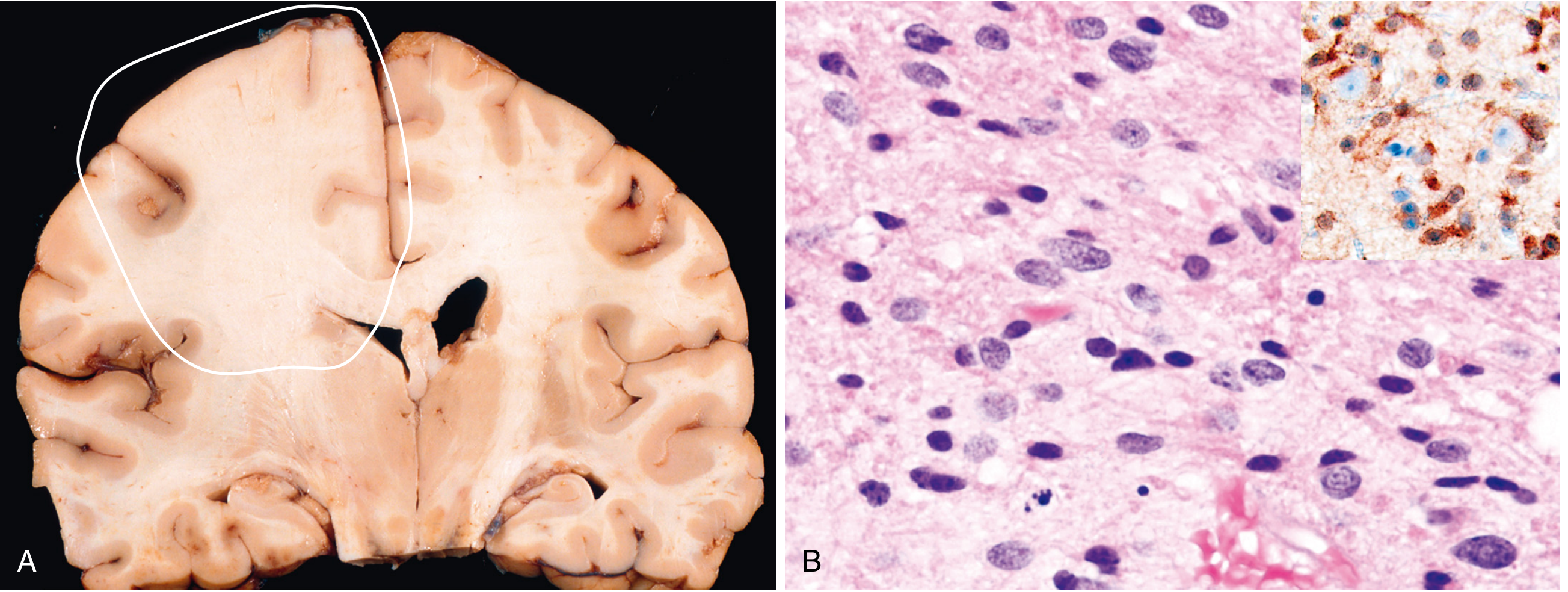
The sneaky infiltrator. These tumors expand and distort brain without forming a discrete mass - no clean edges, no capsule. On MRI they look like "edema" - but that "edema" often contains tumor cells infiltrating centimeters beyond the visible lesion. The corticomedullary junction gets blurred on imaging (see image below). - Robbins Basic Pathology, p. 858
Astrocytoma IDH-mutant: coronal section showing expanded left frontal white matter with blurred corticomedullary junction, and histology showing enlarged irregular nuclei in the fibrillary matrix
Astrocytoma, IDH-mutant. (A) Left frontal white matter expanded, blurring the corticomedullary junction. (B) Histology: enlarged hyperchromatic nuclei embedded in native fibrillary matrix. Inset: IDH1 immunostain positive in tumor cells wrapping around normal neurons ("perineuronal satellitosis") - the tumor cells literally hug neurons.
Weird molecular trick: Nearly all IDH-mutant astrocytomas also show ATRX loss - detectable by simple immunohistochemistry - and p53 overexpression.
Progression is inevitable but slow. Grade 2 can sit quietly for years, then suddenly a more aggressive clone emerges. The tumor grade jumps, the patient deteriorates rapidly, and survival falls from >10 years to 3 years (grade 4). - Robbins Basic Pathology, p. 858

Glioblastoma (GBM), IDH-Wildtype - The Monster

Accounts for 50% of all primary malignant brain tumors in adults and ~14% of ALL primary CNS tumors. This is the one everyone fears.
Crazy genetic features (Robbins lists these explicitly):
  1. Telomere escape: Either TERT promoter mutations or alternative lengthening of telomeres - tumor cells become immortal
  2. CDKN2A deletion (biallelic) - loses p16, escapes senescence completely
  3. EGFR or PDGFR amplification - growth factor signaling goes haywire
  4. TP53 mutation - apoptosis resistance
  5. MGMT promoter methylation - this one is GOOD for the patient; it means the DNA repair gene is silenced, making the tumor sensitive to temozolomide chemotherapy. If MGMT is methylated = better chemo response. - Robbins Basic Pathology, p. 859
The gross appearance is horrifying AND informative: Cut sections show multicolored zones - gray-white (firm tumor), yellow (necrosis), red (hemorrhage). No other primary brain tumor looks this chaotic from the outside.
The "butterfly" pattern: GBM loves to cross the corpus callosum and involve both hemispheres bilaterally - creating a "butterfly glioma" on imaging. It literally bridges the two halves of the brain.
Ring enhancement on MRI: The necrotic core does not enhance (dead cells). The actively growing, hypervascular rim does. This creates the classic ring-enhancing lesion - a signature so characteristic it essentially clinches the diagnosis. - Robbins & Cotran, p. 1194
Pseudopalisading necrosis: Under the microscope, tumor cells line up in neat rows around serpentine areas of necrosis - like soldiers lined up around a battlefield. The cells near necrosis are actually fleeing hypoxia, piling up at the edges. VEGF produced by these hypoxic malignant astrocytes drives the wild, abnormal vessel formation (microvascular proliferation) - the vasculature literally bulges into vessel lumens in tufts. - Robbins & Cotran, p. 1194
The MGMT twist explained simply: MGMT is a DNA repair enzyme. GBM uses it to fix chemotherapy-induced DNA damage. If the MGMT promoter is methylated (silenced), the tumor cannot repair the damage from temozolomide - chemo works. If MGMT is unmethylated and active, the tumor repairs itself and chemo fails. A tumor's genetic methylation status predicts its own treatability.

Oligodendroglioma - The "Fried Egg" Tumor With Chicken Wire

The best prognosis among all diffuse gliomas. Median survival 10-20 years for grade 2. Why? Because of a specific chromosomal deletion: codeletion of 1p and 19q, always paired with IDH mutation. This combination makes the tumor exquisitely sensitive to both chemotherapy and radiotherapy.
The histology is unforgettable:
  • Sheets of regular cells with round nuclei and clear halos of cytoplasm - "fried egg" appearance
  • A delicate network of fine anastomosing capillaries looking exactly like "chicken wire"
  • Calcification in up to 90% of cases - from microscopic flecks to massive calcium deposits. No other common brain tumor calcifies this frequently.
  • Seizures for years before diagnosis - patients often live with the tumor unknowingly for a long time - Robbins Basic Pathology, p. 860
Why fried egg? It is a fixation artifact - the cells swell during tissue processing, pushing the nucleus to the center and creating a clear halo. The living tumor does not look like this. It is one of the most famous artifacts in all of histopathology.

Pilocytic Astrocytoma - The Well-Behaved Child's Tumor

WHO grade 1 - potentially curable with surgery alone. Mostly found in children and young adults. Its favorite location is the cerebellum; it can also involve the optic pathways, hypothalamus, brainstem, and spinal cord.
The cyst trick: These tumors often have an associated cyst. After surgery, if symptoms return, it is often the cyst growing back rather than tumor recurrence. This is a famous clinical trap.
BRAF drives it: Most pilocytic astrocytomas have activating BRAF mutations or translocations (KIAA1549-BRAF fusion is the most common). This activates the MAPK pathway. Importantly, pilocytic astrocytomas do NOT have IDH mutations - completely separate biology from diffuse astrocytomas. - Robbins Basic Pathology, p. 860
Biphasic histology: Areas of compacted bipolar "hair-like" (pilocytic = "hair-like") cells with Rosenthal fibers - elongated, glassy, corkscrew eosinophilic structures that look like cigars. Alternating with loose, spongy areas.

Ependymoma - The Rosette Maker

These arise from the ependymal cells lining the ventricles and central canal of the spinal cord. They tend to grow into the ventricular lumen.
Location by age:
  • Children: posterior fossa / 4th ventricle (PFA subtype - infants, very aggressive)
  • Adults: spinal cord (better prognosis)
The signature histologic pattern: two kinds of rosettes
  1. True ependymal rosette - tumor cells arranged around a central lumen (rare but diagnostic)
  2. Perivascular pseudorosette - cells radiate outward from a central blood vessel with a clear anuclear zone between cells and vessel. This is the common finding.
CSF spread is uncommon despite being adjacent to the ventricular system - which seems counterintuitive. The tumor tends to stay local, not seed the CSF the way medulloblastoma does. - Robbins & Cotran, p. 1198
Myxopapillary ependymoma: A special variant that sits at the filum terminale of the spinal cord (cauda equina region). Cuboidal cells arranged around mucin-filled fibrovascular cores. WHO grade 2. Often presents with low back pain. Being at the very bottom of the cord makes surgical access tricky.
Subependymoma: WHO grade 1. Usually an incidental autopsy finding - asymtomatic, protruding into lateral or 4th ventricle. Only becomes a problem if it causes obstructive hydrocephalus. - Robbins & Cotran, p. 1198

PART 2: EMBRYONAL TUMORS - Small Blue Cell Warriors

Medulloblastoma - The Fastest, Deadliest, Most Curable Paradox

The most paradoxical tumor in the CNS. It is the most common malignant brain tumor of children. It is highly malignant. But it is also exquisitely sensitive to radiation and chemotherapy - and some subtypes have nearly 100% 5-year survival.
Location: Almost exclusively in the cerebellum (posterior fossa), usually arising from the vermis in children. Tendency to fill the 4th ventricle, causing obstructive hydrocephalus.
CSF seeding: When the tumor reaches the subarachnoid space, it drops "seeds" through the CSF - disseminating to distant spinal cord sites. This is why spinal MRI and lumbar puncture are part of staging.
The 4 molecular subtypes - survival ranges wildly:
  1. WNT-activated: ~100% 5-year survival. The best subtype. Histologically often "classic" type.
  2. SHH-activated: Intermediate prognosis. Related to PTCH1 mutations (same pathway as Gorlin syndrome).
  3. Group 3: Worst prognosis, 20-30% survival. Often amplifies MYC oncogene.
  4. Group 4: Intermediate.
The clinical trials approach: Because outcomes vary so dramatically by molecular subtype, current trials are exploring de-escalating therapy (less radiation, less chemo) for WNT-activated tumors to reduce long-term side effects, while intensifying treatment for Group 3. A single histologic diagnosis now leads to completely different treatment plans. - Robbins & Cotran, p. 1199

PART 3: MENINGIOMA - The Dura Squatter
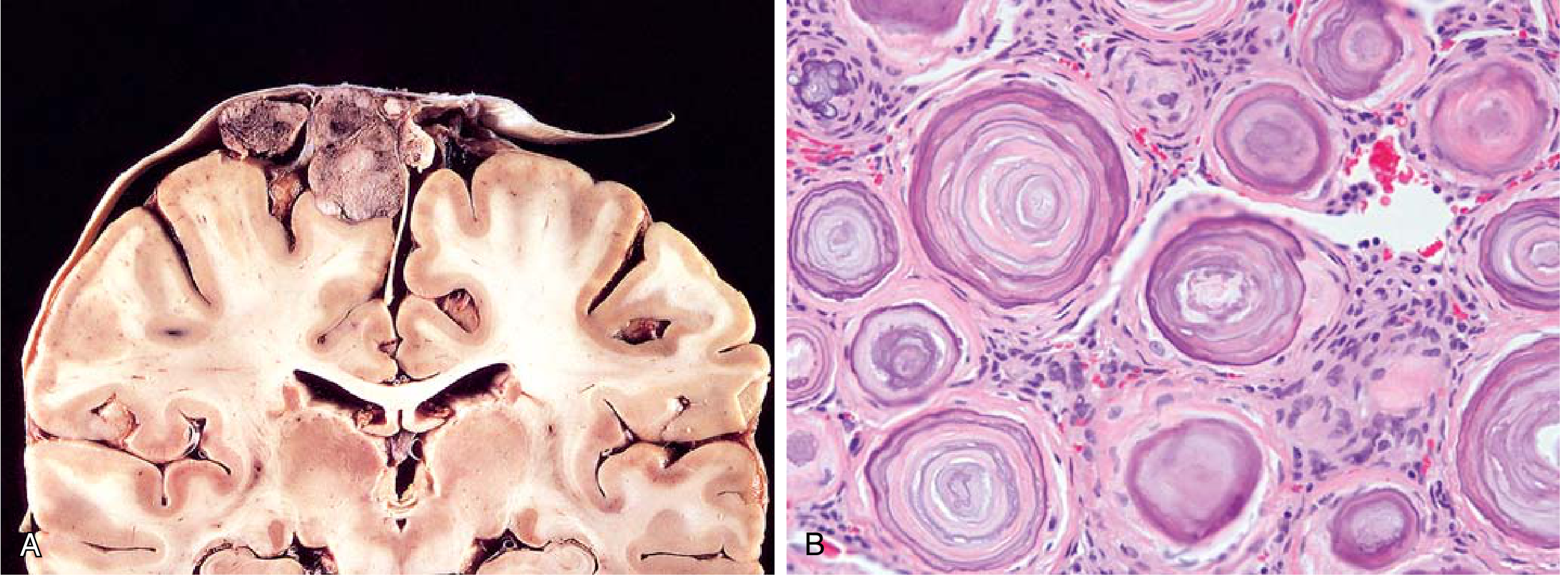
Most common primary brain tumor - arises from meningothelial (arachnoid cap) cells of the arachnoid, usually attached to the dura. Can grow anywhere along the external brain surface or inside ventricles (from choroid plexus stroma).
Meningioma: parasagittal multilobular mass attached to dura compressing underlying brain (A), and whorled pattern with classic psammoma body calcifications (B)
Meningioma. (A) Parasagittal multilobular meningioma attached to dura compressing the underlying brain. (B) Classic whorled pattern of meningothelial cells with concentric psammoma bodies - calcified "onion rings".
Psammoma bodies - the signature feature: Concentric laminated calcifications that look like layered onion rings under the microscope. They develop from progressive calcification of dead tumor cells within whorls. The word "psammoma" comes from Greek for "sandy" - feel the cut surface and it is literally gritty.
Meningioma histology: whorled meningothelial cells with abundant psammoma bodies
Radiation risk: Prior radiation to the head and neck (sometimes decades earlier) is a documented risk factor. A patient who had childhood head radiation is at higher meningioma risk 30-40 years later. - Robbins & Cotran, p. 1199
The most common mutation: NF2 gene mutations are found in many sporadic meningiomas. This is also the gene mutated in Neurofibromatosis type 2 (see familial syndromes below).
Histologic variants (9 benign subtypes plus anaplastic):
  • Syncytial/meningothelial: sheets of cells with indistinct borders
  • Fibrous: spindle cells in collagen bundles
  • Psammomatous: loaded with psammoma bodies
  • Secretory: contains gland-like structures
  • Anaplastic (grade 3): high mitotic activity, necrosis, brain invasion
The "good" tumor that can still kill: A parafalcine or parasagittal meningioma compresses the medial hemispheres. A tentorial meningioma can compress the midbrain. The cranial nerve foramen meningiomas (optic canal, cavernous sinus) cause progressive cranial nerve deficits without technically being "malignant."

PART 4: PRIMARY CNS LYMPHOMA - The Immune System Gone Wrong

The rarest situation that is paradoxically the most common tumor in immunosuppressed patients. Primary CNS lymphoma accounts for 2% of extranodal lymphomas and 1% of intracranial tumors - but in HIV/AIDS patients and transplant recipients, it becomes the most common CNS neoplasm. - Robbins & Cotran, p. 1199
The EBV connection: In immunosuppressed patients, the malignant B cells are latently infected by Epstein-Barr virus. EBV drives the proliferation. This is why AIDS-related CNS lymphoma is strongly EBV-associated - detectable by in situ hybridization for EBERs (small EBV-encoded RNAs).
In immunocompetent patients: Usually diffuse large B-cell lymphoma type, often with PDL1 gene amplification - the tumor hijacks the immune checkpoint to hide from T cells. Trials of checkpoint inhibitors are now ongoing for these patients.
Pattern of involvement: Single or multiple soft gray-white masses in deep subcortical/periventricular locations. The malignant cells cluster around blood vessels (angiocentric pattern) and express CD20. Necrosis is prominent in immunosuppressed cases.
Perverse behavior: Primary CNS lymphoma can involve the eye AND the brain, yet it almost never spreads to lymph nodes or bone marrow (rare, late complication). Conversely, systemic lymphoma rarely invades the brain parenchyma - it prefers the meninges and CSF. These two compartments do not talk to each other the way you might expect.

PART 5: METASTATIC TUMORS - The Most Common CNS Tumor Overall

Here is the fact that trips everyone up: Metastases are the most common tumors involving the CNS overall - more common than ALL primary CNS tumors combined. - Robbins & Cotran, p. 1200
The top 5 sources:
  1. Lung (most common - seeds hematogenously)
  2. Breast
  3. Melanoma (highest propensity to metastasize to brain per primary)
  4. Kidney (renal cell carcinoma)
  5. Colon
The gray-white junction trick: Metastases preferentially lodge at the gray-white matter junction - where blood vessel caliber suddenly narrows, trapping circulating tumor emboli. This creates sharply demarcated masses with surrounding edema and reactive gliosis. The sharp border is the opposite of GBM's infiltrative, indistinct margins.
Paraneoplastic syndromes: Beyond direct tumor compression, systemic cancers can damage the nervous system remotely through paraneoplastic syndromes - the tumor expresses antigens that trigger an immune response that attacks the brain. Patterns include:
  • Cerebellar degeneration (ataxia)
  • Limbic encephalitis (dementia, personality change)
  • Sensory neuropathy
Sometimes the neurological syndrome appears before the primary cancer is diagnosed. - Robbins Basic Pathology, p. 861

PART 6: FAMILIAL TUMOR SYNDROMES - Weird Genetics in Weird Tumors

Tuberous Sclerosis - Potato Tubers in the Brain

The name means "hard tubers" - knobby potato-like hamartomas in the cortex. Autosomal dominant, 1 in 5,000-10,000 births.
Two genes, same pathway:
  • TSC1 encodes hamartin
  • TSC2 encodes tuberin
  • Together they normally suppress mTOR (the cell's nutrient-sensing master growth regulator)
  • Loss of either protein → mTOR goes unchecked → cells keep growing regardless of nutrient status → hamartomas everywhere - Robbins Basic Pathology, p. 862
Clinical CHAOS from one pathway:
  • Brain: cortical tubers (seizures, often intractable), subependymal hamartomas
  • SEGA (subependymal giant cell astrocytoma): A real tumor, near the foramen of Monro - can cause sudden obstructive hydrocephalus. Treatable with mTOR inhibitors (everolimus) - a beautiful example of targeted molecular therapy
  • Kidney: angiomyolipomas
  • Lung: lymphangiomyomatosis (women)
  • Heart: cardiac rhabdomyomas (found prenatally on fetal echo)
  • Skin: ash leaf patches (hypopigmented, elliptical spots visible under Wood's lamp), shagreen patches (rough, leathery skin over lower back), angiofibromas on face, subungual fibromas
The classic exam teaching case: A neonate with a cardiac rhabdomyoma on echo - think tuberous sclerosis and check for brain tubers.

Von Hippel-Lindau (VHL) Disease - Erythropoietin Factory

Autosomal dominant. The VHL protein normally targets HIF-1alpha for destruction. Mutant VHL cannot do this → HIF-1alpha accumulates → drives VEGF expression → hemangioblastomas (tumors made of abnormal blood vessels). - Robbins Basic Pathology, p. 862
Tumor locations:
  • Cerebellar hemisphere hemangioblastomas (most common CNS site)
  • Retinal hemangioblastomas (early clue)
  • Brainstem, spinal cord
  • Renal cell carcinoma (clear cell type - VHL mutation is also the driver of sporadic renal cell carcinoma)
  • Pheochromocytoma
  • Pancreatic cysts
The weird polycythemia connection: Cerebellar hemangioblastomas produce erythropoietin. A patient with hemangioblastoma can develop polycythemia (excess red cells) from a brain tumor - the tumor acts as an ectopic kidney. When you remove the hemangioblastoma, the polycythemia resolves.

Neurofibromatosis Type 2 (NF2) - The Bilateral Acoustic Schwannoma Trap

NF2 mutation (on chromosome 22q12) causes bilateral vestibular schwannomas (old term: acoustic neuromas). Young adults presenting with bilateral hearing loss = NF2 until proven otherwise. Also associated with meningiomas and ependymomas. - Robbins & Cotran, p. 1201

PART 7: CHOROID PLEXUS TUMORS - The CSF Overproducers

Choroid plexus papillomas (grade 1) are intraventricular tumors that look just like normal choroid plexus - papillary fronds covered by epithelioid cells. They cause hydrocephalus by two mechanisms:
  1. Physically blocking CSF flow
  2. Overproducing CSF at a rate faster than it can be reabsorbed - a completely unique mechanism of hydrocephalus not seen with other tumors
Location age rule: lateral ventricle in children, 4th ventricle in adults. - Robbins & Cotran, p. 1198
Choroid plexus carcinoma (grade 3): Almost exclusively in young children, poorly differentiated, aggressive.

PART 8: NEURONAL TUMORS - When Neurons Go Wrong

Ganglioglioma - The Epilepsy Tumor

Mixed tumor: mature neurons + glial cells. Most common location: temporal lobe. Most common presentation: medically refractory epilepsy (years of uncontrolled seizures from this slow-growing benign tumor). Surgical resection often cures both the tumor AND the epilepsy simultaneously.
The neurons are bizarre: Irregularly clustered, randomly oriented processes, dysmorphic binucleate forms (neurons with two nuclei - something normal neurons almost never have). BRAF V600E mutation in 20-50% - potential target for vemurafenib-type therapy. - Robbins & Cotran, p. 1199

QUICK-FIRE WEIRD FACTS (Robbins)

FactTumor
"Fried egg" histologyOligodendroglioma (fixation artifact)
"Chicken wire" capillariesOligodendroglioma
"Pseudopalisading" necrosisGlioblastoma (GBM)
"Butterfly glioma"GBM crossing corpus callosum
"Ring enhancement" on MRIGBM (necrotic core + hypervascular rim)
Calcification in 90% of casesOligodendroglioma
Rosenthal fibers (glassy cigar-shaped inclusions)Pilocytic astrocytoma
Psammoma bodies (gritty calcifications)Meningioma (also papillary thyroid Ca, ovarian serous tumors)
Hair-like cells = "pilocytic"Pilocytic astrocytoma
Causes polycythemiaHemangioblastoma (VHL) - makes EPO
Most common brain tumor overallMetastases (not primary tumors!)
Most common PRIMARY brain tumorMeningioma
Most common malignant primary brain tumorGBM
Most common CNS tumor in AIDSPrimary CNS lymphoma (EBV-driven)
CSF seeding "drop metastases"Medulloblastoma
Nearly 100% 5-year survivalWNT-activated medulloblastoma
mTOR inhibitor works against itSEGA in tuberous sclerosis
MGMT methylation = chemo sensitivityGBM (temozolomide works better)
1p/19q codeletionOligodendroglioma (chemo/RT sensitive)
IDH mutation = oncometabolite 2-HGAll IDH-mutant gliomas
BRAF V600EPilocytic astrocytoma & ganglioglioma
Grows at gray-white junctionMetastases
Cyst may recur after surgery (not tumor)Pilocytic astrocytoma
Bilateral vestibular schwannomasNF2 (chromosome 22)
Ash leaf patches + seizuresTuberous sclerosis

The Oncology Exception: Brain Tumors Rarely Leave the Brain

Even the most malignant glioma - GBM - almost never metastasizes outside the CNS. The mechanism is partly the blood-brain barrier, partly the lack of lymphatics in the brain parenchyma. GBM cells are vicious infiltrators locally but horrible at traveling through the bloodstream. The rare cases of extracranial GBM metastases are documented curiosities in the literature. So the tumor kills by local destruction and herniation, not by spreading to distant organs. This is both the tragedy (you cannot simply "amputate" the brain) and a partial protection (the rest of the body stays relatively intact until the very end).

Sources

  • Robbins & Cotran Pathologic Basis of Disease, 10th Edition (ISBN 9780443264528), pp. 1193-1202
  • Robbins & Kumar Basic Pathology (ISBN 9780323790185), pp. 857-862
This is a shared conversation. Sign in to Orris to start your own chat.