
How a huamn eye work
The human eye is a complex optical organ that converts light into electrical signals the brain interprets as vision. Light enters through the cornea, passes through the pupil (regulated by the iris), is focused by the lens onto the retina, where photoreceptors (rods and cones) convert it to nerve impulses sent via the optic nerve to the brain.
Searching sources...
Reading File
Finding Sources
Searching the Web
how does the human eye work anatomy physiology vision
Finding Sources
Finding Sources
Finding Sources
Finding Sources
Finding Sources
Finding Sources
Excellent - found very relevant content in Guyton & Hall. Let me read the key sections.
Reading File
Reading File
Reading File
Now I have comprehensive information from Guyton & Hall. Let me fetch the key diagram images to include in my answer.
Loading Image
Loading Image
Loading Image
I now have everything needed for a thorough, well-illustrated answer. Here is the complete explanation:
How the Human Eye Works
Source: Guyton and Hall Textbook of Medical Physiology, 14th Ed.
1. Overview - The Eye as an Optical System
The eye works like a high-precision camera. Light from the outside world passes through a series of transparent structures that refract (bend) it and focus it onto a light-sensitive surface (the retina), where it is converted into nerve signals and sent to the brain for interpretation.
2. The Path of Light Through the Eye
Step 1 - The Cornea (The Main Lens)
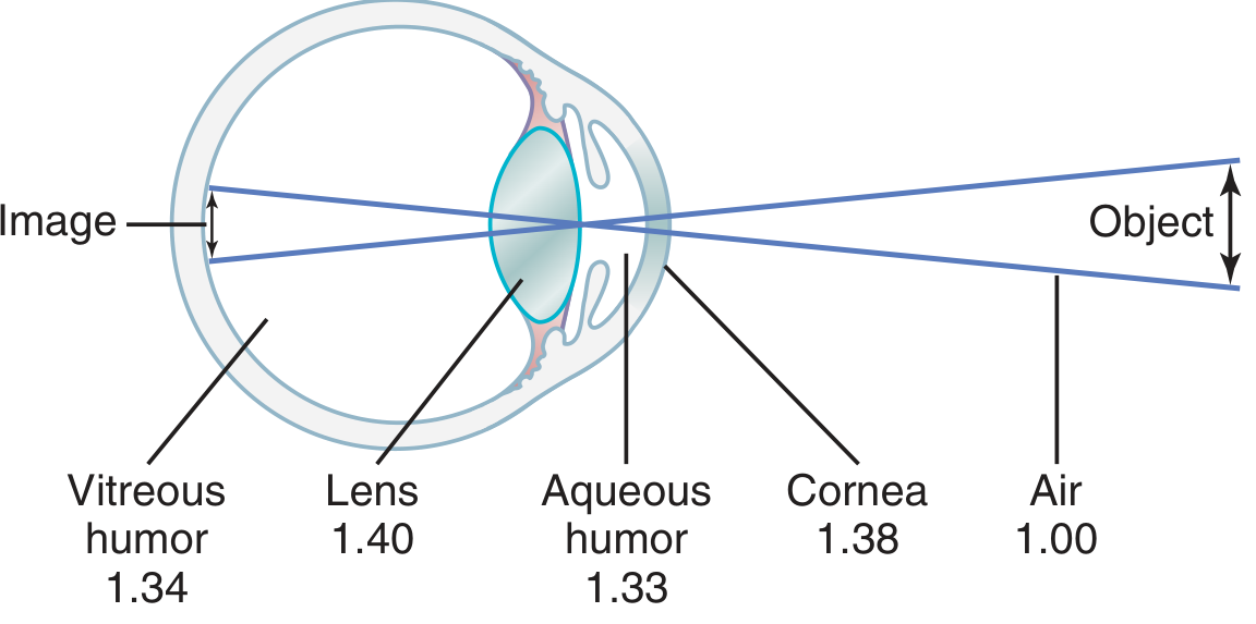
Light first strikes the cornea - the clear, dome-shaped outer surface. The cornea provides about two-thirds of the eye's total focusing power (~40 of the 59 diopters total). The reason it is so powerful is that the refractive index of the cornea (1.38) is markedly different from that of air (1.00), causing significant bending of incoming light rays.
Step 2 - The Pupil and Iris
Behind the cornea is the iris - the colored ring that acts like a camera aperture. The central opening, the pupil, widens (dilates) in dim light and narrows (constricts) in bright light to control how much light enters. This is controlled by smooth muscle in the iris via the autonomic nervous system.
Step 3 - The Crystalline Lens (Fine-Tuning Focus)
The lens sits behind the pupil and adds the remaining ~20 diopters of focusing power. Its unique feature is accommodation - it can change its shape to focus on objects at different distances:
- For distant objects: the ciliary muscles relax, suspensory ligaments (zonules) pull tight, and the lens becomes flatter (less curved)
- For near objects: the ciliary muscles contract, zonules loosen, and the lens becomes rounder (more curved), increasing its power
The total refractive power of the eye system sits at 59 diopters for distant vision (up to 70+ diopters when maximally accommodated for close objects).
Step 4 - The Aqueous and Vitreous Humors
Light then passes through:
- Aqueous humor (refractive index 1.33) - the watery fluid in front of the lens, which also nourishes the cornea
- Vitreous humor (refractive index 1.34) - the gel-like substance filling the large interior of the eyeball, maintaining its spherical shape
These fluids are optically transparent and contribute minor refractive power.
3. The Retina - Where Light Becomes a Signal
The retina is the innermost layer lining the back of the eyeball. Light hits the retina as a small, inverted image (upside-down and reversed, just like a camera). The brain automatically corrects for this.
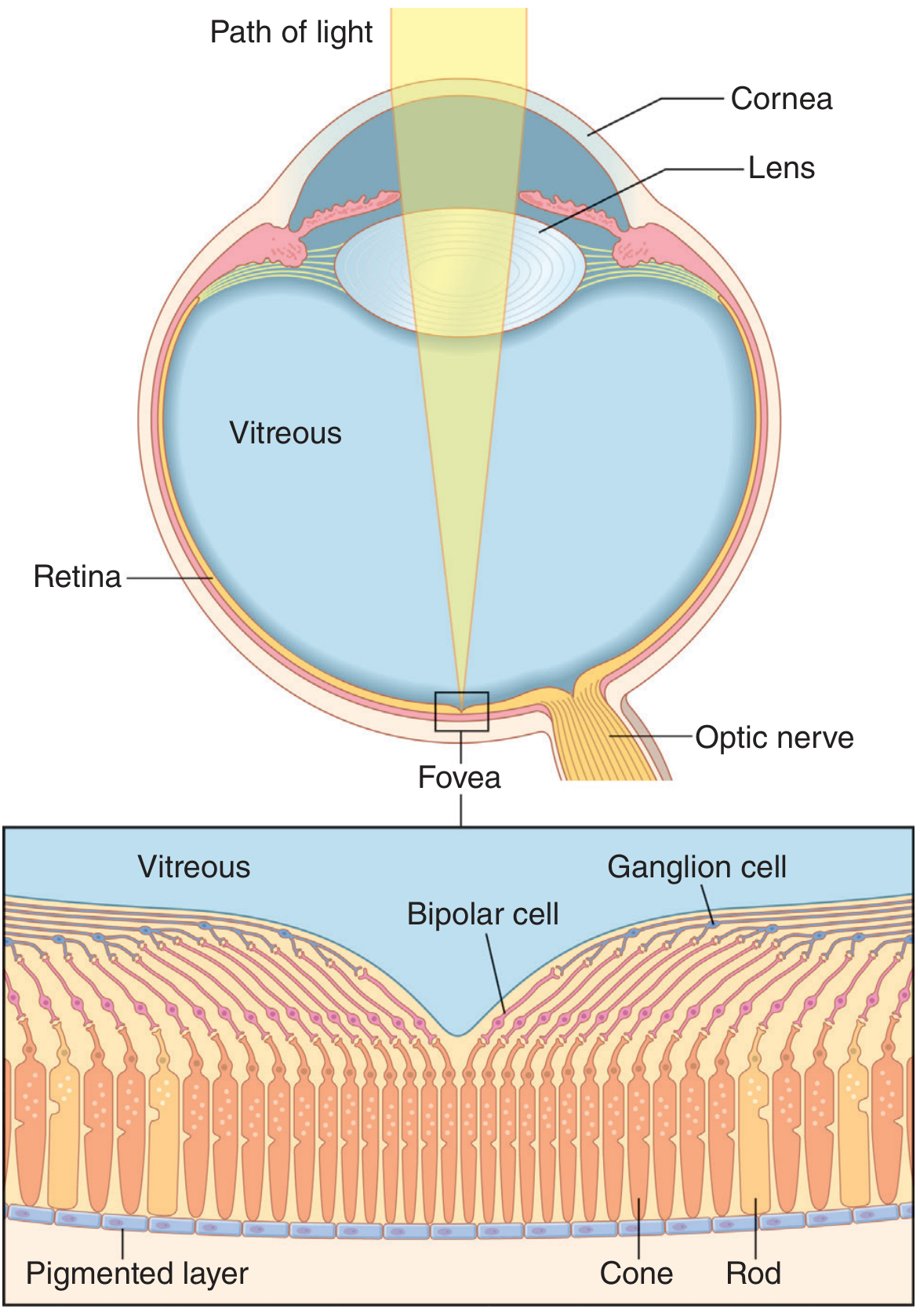
The retina contains 10 distinct layers. The key cell types are:
| Cell Type | Function |
|---|---|
| Rods (~120 million) | Detect dim light; black-and-white vision; peripheral vision |
| Cones (~6 million) | Color vision; fine detail; concentrated in the fovea |
| Bipolar cells | Relay signals from rods/cones inward |
| Ganglion cells | Collect signals and send them via the optic nerve |
| Horizontal & Amacrine cells | Lateral signal processing (contrast, edge detection) |
The Fovea - Your High-Definition Zone
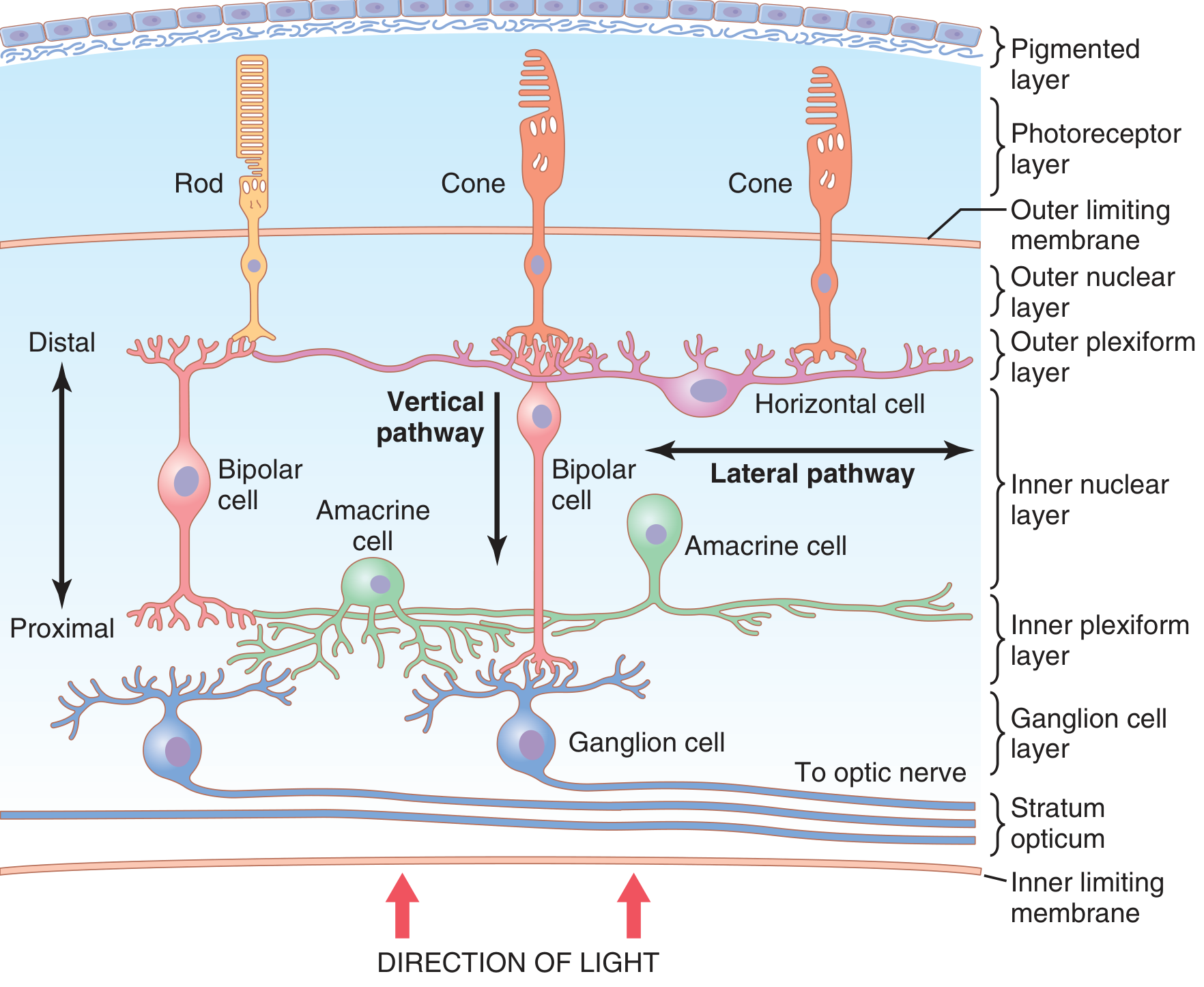
The fovea is a tiny pit (only ~0.3 mm in diameter) at the center of the retina. It contains almost exclusively cones, and the overlying cell layers are pushed aside so light hits the cones directly without obstruction. This is why you see fine detail best when you look directly at something.
Photochemistry - How Light is Detected
Inside each photoreceptor are visual pigments:
- Rods contain rhodopsin (retinal + opsin protein)
- Cones contain three types of photopsins, each sensitive to red, green, or blue wavelengths
When a photon of light hits a visual pigment molecule, retinal changes its shape (from 11-cis to all-trans), triggering a chemical cascade that hyperpolarizes the photoreceptor cell. This change in membrane potential is the signal that gets passed down the retinal layers.
4. From Retina to Brain
Signals flow through bipolar cells to ganglion cells. The axons of all ganglion cells bundle together to form the optic nerve, which exits the eye at the optic disc (the "blind spot" - no photoreceptors here).
The two optic nerves meet at the optic chiasm, where:
- Fibers from the nasal (inner) half of each retina cross to the opposite side
- Fibers from the temporal (outer) half stay on the same side
This means the left visual cortex processes the right visual field, and vice versa. Signals ultimately reach the primary visual cortex (V1) in the occipital lobe, where they are processed into the conscious experience of sight.
Quick Summary
Light → Cornea (bends light ~40D)
      → Pupil (controls amount of light)
      → Lens (fine-focuses ~20D, adjusts for distance)
      → Vitreous humor
      → Retina (rods & cones convert light to nerve signals)
      → Bipolar cells → Ganglion cells
      → Optic nerve → Optic chiasm
      → Visual cortex (brain sees the image)
For a great animated walkthrough, this video from Alila Medical Media covers the anatomy, retinal signal processing, and visual pathways:
This is a shared conversation. Sign in to Orris to start your own chat.