Can you explain me about oral cavity
Reading File
Finding Sources
Finding Sources
Searching Images
oral cavity anatomy diagram
Reading File
Reading File
Reading File
Reading File
Loading Image
Loading Image
Finding Sources
Finding Sources
Finding Sources
Finding Sources
Finding Sources
Reading File
Reading File
I now have comprehensive information to write a thorough answer. Let me compile it:
The Oral Cavity
Definition and Overview
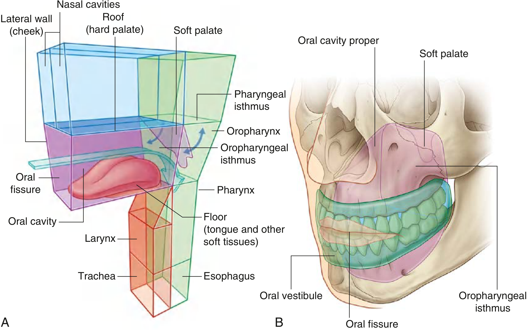
The oral cavity is the first part of the digestive tract. It lies inferior to the nasal cavities, opens onto the face through the oral fissure anteriorly, and communicates posteriorly with the pharynx through the oropharyngeal isthmus.
Boundaries
| Wall | Structure |
|---|---|
| Roof | Hard palate (anterior) + Soft palate (posterior) |
| Floor | Tongue + muscular diaphragm (mylohyoid muscle) |
| Lateral walls | Cheeks (buccinator muscle) |
| Anterior opening | Oral fissure (lips) |
| Posterior opening | Oropharyngeal isthmus |
Two Compartments
The dental arches (teeth + alveolar bone) divide the oral cavity into two regions:
- Oral Vestibule — the horseshoe-shaped space between the dental arches and the inner surface of the cheeks/lips. The oral fissure opens into it.
- Oral Cavity Proper — the space enclosed by the dental arches. The degree of opening between the arches is controlled by elevation/depression of the mandible at the temporomandibular joint (TMJ).
Functions
- Mastication: initial mechanical breakdown of food, aided by saliva
- Deglutition: initiates swallowing
- Speech: modifies sounds produced by the larynx
- Breathing: can serve as an accessory airway (shared pathway into the pharynx)
- Taste and sensation
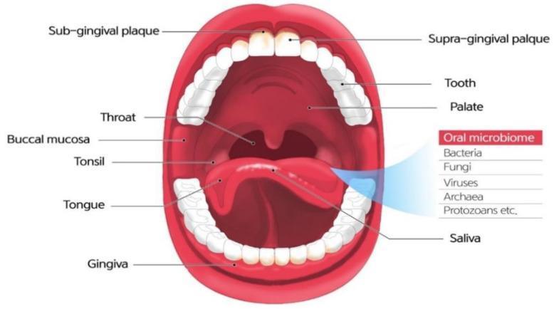
Key Structures
1. The Lips and Cheeks
- The lips surround the oral fissure; they are opened/closed by muscles of facial expression (orbicularis oris).
- The buccinator muscle forms the muscular core of the cheeks and keeps food between the teeth during chewing.
- The parotid (Stensen's) duct opens into the buccal mucosa opposite the upper 2nd molar tooth.
- Clinically important findings on the buccal mucosa:
- Fordyce spots — small yellow sebaceous glands (normal variant)
- Leukoplakia (white plaques that cannot be scraped off) vs. oral thrush (white plaques that can be scraped off)
- Erythroplakia — red lesion, high malignant potential → requires biopsy
2. The Palate
Hard palate (anterior 2/3)
- Formed by the palatine processes of the maxillae and the horizontal plates of the palatine bones.
- Covered by firmly bound mucoperiosteum.
- A midline bony overgrowth, torus palatinus, is a benign finding; midline location is reassuring, off-midline lesions require further evaluation.
Soft palate (posterior 1/3)
- A fibromuscular structure extending posteriorly from the hard palate.
- Ends in the uvula.
- Key muscles: tensor veli palatini, levator veli palatini, palatoglossus, palatopharyngeus, musculus uvulae.
- Closes the nasopharynx during swallowing and speech.
3. The Tongue
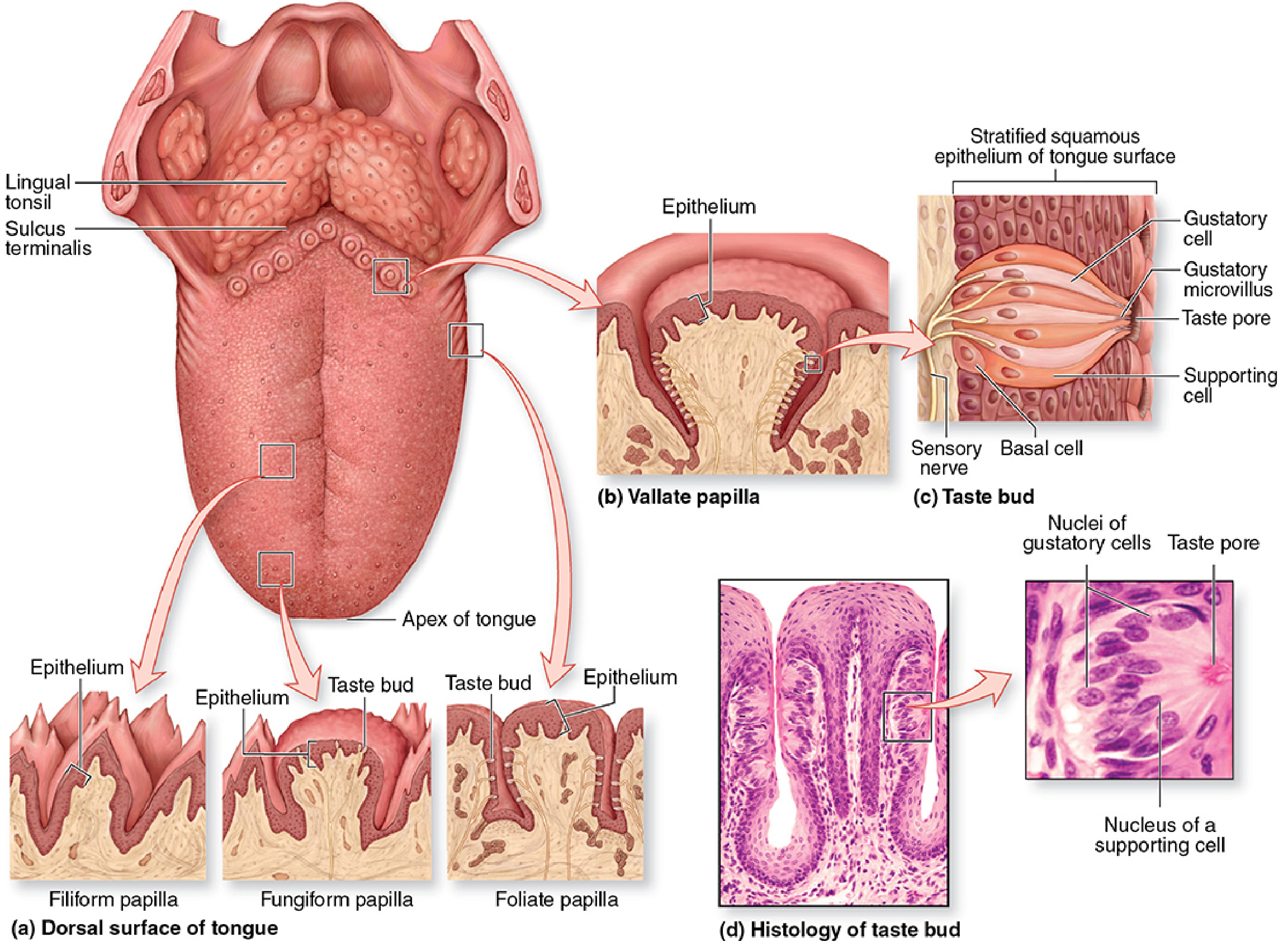
The tongue is a muscular structure forming the floor of the oral cavity and part of the anterior oropharyngeal wall. It is divided by the terminal sulcus (a V-shaped groove) into:
- Oral part (anterior 2/3) — lies in the oral cavity, horizontal orientation
- Pharyngeal part (posterior 1/3) — descends into the oropharynx; bears lymphoid nodules = lingual tonsil
The foramen cecum marks the apex of the terminal sulcus — the embryological site of thyroid gland descent.
Tongue Papillae (on dorsal surface of oral part)
| Type | Description | Taste Buds? |
|---|---|---|
| Filiform | Numerous, cone-shaped, keratinized; friction for food movement | ✗ No |
| Fungiform | Mushroom-shaped, scattered, less numerous | ✓ Yes |
| Vallate (circumvallate) | Largest (1–3 mm); 8–12 in a V-line just anterior to sulcus terminalis | ✓ Yes (most) |
| Foliate | Parallel ridges on lateral tongue; rudimentary in adults | ✓ Yes |
Tongue Muscles
- Intrinsic muscles (superior/inferior longitudinal, transverse, vertical) — change shape
- Extrinsic muscles — move the tongue as a whole:
- Genioglossus (protrudes tongue; CN XII)
- Hyoglossus (depresses)
- Styloglossus (retracts/elevates)
- Palatoglossus (elevates posterior tongue / closes oropharyngeal isthmus)
- All tongue muscles are innervated by CN XII (hypoglossal nerve) except palatoglossus (CN X via pharyngeal plexus).
Undersurface of Tongue
- Frenulum linguae — midline mucosal fold tethering tongue to floor of mouth
- Wharton's ducts (submandibular gland) open on either side of the frenulum
4. Floor of the Mouth
- Formed by the mylohyoid muscle (muscular diaphragm), spanning between the two sides of the mandible.
- Covered by mucosa continuous with the tongue undersurface.
- Houses the sublingual glands and portions of the submandibular gland/duct.
5. Teeth and Alveolar Ridges
- Adults have 32 permanent teeth (8 incisors, 4 canines, 8 premolars, 12 molars including 4 wisdom teeth).
- Each tooth consists of crown (enamel-covered), neck (at gingival margin), and root (embedded in alveolar bone).
- The retromolar trigone is the mucosa posterior to the last lower molar — an important site for early asymptomatic oral cancers.
Innervation
| Region | Nerve |
|---|---|
| Upper palate, upper teeth | CN V2 (maxillary nerve) branches |
| Lower teeth, floor of mouth | CN V3 (mandibular nerve) branches (lingual + inferior alveolar nerves) |
| Taste — anterior 2/3 tongue | Chorda tympani (CN VII) → travels with lingual nerve (CN V3) |
| Taste — posterior 1/3 tongue | CN IX (glossopharyngeal nerve) |
| General sensation — posterior tongue/pharynx | CN IX |
| All tongue muscles | CN XII (except palatoglossus → CN X) |
| Parasympathetic (salivary glands) | CN VII (submandibular, sublingual via chorda tympani → submandibular ganglion); CN IX (parotid via otic ganglion) |
Salivary Glands
Three major pairs open into the oral cavity:
| Gland | Duct | Opens at | Secretion |
|---|---|---|---|
| Parotid | Stensen's duct | Opposite upper 2nd molar | Serous |
| Submandibular | Wharton's duct | Floor of mouth, sublingual papilla | Mixed (mostly serous) |
| Sublingual | Multiple small ducts | Floor of mouth | Mucous |
Saliva functions: lubrication, amylase (begins starch digestion), antimicrobial proteins (IgA, lysozyme), buffering, and facilitation of taste.
Blood Supply
- Upper oral cavity: branches of the maxillary artery (greater palatine, sphenopalatine)
- Tongue: lingual artery (from external carotid), with contributions from dorsal lingual branches
- Lower oral cavity/floor: lingual artery, submental branch of facial artery
- Venous drainage → lingual veins and facial veins → internal jugular vein
Lymphatic Drainage
- Tip of tongue → submental nodes
- Lateral tongue, floor of mouth → submandibular nodes → deep cervical chain
- Posterior tongue → jugulodigastric nodes (most important nodal station for oral cancer spread)
Clinical Highlights
Common Pathology
| Condition | Features |
|---|---|
| Aphthous ulcers (canker sores) | Painful white ulcers on any mucosa; most common on buccal membrane |
| Leukoplakia | White patch not removable; precancerous |
| Erythroplakia | Red patch; higher malignant potential than leukoplakia |
| Oral candidiasis | White plaques that can be scraped off; common in immunocompromised (HIV, transplant patients) |
| Mucocele | Smooth submucosal nodule at lip/cheek; mucus retention cyst |
| Torus palatinus | Benign midline bony protuberance on hard palate |
| Angular stomatitis (cheilosis) | Fissuring at oral commissures |
Oral Cavity Cancer
- Most common type: squamous cell carcinoma (SCC)
- Risk factors: tobacco (smoked or smokeless), alcohol, areca nut (betel quid), poor dentition, HPV (less so for oral cavity than oropharynx)
- Common sites: lateral tongue, floor of mouth
- The retromolar trigone and floor of mouth are high-risk sites that can harbor asymptomatic early lesions
Clinical Examination Protocol
(Cummings Otolaryngology)
- Inspect lips and oral commissures
- Assess mouth opening (trismus = limited opening → concerning for deep space infection or tumor)
- Examine teeth, gingiva, occlusion
- Inspect retromolar trigones bilaterally
- Dorsal, ventral, and lateral tongue surfaces (bimanual palpation of floor of mouth)
- Buccal mucosa (note Stensen's duct)
- Hard palate (note torus palatinus if present)
- All red lesions and most white lesions should be biopsied
Sources:
- Gray's Anatomy for Students, Chapter 8 (Head & Neck), p. 1227–1235
- Cummings Otolaryngology: Head and Neck Surgery, p. 85–86
- Junqueira's Basic Histology: Text and Atlas, 17e, p. 738–740
This is a shared conversation. Sign in to Orris to start your own chat.