Hi
Hi! How can I help you today?
Olfactory nerve pathway
Reading File
Finding Sources
Searching the Web
olfactory nerve pathway anatomy cranial nerve I
Finding Sources
Reading Web Page
https://teachmeanatomy.info/head/cranial-nerves/olfactory-cn…
Reading File
Reading File
Loading Image
Reading File
Here is a complete breakdown of the olfactory nerve pathway (CN I):
Olfactory Nerve Pathway (CN I)
CN I is a special visceral afferent nerve - the shortest cranial nerve. Uniquely, it is the only sensory pathway that does NOT relay through the thalamus before reaching the primary cortex.
Step 1 - Olfactory Epithelium (1st-order neurons)
- Located in the roof of the nasal cavity (lateral and septal walls)
- Composed of pseudostratified columnar epithelium containing three cell types:
- Olfactory receptor neurons (bipolar neurons) - the actual sensory cells; each has cilia bearing G protein-coupled receptor proteins (>1000 types) that bind odorant molecules
- Sustentacular cells - supporting cells
- Basal cells - stem cells that regenerate receptor neurons throughout life
- Bowman's glands secrete mucus to dissolve odorant molecules
Transduction: Odorant → receptor protein → Golf (G protein) → adenylyl cyclase → cAMP↑ → cation channels open (Na+, K+, Ca2+) → depolarization → action potential
Step 2 - Cribriform Plate
- Axons of olfactory receptor neurons (unmyelinated, called fila olfactoria) bundle together
- They pass through the foramina of the cribriform plate of the ethmoid bone to enter the anterior cranial fossa
Step 3 - Olfactory Bulb (2nd-order neurons)
- Axons synapse onto mitral cells in structures called glomeruli
- ~1000 olfactory receptor axons converge onto 1 mitral cell (massive convergence)
- Also contains periglomerular cells and granule cells (interneurons for lateral inhibition and processing)
- The olfactory bulb lies in the olfactory groove of the anterior cranial fossa
Step 4 - Olfactory Tract
- Mitral cell axons travel posteriorly along the inferior surface of the frontal lobe as the olfactory tract
- At the anterior perforated substance (near the optic chiasm), the tract divides into two striae:
| Stria | Destination | Function |
|---|---|---|
| Lateral olfactory stria | Primary olfactory cortex (piriform cortex), olfactory tubercle, amygdala | Conscious smell perception and emotional/memory associations |
| Medial olfactory stria | Septal area (via diagonal band); crosses midline via anterior commissure to contralateral olfactory bulb | Inhibits contralateral mitral cells; enhances localization of smell |
Step 5 - Primary Olfactory Cortex (no thalamic relay!)
- Piriform (prepiriform) cortex - main conscious olfactory perception
- Amygdala - emotional responses to smell, fear/memory associations
- Olfactory tubercle - integration
- Entorhinal cortex - connects to hippocampus for memory
Step 6 - Secondary Connections
- The medial forebrain bundle (MFB) connects olfactory cortex to the hypothalamus and brainstem
- Regulates autonomic responses: salivation, gastric contraction, arousal via the reticular formation
Key Distinguishing Features
| Feature | Detail |
|---|---|
| Fiber type | Unmyelinated (slow conduction) |
| No thalamic relay | Direct cortical projection - unique among sensory systems |
| Embryological origin | Olfactory placode (CNS-derived, not PNS) |
| Regeneration | Olfactory receptor neurons can regenerate from basal cells |
Clinical Correlate - Anosmia
- Loss of smell (anosmia) most commonly results from: head trauma shearing the fila olfactoria at the cribriform plate, viral infections (e.g., post-COVID), nasal polyps, or anterior cranial fossa tumors (e.g., olfactory groove meningioma)
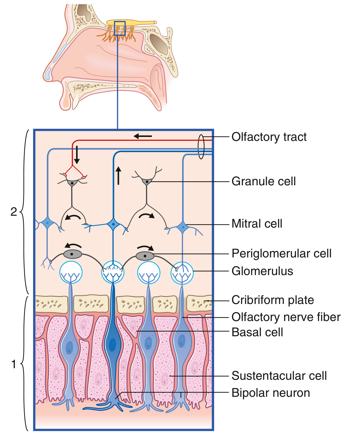
Cellular connections of the olfactory epithelium - Gray's Anatomy for Students
Sources: Costanzo Physiology 7th Ed., p. 105-106 | Gray's Anatomy for Students, p. 1360
I am asking simple but accurate pathway scheme
Olfactory Nerve Pathway
Odorant molecules
        ↓
Olfactory receptor neurons (bipolar cells)
— in olfactory epithelium (roof of nasal cavity)
        ↓
Fila olfactoria (unmyelinated axons)
— pierce cribriform plate of ethmoid bone
        ↓
Olfactory bulb → synapse on Mitral cells (glomeruli)
— in anterior cranial fossa
        ↓
Olfactory tract
— runs on inferior surface of frontal lobe
        ↓
        ├── Lateral stria ──────────────────────────────→ Piriform cortex (primary olfactory cortex)
        │                                                  Amygdala, Olfactory tubercle
        │
        └── Medial stria ──→ Septal nuclei
                           → Anterior commissure → Contralateral olfactory bulb
Key points to remember:
- 1st neuron: Bipolar receptor cell (nasal epithelium)
- 2nd neuron: Mitral cell (olfactory bulb)
- No thalamic relay - unique among all sensory pathways
- Fibers are unmyelinated
- Olfactory receptor neurons are CNS-derived and can regenerate
This is a shared conversation. Sign in to Orris to start your own chat.