What causes S wave in ecg in V1 and in V5

Reading File
Reading File
Finding Sources
Searching Images

ECG normal precordial lead progression V1 V5 R wave S wave diagram

Searching PubMed

electrocardiography AND precordial leads

Searching the Web

ECG precordial leads V1 V5 normal R wave S wave progression explanation

An S wave is a negative QRS deflection that occurs after an R wave. It appears when ventricular depolarization is moving away from the positive electrode of that lead.
  • In V1: The main ventricular activation is dominated by the much larger left ventricle and travels leftward and posteriorly, away from the right-anterior V1 electrode. Therefore V1 normally shows a small r wave followed by a deep S wave.
  • In V5: V5 lies over the left lateral chest, so the main leftward ventricular activation moves toward it, producing a large R wave. A small S wave can still occur because the last areas to depolarize, especially basal/posterior ventricular regions, have a vector directed away from V5. Thus, in a normal ECG, V5 usually has a tall R with only a small terminal S.
This is normal R-wave progression: from V1 to V5, R waves generally become taller while S waves become smaller. The ECG QRS explanation describes this leftward dominant ventricular vector and the small terminal vector causing the V5 S wave.
A deep S in V1 is usually normal. A prominent or persistent S in V5/V6, particularly if R/S is less than 1, is not typical and can reflect delayed rightward/posterior terminal forces, such as right bundle-branch block, right ventricular hypertrophy, lung disease, or occasionally incorrect chest-lead placement.

Why does left bundle branch block cause leftward axis in ecg?

Reading File
Finding Sources
Searching Images

left bundle branch block ECG diagram QRS vectors limb leads axis deviation

Searching PubMed

left bundle branch block electrocardiography

Searching the Web

left bundle branch block left axis deviation why delayed left ventricular activation ECG vector

Reading File
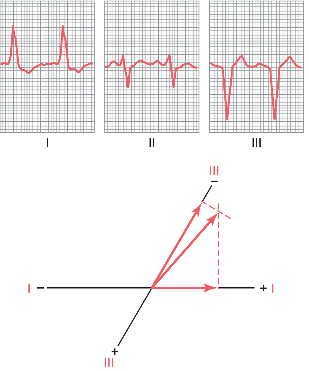
Left bundle branch block (LBBB) can cause a leftward QRS axis because left-ventricular activation is delayed. But an important correction: isolated LBBB does not always produce left-axis deviation. The frontal QRS axis may be normal or leftward. Marked left-axis deviation often suggests associated left anterior fascicular block or underlying structural heart disease.

Mechanism

Normally, both ventricles depolarize nearly simultaneously through their bundle branches, so many opposing electrical forces partly cancel.
In LBBB:
  1. The impulse travels down the right bundle branch, activating the right ventricle first.
  2. It then spreads slowly, cell-to-cell, across the septum to activate the left ventricle.
  3. For much of the QRS, the right ventricle is already depolarized while a large part of the left ventricle is still depolarizing.
  4. The net, later and larger electrical force points toward the left ventricle. In the frontal plane this may be directed leftward, causing left-axis deviation.
This same delayed right-to-left activation explains the typical LBBB pattern:
  • Broad/notched R waves in lateral leads I, aVL, V5-V6
  • Deep broad S or QS complexes in V1-V3
  • Wide QRS, usually at least 120 ms
Guyton describes the vector as pointing from the already-depolarized right ventricle toward the still-activating left ventricle, which can produce an axis around -50°. Guyton and Hall Textbook of Medical Physiology, p. 157.
Vector diagram of left-axis deviation caused by LBBB
Key distinction: LBBB primarily creates a right-to-left activation sequence, most obvious in the horizontal/precordial leads. A strongly superior-left frontal axis is more characteristic when LBBB coexists with left anterior fascicular block, because activation is then relatively directed inferior-to-superior as well.

I didnt understand, if there is left bundle branch block then shouldnt the qrs vector should be directed towards the right

Reading File
Finding Sources
Searching PubMed

left bundle branch block electrocardiography

Searching the Web

LBBB vector why leftward net QRS vector right ventricle activates first explanation

You are thinking about it correctly at first: the impulse begins in the right ventricle, so the very early activation is toward the right.
But the ECG QRS axis reflects the net (average) vector during the entire ventricular depolarization, not just where activation starts.

In LBBB, sequence is:

  1. Right ventricle depolarizes first through the intact right bundle.
    • This produces a small, early rightward force.
  2. The large left ventricle is still not depolarized because its bundle is blocked.
  3. The impulse then slowly spreads from the RV across the septum into the LV.
    • The advancing depolarization front now travels right to left.
    • Since the LV has much more muscle mass than the RV, this delayed LV activation creates the dominant QRS force.
So the larger, longer-lasting vector is directed toward the left, not right.
A useful way to picture it:
  • The RV is already electrically “finished.”
  • The LV is the big area that remains to be activated.
  • Therefore, most of the electrical activity recorded during the widened QRS is the wave moving into the LV, from right to left.
This is why LBBB produces:
  • Deep negative QRS in V1 on the right chest, because the major force moves away from V1
  • Broad positive R wave in I, aVL, V5, and V6, because the force moves toward the left-lateral leads
Important correction: isolated LBBB does not necessarily cause frontal-plane left-axis deviation. It mainly causes a major right-to-left horizontal-plane vector. The frontal axis can be normal or leftward; marked left-axis deviation often implies associated left anterior fascicular block.
The physiology text describes the same idea in charge terms: while the RV is already depolarized, much of the LV remains to be depolarized, producing a net vector toward the LV. Guyton and Hall Textbook of Medical Physiology, p. 157. The LBBB electrophysiology explanation also illustrates this right-to-left septal and LV activation sequence.

Is P pulmonale is usually lead 2, 3 and AVF?

Reading File
Finding Sources
Searching PubMed

P pulmonale electrocardiography

Searching the Web

P pulmonale ECG peaked P waves lead II III aVF criteria

Yes. P pulmonale is best seen in the inferior leads: II, III, and aVF, because right atrial depolarization is directed inferiorly toward these leads.
ECG criterion:
  • Tall, narrow, peaked P wave > 2.5 mm (0.25 mV) in lead II.
  • It may also be prominent in III and aVF.
  • In V1, a positive P-wave component > 1.5 mm can support right atrial enlargement.
“P pulmonale” is the ECG pattern of right atrial enlargement, commonly associated with pulmonary hypertension, chronic lung disease/cor pulmonale, pulmonary embolism, or some congenital heart diseases. It is an ECG clue, not by itself a definitive diagnosis of right atrial enlargement.
Rosen’s Emergency Medicine states the usual description as peaked P waves >2.5 mm in leads II, III, and aVF.
This is a shared conversation. Sign in to Orris to start your own chat.