
What is headache? Uh, in the, in, uh, high secretion of HCl, uh, I mean, uh, there is a-- sometimes there is a headache with the acidity condition, like physiological answer
physiological mechanism headache high gastric acid secretion HCl acidity
why does acidity cause headache gut brain axis vagus nerve histamine mechanism physiology
Headache Associated with High HCl (Gastric Acid Hypersecretion) - Physiological Explanation
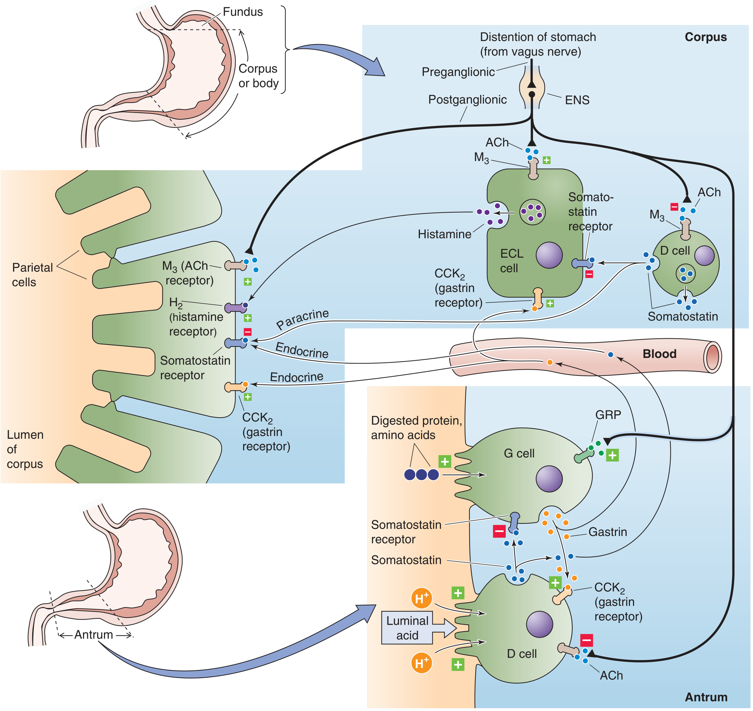
1. Histamine - The Central Link
- H2 receptors on parietal cells - stimulating HCl secretion (gastric effect)
- H1 receptors on cranial blood vessels - causing vasodilation of cerebral vessels
2. The Gut-Brain Axis via the Vagus Nerve
- Afferent vagal fibers detect the chemical disturbance
- They transmit signals to the brainstem (nucleus tractus solitarius)
- This triggers autonomic responses including changes in vascular tone, nausea, and pain referral - which can manifest as a headache
3. Alkaline Tide Effect
- Slight cerebral vasoconstriction/vasodilation shifts
- Hypocapnia-like effects
- Contributes to the sense of fullness, light-headedness, and headache
4. Referred Pain / Visceral Hypersensitivity
5. Serotonin Pathway
Summary Table
| Mechanism | How it causes headache |
|---|---|
| Excess histamine (from ECL cells) | H1-mediated cranial vasodilation + nitric oxide release |
| Vagus nerve afferents | Gut signals reach brainstem, trigger vascular/pain responses |
| Alkaline tide (HCO3- release) | Transient blood alkalosis alters cerebrovascular tone |
| Esophageal acid irritation | Referred pain via visceral afferent pathways |
| Gut serotonin release | Activates trigeminovascular system |
Types of genes
types of genes classification structural regulatory housekeeping oncogenes genetics
Types of Genes
1. Based on What the Gene Encodes
A. Protein-Coding Genes
- Enzymes
- Structural proteins
- Receptor proteins
- Regulatory proteins (transcription factors, signaling molecules)
B. Non-Coding RNA (ncRNA) Genes
| ncRNA Type | Size | Function |
|---|---|---|
| rRNA (ribosomal RNA) | Large | Structural/catalytic core of ribosomes |
| tRNA (transfer RNA) | ~70-90 nt | Carries amino acids during translation |
| miRNA (microRNA) | ~22 bases | Silences target mRNAs; controls ~30% of all protein-coding genes |
| lncRNA (long ncRNA) | >200 nt | Gene regulation, gene silencing, X-chromosome dosage |
| snoRNA (small nucleolar RNA) | Small | Modifies rRNA; deletions cause Prader-Willi syndrome |
| snRNA (small nuclear RNA) | Small | Controls RNA splicing |
- Thompson & Thompson Genetics and Genomics in Medicine, 9th Ed
2. Based on Function / Expression Pattern
A. Structural Genes
B. Regulatory Genes
C. Housekeeping Genes
D. Tissue-Specific (Luxury) Genes
E. Inducible Genes
3. Based on Role in Cancer (Clinically Important)
A. Proto-Oncogenes
- Normal genes that regulate cell growth and proliferation
- When mutated or overexpressed, they become oncogenes
- Their protein products include: growth factors, growth factor receptors, signal transduction proteins (RAS), transcription factors (MYC)
- Mutations cause gain-of-function - only one allele needs to be mutated (dominant)
- Example: RAS proto-oncogene → mutant ras oncogene (found in lung, colon, bladder cancers)
B. Oncogenes
- Mutated forms of proto-oncogenes that drive uncontrolled cell proliferation
- Activation mechanisms: point mutation, gene amplification, chromosomal translocation
- "A transforming mutation in a proto-oncogene increases the activity or amount of the gene product - a gain-of-function mutation." - Basic Medical Biochemistry, 6th Ed
C. Tumor Suppressor Genes
- Normal genes that suppress cell proliferation and promote apoptosis
- Both alleles must be inactivated for cancer to result ("two-hit hypothesis")
- Loss-of-function mutations
- Examples: TP53 (p53), RB (retinoblastoma), APC, BRCA1/2
- "Tumor-suppressor genes contribute to the development of cancer when both copies of the gene are inactivated." - Basic Medical Biochemistry, 6th Ed
D. DNA Repair Genes (Caretaker Genes)
- Maintain genomic integrity by repairing damaged DNA
- Loss leads to increased mutation rate throughout the genome
- Examples: MLH1, MSH2 (mismatch repair genes - mutated in Lynch syndrome), BRCA1/2
4. Based on Inheritance Pattern
| Gene Type | Pattern |
|---|---|
| Autosomal dominant | One mutant allele causes disease (e.g., FGFR3 in achondroplasia) |
| Autosomal recessive | Both alleles must be mutant (e.g., CFTR in cystic fibrosis) |
| X-linked | Gene on X chromosome (e.g., F8 in hemophilia A) |
| Mitochondrial | Encoded by mitochondrial DNA; maternally inherited |
| Imprinted | Only the maternal or only the paternal copy is expressed (e.g., IGF2) |
5. Based on Gene Family / Evolutionary Relationship
- Multigene families: Groups of genes with similar sequences and related functions (e.g., globin gene family - α-globin and β-globin clusters)
- Pseudogenes: Non-functional gene copies that have lost their ability to be expressed (due to accumulated mutations)
- Paralogues: Genes related by duplication within the same species
- Orthologues: Genes in different species that evolved from a common ancestor
Summary Diagram (Conceptual)
GENES
├── By product
│   ├── Protein-coding (~20,000 genes)
│   └── Non-coding RNA (rRNA, tRNA, miRNA, lncRNA, snoRNA)
├── By expression
│   ├── Housekeeping (constitutive)
│   ├── Tissue-specific (luxury)
│   └── Inducible
├── By cancer role
│   ├── Proto-oncogenes → Oncogenes (gain-of-function)
│   ├── Tumor suppressor genes (loss-of-function)
│   └── DNA repair / caretaker genes
└── By inheritance
    ├── Autosomal dominant / recessive
    ├── X-linked
    ├── Mitochondrial
    └── Imprinted
- Thompson & Thompson Genetics and Genomics in Medicine, 9th Ed - gene structure, ncRNA types
- Basic Medical Biochemistry (Lippincott), 6th Ed - oncogenes, tumor suppressors
- Harper's Illustrated Biochemistry, 32nd Ed - housekeeping/constitutive genes
- Robbins & Kumar Basic Pathology - protein-coding vs non-coding DNA
Monogenic
monogenic disorders definition types autosomal dominant recessive X-linked examples genetics
Monogenic Disorders
Definition
"Single-gene or monogenic disorders result from mutations that have large functional effects and manifest in families with Mendelian inheritance patterns."
- Robbins, Cotran & Kumar Pathologic Basis of Disease
Types of Monogenic (Single-Gene) Disorders by Inheritance Pattern
1. Autosomal Dominant (AD)
- Mutation in one allele is sufficient to cause disease (heterozygous state)
- Both males and females are equally affected and can transmit
- Each child of an affected parent has a 50% chance of inheriting the disease
- Typically shows vertical transmission across generations
| Mechanism | Explanation | Example |
|---|---|---|
| Haploinsufficiency | One normal allele cannot produce enough protein | Familial hypercholesterolemia (LDL receptor) |
| Dominant negative | Mutant protein interferes with normal protein (esp. structural proteins) | Osteogenesis imperfecta (collagen) |
| Gain-of-function | Mutant protein has new or excessive activity | Achondroplasia (FGFR3), Huntington disease |
- Incomplete penetrance: some people carry the gene but show no symptoms (e.g., 50% penetrance = only 50% of carriers are affected)
- Variable expressivity: all carriers are affected, but to different degrees (e.g., neurofibromatosis type 1 - ranges from skin spots to tumors)
- De novo mutations: new germline mutations can cause AD disease with no family history, especially with advanced paternal age
- Huntington disease (HTT gene - CAG repeat expansion)
- Marfan syndrome (FBN1 gene - fibrillin)
- Familial hypercholesterolemia (LDLR gene)
- Neurofibromatosis type 1 (NF1 gene)
- Achondroplasia (FGFR3 gene)
2. Autosomal Recessive (AR)
- Both alleles must be mutated for disease to manifest
- Parents are typically unaffected carriers (heterozygotes)
- Risk in offspring of two carriers: 25% affected, 50% carrier, 25% normal
- Often appears horizontally in a pedigree (siblings affected, parents not)
- More common with consanguinity
| Disease | Gene | Defect |
|---|---|---|
| Cystic fibrosis | CFTR | Cl- channel dysfunction |
| Sickle cell anemia | HBB | β-globin point mutation (Glu→Val) |
| Tay-Sachs | HEXA | Hexosaminidase A deficiency |
| Phenylketonuria (PKU) | PAH | Phenylalanine hydroxylase deficiency |
| Wilson's disease | ATP7B | Copper transport defect |
3. X-Linked Recessive (XLR)
- Gene is on the X chromosome; males (XY) are hemizygous - one mutant copy is enough to cause disease
- Females (XX) are usually carriers - protected by their second normal X allele
- No father-to-son transmission (fathers give Y to sons)
- Affected males transmit the carrier state to all daughters, but to no sons
| System | Disease |
|---|---|
| Musculoskeletal | Duchenne muscular dystrophy (DMD) |
| Hematopoietic | Hemophilia A (F8), Hemophilia B (F9) |
| Hematopoietic | G6PD deficiency |
| Immune | Agammaglobulinemia, Wiskott-Aldrich syndrome |
| Metabolic | Lesch-Nyhan syndrome, Diabetes insipidus |
| Nervous | Fragile X syndrome |
- Robbins, Cotran & Kumar Pathologic Basis of Disease
4. X-Linked Dominant (XLD)
- Mutation on X chromosome causes disease in both males and females in heterozygous state
- Affected father → all daughters affected, no sons affected
- Affected mother → 50% daughters and 50% sons affected
- Pedigree resembles AD but with no father-to-son transmission
- Vitamin D-resistant rickets (X-linked hypophosphatemia)
- Alport syndrome (some forms)
- Rett syndrome (MECP2 - lethal in males, affects girls)
5. Mitochondrial Inheritance
- Genes encoded in mitochondrial DNA (mtDNA)
- Transmitted exclusively through the mother (maternal inheritance) - only egg contributes mitochondria
- Affects both sexes, but an affected father CANNOT pass it to children
- Affects tissues with high energy demand - muscle, brain, eye
- MELAS (mitochondrial encephalomyopathy, lactic acidosis)
- Leber's hereditary optic neuropathy (LHON)
- MERRF (myoclonic epilepsy with ragged-red fibers)
Summary: Key Distinguishing Features
| Feature | AD | AR | XLR | XLD | Mitochondrial |
|---|---|---|---|---|---|
| Alleles needed | 1 | 2 | 1 (in males) | 1 | Mutation in mtDNA |
| Males vs females | Equal | Equal | Males >> Females | Both affected | Both (maternal line) |
| Carrier state | No (usually affected) | Yes | Females can be carriers | No | No |
| Father to son | Yes | Yes | No | No | No |
| Maternal inheritance | No | No | No | No | Yes (exclusively) |
Related Concepts
- Codominance: Both alleles contribute to phenotype (e.g., ABO blood groups, HbAS in sickle cell trait)
- Anticipation: Severity increases / onset decreases in successive generations due to expanding trinucleotide repeats (e.g., Huntington, Myotonic dystrophy)
- Imprinting: Only one parental allele is expressed; disruption causes disease depending on which parent's copy is affected (e.g., Prader-Willi vs. Angelman syndrome, both involve chromosome 15)
Types of genes
Types of Genes
1. Based on What the Gene Encodes
A. Protein-Coding Genes
- Transcribed into mRNA → translated into protein
- ~20,000 protein-coding genes in the human genome
- Products: enzymes, structural proteins, receptors, regulatory proteins
- Through alternative splicing and post-translational modification, these ~20,000 genes produce hundreds of thousands of different proteins (the proteome)
B. Non-Coding RNA (ncRNA) Genes
| ncRNA Type | Size | Function | Disease link |
|---|---|---|---|
| rRNA | Large | Ribosome structure/catalysis | - |
| tRNA | ~70-90 nt | Carries amino acids to ribosome | - |
| miRNA | ~22 bases | Silences target mRNAs; controls ~30% of all protein-coding genes | Cancer, developmental disorders |
| lncRNA | >200 nt | Gene regulation, gene silencing, X-chromosome dosage | HELLP syndrome, telomere disorders |
| snoRNA | Small | Modifies rRNA | Prader-Willi syndrome (chromosome 15 deletion) |
| snRNA | Small | Controls RNA splicing | - |
- Thompson & Thompson Genetics and Genomics in Medicine, 9th Ed
2. Based on Expression Pattern
| Type | Description | Example |
|---|---|---|
| Housekeeping genes | Expressed constitutively in all cells - needed for basic survival | Glycolytic enzyme genes, ribosomal genes |
| Tissue-specific (luxury) genes | Expressed only in certain cell types | Insulin gene (pancreatic β-cells only), hemoglobin genes (erythroid cells) |
| Inducible genes | Normally silent; switched on by specific stimuli | Heat shock protein genes, cytokine genes |
- Harper's Illustrated Biochemistry, 32nd Ed
3. Based on Function
| Type | Description |
|---|---|
| Structural genes | Encode proteins giving physical shape/support - collagen, actin, keratin, tubulin |
| Regulatory genes | Encode transcription factors, repressors, activators that control other genes |
| Enzyme-coding genes | Encode metabolic enzymes (majority of AR inborn errors of metabolism) |
| Receptor genes | Encode cell surface or nuclear receptors |
4. Based on Role in Cancer (Clinically Most Important)
| Gene Type | Normal Role | Mutation Effect | Alleles Needed | Example |
|---|---|---|---|---|
| Proto-oncogene | Promotes normal cell growth | Gain-of-function → oncogene | 1 (dominant) | RAS, MYC, HER2 |
| Oncogene | Mutated proto-oncogene driving uncontrolled growth | Already activated | - | ras, BCR-ABL |
| Tumor suppressor gene | Brakes on cell cycle / promotes apoptosis | Loss-of-function | 2 (both alleles - "two-hit") | TP53, RB, APC, BRCA1/2 |
| DNA repair / caretaker genes | Fix DNA damage | Loss → genome instability, more mutations | 2 | MLH1, MSH2 (Lynch syndrome) |
"A transforming mutation in a proto-oncogene increases the activity or amount of the gene product - a gain-of-function mutation. Tumor-suppressor genes... a transforming mutation results in a loss of activity."
- Basic Medical Biochemistry (Lippincott), 6th Ed
5. Based on Inheritance Pattern
| Type | Pattern | Example |
|---|---|---|
| Autosomal dominant | One mutant allele causes disease | Huntington disease (HTT), achondroplasia (FGFR3) |
| Autosomal recessive | Both alleles must be mutant | Cystic fibrosis (CFTR), PKU (PAH) |
| X-linked | Gene on X chromosome | Hemophilia A (F8), DMD (DMD) |
| Mitochondrial | mtDNA gene, maternally inherited | MELAS, LHON |
| Imprinted | Only maternal or paternal copy expressed | IGF2, SNRPN (Prader-Willi/Angelman) |
6. Based on Gene Family / Evolutionary Relationship
| Type | Description |
|---|---|
| Multigene families | Related genes with similar sequences/functions - e.g., α-globin and β-globin gene clusters |
| Pseudogenes | Non-functional gene copies that have lost expression due to accumulated mutations |
| Paralogues | Genes related by duplication within the same species |
| Orthologues | Genes in different species descended from the same ancestral gene |
Quick Visual Summary
GENES
├── By product
│   ├── Protein-coding (~20,000)
│   └── Non-coding RNA (rRNA, tRNA, miRNA, lncRNA, snoRNA, snRNA)
├── By expression
│   ├── Housekeeping (always ON)
│   ├── Tissue-specific (cell-type restricted)
│   └── Inducible (stimulus-dependent)
├── By cancer role
│   ├── Proto-oncogenes → Oncogenes (gain-of-function, 1 hit)
│   ├── Tumor suppressor genes (loss-of-function, 2 hits)
│   └── DNA repair / caretaker genes
└── By inheritance
    ├── Autosomal dominant / recessive
    ├── X-linked (recessive / dominant)
    ├── Mitochondrial
    └── Imprinted
Autosomal recessive disease
Autosomal Recessive (AR) Disease
Definition
"Both alleles of an autosomal gene must be mutated in order to produce a phenotypic manifestation."
- Robbins, Cotran & Kumar Pathologic Basis of Disease
Key Characteristics
| Feature | Detail |
|---|---|
| Alleles needed | Both alleles mutated (homozygous OR compound heterozygous) |
| Parents | Usually unaffected carriers (heterozygotes) |
| Recurrence risk | 25% affected, 50% carrier, 25% completely normal (each birth) |
| Sex | Males and females equally affected |
| Pedigree pattern | Horizontal - siblings affected, parents not; "skips generations" |
| Penetrance | Complete penetrance is common |
| Onset | Frequently early in life |
| Consanguinity | Increases risk (both parents more likely to share the same rare allele) |
Pedigree Pattern (Horizontal Transmission)
   Cc  ×  Cc       (C = normal allele, c = mutant allele)
 (carrier) (carrier)

Offspring:
  CC (25%) - normal
  Cc (50%) - carrier (unaffected)
  cc (25%) - AFFECTED
- Parents are clinically normal
- Affected individuals appear in one generation (horizontal), often separated by unaffected generations
- No vertical (generation-to-generation) transmission like in AD
Molecular Basis
Why does one normal allele compensate?
"Many of the mutated genes encode enzymes. In heterozygote carriers, cells with half the usual complement of the enzyme function normally."
- Robbins Pathology
Genotype possibilities in affected individuals
- Homozygous: Inherited the same mutant allele from both parents (common when consanguinity is present or allele frequency is high)
- Compound heterozygous: Inherited two different loss-of-function mutations in the same gene, one from each parent (e.g., one ΔF508 + one other CFTR mutation in cystic fibrosis)
Distinguishing Features vs. Autosomal Dominant
| Feature | Autosomal Recessive | Autosomal Dominant |
|---|---|---|
| Alleles needed | 2 (both mutant) | 1 (heterozygous) |
| Parents affected? | Usually NO (carriers) | Usually YES |
| Pedigree | Horizontal (siblings) | Vertical (generations) |
| Penetrance | Usually complete | Often incomplete |
| Expressivity | More uniform | More variable |
| Onset | Often early in life | May be delayed (e.g., Huntington's) |
| Mutation type | Usually loss-of-function (enzyme) | Dominant negative, haploinsufficiency, gain-of-function |
| Consanguinity | Relevant | Less relevant |
Major Categories and Examples
A. Inborn Errors of Metabolism (most common category)
| Disease | Gene / Enzyme Defect | Key Feature |
|---|---|---|
| Phenylketonuria (PKU) | PAH - phenylalanine hydroxylase | Mental disability, seizures, hypopigmentation; treatable with dietary restriction |
| Galactosemia | Galactose-1-phosphate uridylyltransferase | Jaundice, liver damage, cataracts, E. coli sepsis in neonates |
| Tay-Sachs | HEXA - hexosaminidase A | GM2 ganglioside accumulation in CNS; blindness, motor weakness, death by 2-3 years |
| Wilson's disease | ATP7B - copper transporter | Copper accumulation in liver, brain, cornea (Kayser-Fleischer rings) |
| Glycogen storage disease type II (Pompe) | Acid alpha-glucosidase | Glycogen in lysosomes; cardiomegaly, hypotonia |
- Basic Medical Biochemistry (Lippincott), 6th Ed; Bradley & Daroff's Neurology
B. Lysosomal Storage Diseases (a subset of above)
| Disease | Deficient Enzyme | Accumulates | Key Finding |
|---|---|---|---|
| Tay-Sachs | Hexosaminidase A | GM2 ganglioside | Cherry-red spot on macula |
| Niemann-Pick A/B | Sphingomyelinase | Sphingomyelin | Hepatosplenomegaly; neurodegeneration (type A) |
| Niemann-Pick C | Cholesterol transport (NPC1) | Cholesterol + gangliosides | Ataxia, dysarthria, psychomotor regression |
| Gaucher's | Glucocerebrosidase | Glucocerebroside | Bone pain, hepatosplenomegaly |
- Robbins & Kumar Basic Pathology
C. Ion Channel / Transporter Defects
- Gene: CFTR (chromosome 7)
- Most common: ΔF508 deletion mutation
- Defect: CFTR protein is a cAMP-activated Cl⁻/HCO₃⁻ channel - loss causes viscous secretions in lung, pancreas, GI tract
- Features: Recurrent pulmonary infections (Pseudomonas, Burkholderia), bronchiectasis, pancreatic insufficiency, male infertility (absent vas deferens), high sweat chloride
- Death: Most commonly from cardiopulmonary complications
- Treatment: Molecular therapies (CFTR modulators - ivacaftor, elexacaftor-tezacaftor) for certain mutations
D. Hemoglobin Disorders
| Disease | Mutation | Effect |
|---|---|---|
| Sickle cell anemia | HBB Glu→Val (point mutation) | HbS polymerizes when deoxygenated → sickling, hemolysis, vaso-occlusion |
| β-Thalassemia major | HBB - reduced/absent β-globin | Severe hemolytic anemia, iron overload, extramedullary hematopoiesis |
E. Neurological AR Disorders
| Disease | Gene | Mechanism |
|---|---|---|
| Friedreich's ataxia | FXN (frataxin) - GAA repeat expansion | Mitochondrial iron accumulation, neurodegeneration |
| Spinal muscular atrophy (SMA) | SMN1 - exon 7 deletion | Loss of anterior horn cells → progressive weakness |
| Ataxia-telangiectasia | ATM - DNA repair kinase | DNA repair failure, cerebellar ataxia, immunodeficiency, cancer predisposition |
Special Concepts
Consanguinity and Hardy-Weinberg
- When a rare recessive allele exists at low frequency (q), the chance of two unrelated carriers meeting and having an affected child is very low (q²)
- Consanguinity dramatically increases this risk - related individuals are far more likely to share the same rare allele
- A pseudodominant pattern can occur in highly inbred communities where the recessive allele is so common that disease appears in successive generations
Compound Heterozygosity
- Many AR patients are not simple homozygotes; they inherit two different loss-of-function mutations in the same gene
- Example: One parent carries ΔF508 CFTR, the other carries a different CFTR mutation → child is compound heterozygous but still has CF
Severity and Allelic Heterogeneity
- The severity of AR disease often depends on the type of mutation:
- Null/truncating mutations → complete loss of protein → severe phenotype
- Missense mutations → partial function retained → milder phenotype
- Example: Severe GBE1 mutations → glycogen storage disease type IV (fatal in infancy); mild GBE1 mutations → adult polyglucosan body disorder
Summary
AR DISEASE
├── BOTH alleles must be mutated
├── Parents = unaffected carriers
├── 25% risk per child
├── Horizontal pedigree, skips generations
├── Mostly enzyme deficiencies → substrate accumulation
├── More uniform phenotype, complete penetrance, early onset
└── Common categories:
    ├── Inborn errors of metabolism (PKU, galactosemia)
    ├── Lysosomal storage diseases (Tay-Sachs, Gaucher's)
    ├── Ion channel defects (Cystic fibrosis)
    ├── Hemoglobin disorders (Sickle cell, β-thalassemia)
    └── Neurological (Friedreich's ataxia, SMA)
Disease of autosomal recessive
Diseases of Autosomal Recessive Inheritance
1. Inborn Errors of Metabolism
| Disease | Deficient Enzyme / Gene | What Accumulates / Deficiency | Key Clinical Features |
|---|---|---|---|
| Phenylketonuria (PKU) | Phenylalanine hydroxylase (PAH) | Phenylalanine | Mental disability, seizures, fair skin/hair, musty odor; treatable by diet |
| Alkaptonuria | Homogentisate oxidase | Homogentisic acid | Dark urine, ochronosis (dark pigment in cartilage), arthritis |
| Maple syrup urine disease | Branched-chain α-keto acid dehydrogenase | Leucine, isoleucine, valine | Maple syrup odor, encephalopathy, death if untreated |
| Homocystinuria | Cystathionine β-synthase | Homocysteine | Marfanoid habitus, lens dislocation, thrombosis, intellectual disability |
| Galactosemia | Galactose-1-phosphate uridylyltransferase | Galactose-1-phosphate | Jaundice, liver damage, cataracts, E. coli sepsis in neonates |
| Glycogen storage diseases | Various (e.g., glucose-6-phosphatase in type I) | Glycogen | Hepatomegaly, hypoglycemia (type I - Von Gierke), cardiomegaly (type II - Pompe) |
| Wilson's disease | Copper-transporting ATPase (ATP7B) | Copper (liver, brain, cornea) | Liver cirrhosis, neuropsychiatric symptoms, Kayser-Fleischer rings |
2. Lysosomal Storage Diseases
| Disease | Deficient Enzyme | What Accumulates | Key Features |
|---|---|---|---|
| Tay-Sachs | Hexosaminidase A (HEXA) | GM2 ganglioside | Cherry-red spot on macula, progressive neurodegeneration, death by 2-3 years; common in Ashkenazi Jews |
| Gaucher's disease | Glucocerebrosidase (GBA) | Glucocerebroside | Bone pain/fractures, hepatosplenomegaly, Gaucher cells (crumpled tissue paper appearance); most common lysosomal storage disease |
| Niemann-Pick A/B | Sphingomyelinase (SMPD1) | Sphingomyelin | Type A: neurodegeneration + hepatosplenomegaly; Type B: hepatosplenomegaly only |
| Niemann-Pick C | NPC1/NPC2 (cholesterol transport) | Cholesterol + gangliosides | Ataxia, dysarthria, vertical gaze palsy, psychomotor regression |
| Fabry's disease | α-Galactosidase A (GLA) | Globotriaosylceramide | X-linked! - pain crises, angiokeratomas, renal failure, cardiac disease |
| Krabbe disease | Galactocerebrosidase | Galactocerebroside | Infantile onset, rapid neurodegeneration |
| Metachromatic leukodystrophy | Arylsulfatase A | Sulfatides | Demyelination, progressive neurological decline |
| Hurler syndrome (MPS I) | α-L-iduronidase | Heparan/dermatan sulfate | Coarse facies, corneal clouding, hepatosplenomegaly, intellectual disability |
3. Ion Channel / Transporter Defects
| Disease | Gene | Defect | Key Features |
|---|---|---|---|
| Cystic fibrosis | CFTR (Cl⁻ channel) | Viscous secretions in lung, pancreas, GI | Recurrent lung infections (Pseudomonas), bronchiectasis, pancreatic insufficiency, male infertility, high sweat Cl⁻ |
| Bartter syndrome | SLC12A1, KCNJ1, others | Renal tubular Cl⁻/K⁺ transport | Hypokalemia, metabolic alkalosis, polyuria, low BP |
| Hartnup disease | SLC6A19 (amino acid transporter) | Tryptophan malabsorption | Pellagra-like rash, cerebellar ataxia, psychosis |
4. Hemoglobin / Red Cell Disorders
| Disease | Gene | Mutation | Key Features |
|---|---|---|---|
| Sickle cell anemia | HBB (Glu6Val) | HbS polymerization when deoxygenated | Hemolytic anemia, vaso-occlusive crises, splenic infarction, stroke, avascular necrosis |
| β-Thalassemia major (Cooley's anemia) | HBB - absent/reduced β-globin | Ineffective erythropoiesis | Severe hemolytic anemia, iron overload, hepatosplenomegaly, frontal bossing, chipmunk facies |
| G6PD deficiency | G6PD | Impaired RBC redox defense | X-linked (mostly); hemolysis triggered by drugs, infections, fava beans |
| Hereditary spherocytosis | ANK1, SPTB, SLC4A1 | RBC membrane defect | Can be AD or AR; hemolytic anemia, splenomegaly, jaundice, gallstones |
5. Neurological / Neuromuscular Diseases
| Disease | Gene | Defect | Key Features |
|---|---|---|---|
| Spinal muscular atrophy (SMA) | SMN1 - exon 7 deletion | Loss of anterior horn cells | Progressive proximal muscle weakness; type I (Werdnig-Hoffmann) is fatal in infancy |
| Friedreich's ataxia | FXN - GAA trinucleotide repeat | Frataxin deficiency → mitochondrial iron accumulation | Progressive cerebellar ataxia, cardiomyopathy, diabetes, scoliosis |
| Ataxia-telangiectasia | ATM - DNA repair kinase | Impaired DNA double-strand break repair | Cerebellar ataxia, telangiectasias, immunodeficiency, very high cancer risk |
| Wilson's disease | ATP7B | Copper accumulation in brain | Kayser-Fleischer rings, tremor, dysarthria, psychiatric symptoms |
| Neuronal ceroid lipofuscinosis | CLN genes | Lipopigment accumulation in neurons | Progressive neurodegeneration, seizures, visual failure |
| Pompe disease (GSD II) | Acid alpha-glucosidase (GAA) | Glycogen in lysosomes | Cardiomegaly, hypotonia, respiratory failure (infantile form) |
6. Renal / Urinary Diseases
| Disease | Gene | Key Features |
|---|---|---|
| Alport syndrome (AR form) | COL4A3/4 | Hematuria, progressive renal failure, sensorineural deafness |
| Cystinosis | CTNS (cystinosin) | Cystine crystals in lysosomes; Fanconi syndrome, renal failure, photophobia |
| Primary hyperoxaluria | AGXT | Oxalate stones, renal failure |
7. Skeletal / Connective Tissue
| Disease | Gene | Key Features |
|---|---|---|
| Osteogenesis imperfecta (severe AR forms) | COL1A1/2, LEPRE1, others | Severe bone fragility, blue sclerae, deafness |
| Achondrogenesis type II | COL2A1 | Lethal skeletal dysplasia |
8. Endocrine / Reproductive
| Disease | Gene | Key Features |
|---|---|---|
| Congenital adrenal hyperplasia (CAH) | CYP21A2 (21-hydroxylase) - most common | Salt wasting, virilization in females, ambiguous genitalia; most common AR endocrine disorder |
| Congenital hypothyroidism (some forms) | TPO, TG genes | Thyroid dyshormonogenesis; cretinism if untreated |
9. Immune / Blood
| Disease | Gene | Key Features |
|---|---|---|
| Severe combined immunodeficiency (SCID) | ADA (adenosine deaminase), RAG1/2 | Complete T and B cell failure; "bubble boy" disease |
| Chronic granulomatous disease (AR form) | CYBA, NCF1/2 | Phagocytes cannot kill catalase+ organisms; recurrent fungal/bacterial infections |
| Sickle cell anemia | HBB | (see hemoglobin section above) |
10. GI / Hepatic
| Disease | Gene | Key Features |
|---|---|---|
| Wilson's disease | ATP7B | Liver cirrhosis + neuropsychiatric (see above) |
| Hemochromatosis (HFE type) | HFE | Iron overload; liver cirrhosis, diabetes, cardiomyopathy, bronze skin, arthropathy |
| Alpha-1 antitrypsin deficiency | SERPINA1 (ZZ genotype) | Liver disease (PiZ protein misfolding) + panlobular emphysema (lungs) |
Quick Memory Aid - Common AR Diseases
"CF TWIST GAP" mnemonic:
C - Cystic fibrosis
F - Friedreich's ataxia
T - Tay-Sachs
W - Wilson's disease
I - Inborn errors of metabolism (PKU, galactosemia)
S - Sickle cell / SMA
T - Thalassemia (β)
G - Gaucher's disease
A - Ataxia-telangiectasia
P - Pompe / PKU
"Autosomal recessive disorders include almost all inborn errors of metabolism... mutations in genes that encode enzymes."
- Robbins, Cotran & Kumar Pathologic Basis of Disease
X lined dominant and recessive
X-Linked Dominant and X-Linked Recessive Inheritance
Foundation: The X Chromosome and Hemizygosity
- Males are XY - they have only one X chromosome, so any gene on that X has no partner allele. This is called hemizygosity
- Females are XX - they have two X chromosomes, one from each parent
- Since males are hemizygous for X-linked genes, dominance/recessiveness only applies to females - a male with any mutant X-linked allele will always express the disease
"As males only possess a single X chromosome, they are hemizygous for all its genes, and consequently any pathogenic mutation is expressed by default. Because of this, dominance of X-linked genes applies with respect to whether female carriers express disease."
- Bradley & Daroff's Neurology in Clinical Practice
Critical Rule: No Father-to-Son (No Male-to-Male) Transmission
X Inactivation (Lyonization) - Key Concept for Both Types
- All females are naturally mosaic for X-linked genes - some cells express the maternal X, others express the paternal X
- This is why carrier females of XLR conditions are usually unaffected - most of their cells happen to use the normal X as the active one
- However, if X inactivation is skewed (disproportionate activation of the mutant X), even XLR carrier females may show symptoms ("manifesting carriers")
- For XLD: the disease is expressed in heterozygous females, but severity varies depending on the pattern of lyonization
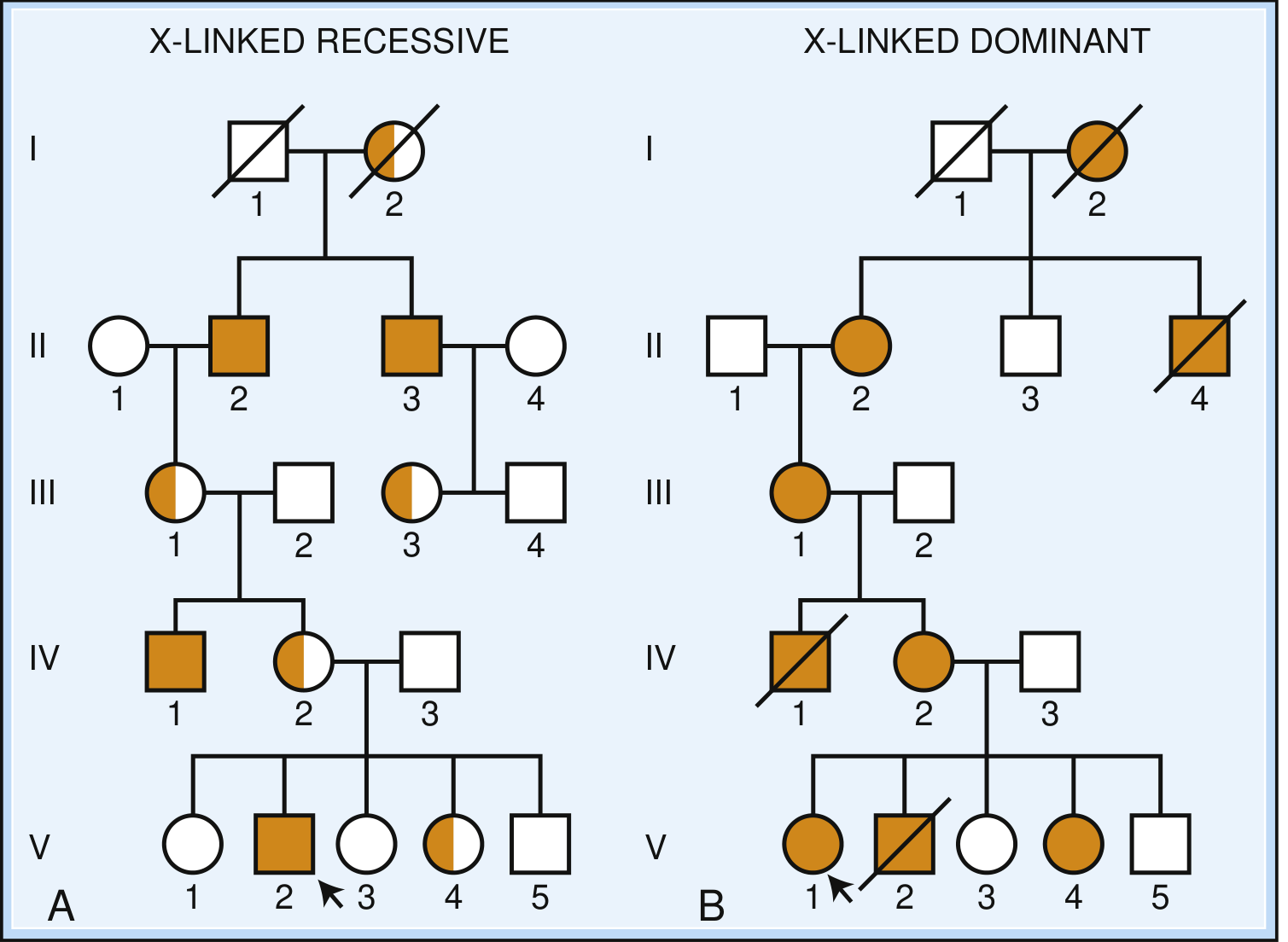
PEDIGREE DIAGRAM
Part 1: X-Linked Recessive (XLR)
Definition
Characteristics
| Feature | Detail |
|---|---|
| Who is affected? | Males predominantly; females are usually unaffected carriers |
| Female carrier risk | Each son: 50% affected; each daughter: 50% carrier |
| Affected male's children | All daughters = obligate carriers; No sons affected |
| Father-to-son transmission | NEVER (fathers give Y to sons) |
| Pedigree pattern | Males affected in alternate generations ("knight's move" pattern) |
| New mutations | Some XLR diseases arise de novo (e.g., ~1/3 of DMD cases) |
Genetic Risk Calculation
Carrier mother (X^A X^a) × Normal father (X^A Y)

Sons:   X^A Y (50% normal)   |   X^a Y (50% AFFECTED)
Daughters: X^A X^A (25% normal) | X^A X^a (25% carrier, unaffected)

Affected father (X^a Y) × Normal mother (X^A X^A)

Sons:   All normal (X^A Y) ← get Y from dad
Daughters: All carriers (X^A X^a) ← all get X^a from dad
When Females Can Be Affected in XLR
- Turner syndrome (45, XO): Only one X chromosome - if it carries the mutant allele, disease is expressed (e.g., females with Turner syndrome + hemophilia or DMD have been reported)
- X-autosome translocation: If the translocation breakpoint disrupts an X-linked gene, the normal X is preferentially inactivated (to preserve autosomal gene dosage), so the mutant allele is always active
- Skewed X inactivation: Random chance results in the mutant X being active in most cells ("manifesting carrier") - causes mild symptoms
- Homozygous female: If both X chromosomes carry the mutant allele (rare, but possible in consanguineous families or high-frequency alleles like G6PD)
Diseases - X-Linked Recessive
Neuromuscular
| Disease | Gene | Key Features |
|---|---|---|
| Duchenne muscular dystrophy (DMD) | DMD (dystrophin) - deletions | Progressive muscle weakness from age 3-5; wheelchair by ~10 years; death in early 20s from respiratory/cardiac failure; Gower's sign |
| Becker muscular dystrophy | DMD (milder mutations) | Same gene as DMD but milder; ambulation preserved beyond age 15 |
| Emery-Dreifuss muscular dystrophy | EMD (emerin) | Muscle wasting + early contractures + cardiac conduction defects |
| X-linked adrenoleukodystrophy (X-ALD) | ABCD1 | Very long chain fatty acid accumulation; cerebral demyelination in boys; adrenal insufficiency |
| Pelizaeus-Merzbacher disease | PLP1 | Hypomyelination; nystagmus, hypotonia, ataxia |
| Menkes disease | ATP7A (copper transport) | Copper deficiency; "kinky hair," progressive neurodegeneration, hypothermia |
Hematological
| Disease | Gene | Key Features |
|---|---|---|
| Hemophilia A | F8 (Factor VIII) | Deficient clotting; bleeding into joints (hemarthrosis), muscles; "royal hemophilia" - Queen Victoria was a carrier |
| Hemophilia B (Christmas disease) | F9 (Factor IX) | Clinically identical to hemophilia A; less common |
| G6PD deficiency | G6PD | Episodic hemolytic anemia triggered by drugs (primaquine, dapsone), infections, fava beans; most common enzyme deficiency worldwide |
| Chronic granulomatous disease (X-linked form) | CYBB (gp91phox) | Recurrent catalase-positive bacterial/fungal infections |
Immune / Metabolic
| Disease | Gene | Key Features |
|---|---|---|
| Wiskott-Aldrich syndrome | WAS | Thrombocytopenia, eczema, immunodeficiency (triad) |
| X-linked agammaglobulinemia (Bruton's) | BTK | Absent B cells; recurrent bacterial infections after 6 months |
| Fabry disease | GLA (α-galactosidase A) | Pain crises, angiokeratomas, renal failure, cardiomyopathy |
| Lesch-Nyhan syndrome | HPRT1 | Hyperuricemia, self-mutilation, choreoathetosis, intellectual disability |
| Diabetes insipidus (nephrogenic) | AVPR2 | Resistance to ADH; polydipsia, polyuria |
Neurological / Developmental
| Disease | Gene | Key Features |
|---|---|---|
| Fragile X syndrome | FMR1 (CGG repeat expansion) | Most common inherited intellectual disability; macroorchidism, long face, large ears, autism features |
| X-linked ichthyosis | STS (steroid sulfatase) | Scaly skin; corneal opacities |
| Ocular albinism | GPR143 | Lack of pigment in eye; nystagmus, reduced visual acuity |
Part 2: X-Linked Dominant (XLD)
Definition
Characteristics
| Feature | Detail |
|---|---|
| Who is affected? | Both males and females (heterozygous females affected) |
| Severity in males | Usually more severe than females; often lethal in males (embryonic death) |
| Affected mother's children | 50% of sons and 50% of daughters affected |
| Affected father's children | ALL daughters affected; NO sons affected |
| Father-to-son transmission | NEVER |
| Pedigree pattern | Resembles autosomal dominant but with no male-to-male transmission; excess of affected females; missing males (if lethal) |
Genetic Risk Calculation
Affected mother (X^A X^a) × Normal father (X^+ Y)

Sons:   50% X^+ Y (normal)  |  50% X^a Y (affected/lethal)
Daughters: 50% X^+ X^A (normal) | 50% X^A X^a (affected)

Affected father (X^a Y) × Normal mother (X^+ X^+)

Sons:   All X^+ Y = all NORMAL (sons get Y from dad)
Daughters: All X^+ X^a = ALL AFFECTED (all get X^a from dad)
Diseases - X-Linked Dominant
| Disease | Gene | Key Features |
|---|---|---|
| X-linked hypophosphatemia (Vitamin D-resistant rickets) | PHEX | Short stature, bowed legs, rickets despite normal vitamin D intake; females milder than males |
| Rett syndrome | MECP2 | Girls: progressive neurological regression, hand-wringing, loss of speech; Males: usually lethal |
| Incontinentia pigmenti | IKBKG | Skin: swirling pigmentation following Blaschko's lines (mosaic pattern in females); usually lethal in males; also eye and CNS involvement |
| X-linked Charcot-Marie-Tooth (CMTX1) | GJB1 (connexin 32) | Hereditary motor-sensory neuropathy; males more severely affected |
| Alport syndrome (X-linked form) | COL4A5 | Hematuria, progressive renal failure, sensorineural deafness; males more severe |
| Periventricular nodular heterotopia | FLNA (filamin A) | Brain malformation; seizures; males usually lethal |
| PCDH19-related epilepsy (Paradoxical XLD) | PCDH19 | Unique reversal: Females severely affected; males completely unaffected - due to "metabolic interference" between normal and mutant protein |
Side-by-Side Comparison
| Feature | XL Recessive | XL Dominant |
|---|---|---|
| Females affected? | Usually NO (carriers) | YES (heterozygous) |
| Males affected? | YES (always, hemizygous) | YES (more severely) |
| Males lethal? | No | Often YES (e.g., Rett, incontinentia pigmenti) |
| Pedigree | Males affected in alternate generations | Both sexes, all generations; excess females |
| Father → daughter | All daughters = carriers | All daughters = affected |
| Father → son | No transmission | No transmission |
| Mother → son | 50% affected | 50% affected (or lethal) |
| Mother → daughter | 50% carriers | 50% affected |
| Examples | Hemophilia, DMD, G6PD, Fragile X | X-ALD rickets, Rett syndrome, Incontinentia pigmenti |
Important Special Note: Dominant vs. Recessive in X-Linked is a Spectrum
"Nearly a third of X-linked disorders are penetrant in some (but not all) female heterozygotes and cannot be classified as either dominant or recessive... they show incomplete penetrance that varies as a function of X inactivation patterns."
- Thompson & Thompson Genetics and Genomics in Medicine, 9th Ed
Vitamin D resistant rickets
vitamin D resistant rickets X-linked hypophosphatemia PHEX FGF23 pathophysiology treatment
Vitamin D-Resistant Rickets
Overview
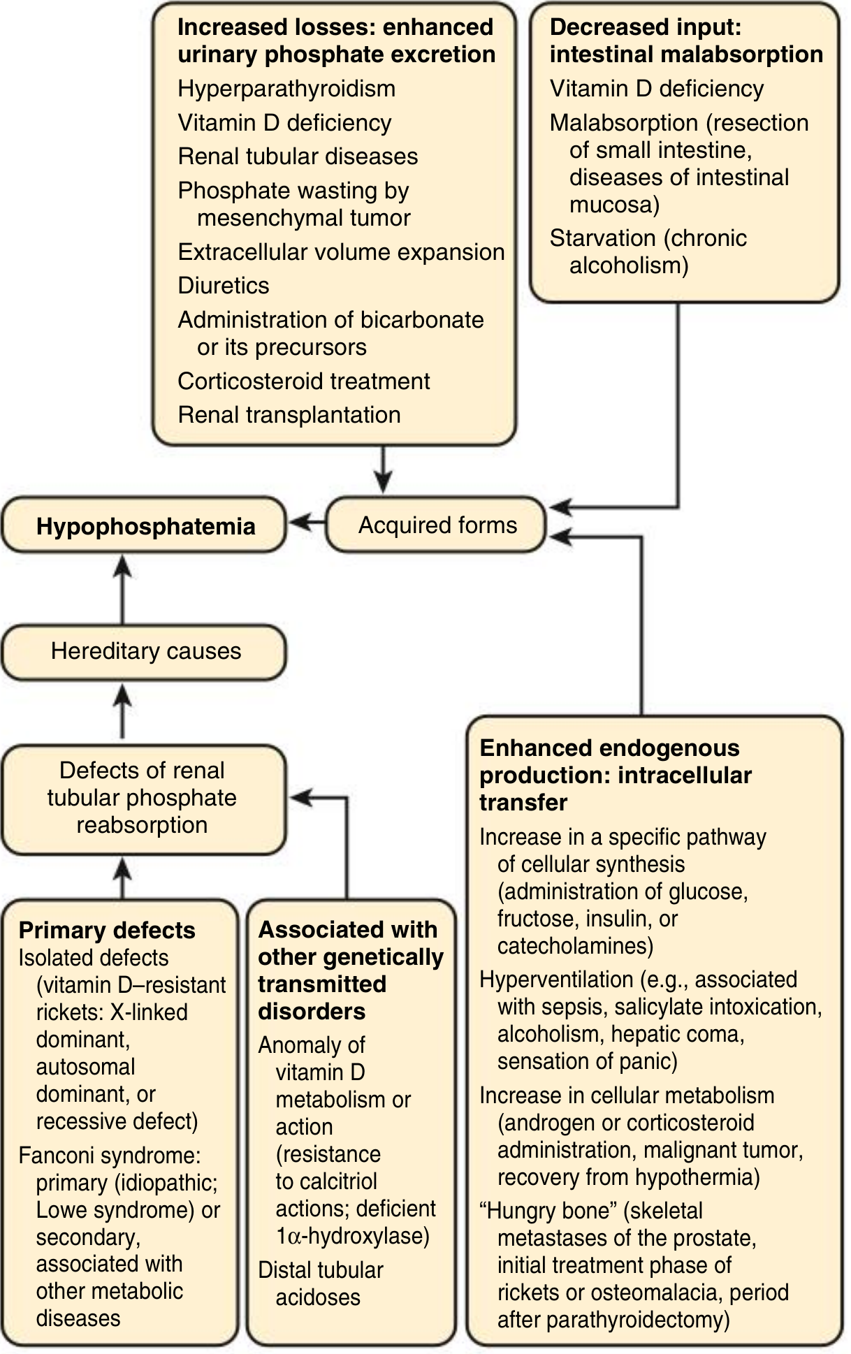
"The most common cause of rickets in the United States is hypophosphatemic osteomalacia (also known as hypophosphatemic vitamin D-resistant rickets). This disorder is an X-linked dominant inherited trait characterized by renal phosphate wasting."
- Tietz Textbook of Laboratory Medicine, 7th Ed
Classification of Vitamin D-Resistant Rickets
| Type | Old Name | Gene Affected | Mechanism |
|---|---|---|---|
| X-linked hypophosphatemia (XLH) | Vitamin D-resistant rickets (classic) | PHEX | Excess FGF23 → renal phosphate wasting |
| Vitamin D-dependent rickets Type 1 (VDDR-1) / Pseudovitamin D-deficiency rickets | PDDR | CYP27B1 (1α-hydroxylase) | Cannot convert 25(OH)D → 1,25(OH)2D |
| Vitamin D-dependent rickets Type 2 (VDDR-2) / Hereditary Vitamin D-resistant rickets | HVDRR | VDR (vitamin D receptor) | End-organ resistance to 1,25(OH)2D |
PART 1: X-Linked Hypophosphatemia (XLH) - The Classic "Vitamin D-Resistant Rickets"
Genetics
- Inheritance: X-linked dominant (XLD)
- Gene: PHEX (Phosphate-Regulating Endopeptidase on the X chromosome)
- Prevalence: 1 in 20,000; high penetrance; both males and females affected
- Males tend to be more severely affected than females
Pathophysiology
PHEX mutation (loss of function)
        ↓
PHEX normally suppresses FGF23 production in osteocytes
(via binding to DMP1 - dentin matrix protein 1)
        ↓
Without PHEX: FGF23 levels rise markedly
        ↓
FGF23 acts on kidney proximal tubules:
  1. Blocks NaPi-IIa/IIc cotransporters → phosphate NOT reabsorbed → phosphaturia
  2. Inhibits CYP27B1 (1α-hydroxylase) → cannot convert 25(OH)D → 1,25(OH)2D
        ↓
Result: LOW serum phosphate + LOW/Normal 1,25(OH)2D
        ↓
Defective bone mineralization → RICKETS in children / OSTEOMALACIA in adults
"Binding of PHEX to DMP1 appears to be critical for the suppression of osteocyte production of FGF-23 in bone. Inactivating mutations in PHEX or DMP1 result in higher circulating levels of FGF-23 and resultant phosphate wasting. Abnormal synthesis of 1,25(OH)2D in XLH can be explained by increased levels of FGF-23, which suppress kidney 1α-hydroxylase activity."
- Brenner & Rector's The Kidney
Key Biochemical Profile
| Lab Parameter | Result in XLH |
|---|---|
| Serum phosphate | ↓↓ (Low) |
| Serum calcium | Normal |
| PTH | Normal |
| 25(OH)D | Normal |
| 1,25(OH)2D | Low or inappropriately normal |
| Alkaline phosphatase (ALP) | ↑ (High) |
| Urine phosphate | ↑↑ (High) - phosphaturia |
| FGF23 | ↑↑ (High) |
Clinical Features
- Children: Rickets - bowing of legs, short stature, widened growth plates
- Skeletal: Short stature, bowed long bones, frontal bossing, rachitic rosary, short bowed legs
- Dental: Dental abscesses due to defective dentin mineralization (spontaneous, without caries)
- Cranial: Premature craniosynostosis (premature fusion of skull sutures)
- Adults: Osteomalacia, enthesopathy (calcification of tendons/ligaments), bone pain, pseudofractures, osteoarthritis, hearing loss
- Key difference from nutritional rickets: Does NOT respond to standard vitamin D doses
Treatment
- Oral phosphate supplements (1-3 g/day in divided doses) to replace lost phosphate
- Calcitriol (1,25(OH)2D - active vitamin D, 0.25-2 mcg/day) to improve calcium/phosphate absorption
- Limitation: Prolonged use leads to secondary/tertiary hyperparathyroidism (phosphate supplements stimulate PTH)
- Burosumab (Crysvita) - a human anti-FGF23 monoclonal antibody
- FDA approved for children ≥1 year and adults
- Blocks excess FGF23 directly → restores renal phosphate reabsorption and normalizes 1,25(OH)2D production
- Dosing: 0.8-2 mg/kg SC every 2 weeks (children); 1 mg/kg up to 90 mg SC every 4 weeks (adults)
- Significantly more effective than conventional therapy with fewer complications
"Burosumab, a human anti-FGF23 monoclonal antibody, has been approved for the treatment of XLH. It corrects hypophosphatemia, improves bone pain, and heals fractures in both children and adults."
- Harrison's Principles of Internal Medicine, 22nd Ed
PART 2: Vitamin D-Dependent Rickets Type 1 (VDDR-1) / Pseudovitamin D-Deficiency Rickets
Genetics
- Inheritance: Autosomal recessive
- Gene: CYP27B1 encoding renal 1α-hydroxylase
Mechanism
- The enzyme that converts 25(OH)D (calcidiol) → 1,25(OH)2D (calcitriol) is absent/deficient
- The body cannot make the active form of vitamin D despite having plenty of 25(OH)D
- Presents in infancy with hypocalcemia, hypophosphatemia, elevated PTH, elevated ALP
Biochemical Profile
| Parameter | VDDR-1 |
|---|---|
| Calcium | ↓ |
| Phosphate | ↓ |
| 25(OH)D | Normal |
| 1,25(OH)2D | ↓↓ (Very low) |
| PTH | ↑ |
| ALP | ↑ |
Treatment
- Calcitriol (1,25(OH)2D) - replaces the missing active vitamin D
- Responds well to physiological doses (0.25-0.5 mcg/day)
PART 3: Vitamin D-Dependent Rickets Type 2 (VDDR-2) / Hereditary Vitamin D-Resistant Rickets (HVDRR)
Genetics
- Inheritance: Autosomal recessive
- Gene: VDR (vitamin D receptor)
Mechanism
- Vitamin D is activated normally - 1,25(OH)2D levels are actually very high (the body tries to compensate)
- But the receptor itself is non-functional → complete end-organ resistance
- Cells cannot respond to vitamin D at all
Biochemical Profile
| Parameter | VDDR-2 |
|---|---|
| Calcium | ↓ |
| Phosphate | ↓ |
| 25(OH)D | Normal |
| 1,25(OH)2D | ↑↑↑ (Very high) - the hallmark |
| PTH | ↑ |
| ALP | ↑ |
Clinical Features
- Similar to VDDR-1 plus a unique feature: Alopecia (hair loss) - the VDR is required in hair follicles
- Alopecia is actually a defining distinguishing feature of VDDR-2
Treatment
- Very difficult - partially functional VDR mutations may respond to very large doses of calcitriol
- Most severe cases: IV calcium and phosphate infusions can correct rickets by bypassing the VDR
- After puberty: requirements often diminish spontaneously
Comparison of All Types - Summary Table
| Feature | XLH (VDRR) | VDDR-1 | VDDR-2 |
|---|---|---|---|
| Inheritance | X-linked dominant | AR | AR |
| Gene | PHEX | CYP27B1 | VDR |
| Mechanism | FGF23 excess → phosphate wasting | No 1α-hydroxylase | No VDR function |
| Serum Ca | Normal | ↓ | ↓ |
| Serum PO4 | ↓↓ | ↓ | ↓ |
| 25(OH)D | Normal | Normal | Normal |
| 1,25(OH)2D | Low/normal (inappropriately) | ↓↓ | ↑↑↑ |
| PTH | Normal | ↑ | ↑ |
| FGF23 | ↑↑ | Normal | Normal |
| Alopecia | No | No | Yes |
| Treatment | Phosphate + calcitriol; Burosumab | Calcitriol | High-dose calcitriol / Ca+PO4 infusions |
Also: Other Causes of Phosphate-Wasting Rickets (same mechanism as XLH)
| Condition | Mutation | Notes |
|---|---|---|
| Autosomal dominant hypophosphatemic rickets (ADHR) | FGF23 gene - activating mutation makes FGF23 resistant to proteolysis | Same phenotype as XLH |
| Autosomal recessive hypophosphatemic rickets (ARHR) | DMP1, ENPP1, or FAM20C | Increased FGF23 production; ENPP1/FAM20C also cause arterial calcifications |
| Tumor-induced osteomalacia (TIO) | Acquired (mesenchymal tumor overproduces FGF23) | Adult-onset; resolves with tumor removal |
| Fanconi syndrome | Multiple proximal tubule defects | Phosphate + glucose + amino acid + bicarbonate wasting |