
Diabetes in CKD pathophysiology, clinical features and management
"diabetes mellitus" AND "chronic kidney disease" AND management
diabetic nephropathy Kimmelstiel-Wilson nodular glomerulosclerosis histology
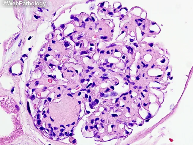
This is a renal biopsy histology slide examined under brightfield light microscopy. The primary subject is diabetic nephropathy with nodular glomerulosclerosis (Kimmelstiel-Wilson nodules) and diffuse mesangial sclerosis within the glomerular tuft of a renal cortex specimen. The glomerulus shows nodular mesangial expansion forming rounded, eosinophilic nodules that disrupt capillary loops, together with diffuse increased mesangial matrix. Capillary lumina are variably compressed, and arterioles frequently exhibit hyaline thickening consistent with diabetic microangiopathy. The cellular morphology is characterized by expanded mesangial cells with increased extracellular matrix and relatively sparse inflammatory infiltrate. The basement membrane appears thickened in cross-sectional elements. In this image, Masson’s trichrome is not visible; a subsequent stain would color collagen in blue, highlighting fibrotic remodeling. Clinically, these histologic features correlate with progressive proteinuria and reduced glomerular filtration rate in long-standing diabetes. Diagnostic significance rests on the combination of nodular sclerosis (Kimmelstiel-Wilson nodules) and diffuse mesangial sclerosis as hallmarks of diabetic nephropathy. Differential considerations include focal segmental glomerulosclerosis and hypertensive nephrosclerosis, but the nodular pattern is characteristic. This image is valuable for pathology education, nephrology training, and research into diabetic kidney disease progression and glycemic control effects. Representative for teaching clinics, this image supports correlating histology with clinical nephrology.
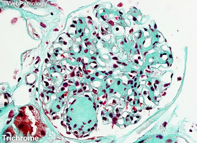
Masson's trichrome-stained renal cortex histology imaged at light microscopy reveals nodular glomerulosclerosis characteristic of diabetic nephropathy. The glomerulus shows circumscribed Kimmelstiel-Wilson nodules-eosinophilic mesangial nodules that expand the tuft and encroach on capillary loops. Diffuse mesangial sclerosis is evident as widespread mesangial matrix expansion, with increased basophilic mesangial tissue and accentuated blue-green collagen deposition in the extracellular matrix on Masson stain. The stain differentiates collagen (blue/green) from cytoplasm and nuclei (red/purple), highlighting the fibrotic nodules and mesangial expansion. Within the mesangium, there is progressive accumulation of matrix with variable cellularity; some capillary lumina appear narrowed due to nodular compression. The surrounding tubulointerstitial tissue may show mild chronic changes, consistent with chronic diabetic nephropathy. The overall pattern is typical of long-standing diabetes mellitus with nephropathy and correlates with progressive proteinuria, reduced glomerular filtration rate, and eventual renal insufficiency. Clinically, this finding supports diagnosis of diabetic kidney disease (diabetic nephropathy) and is essential for grading disease stage and guiding management. Differential diagnoses include nodular glomerulosclerosis due to amyloidosis or hypertensive nephrosclerosis, but the classic KW nodules and mesangial expansion favor diabetes. This image is valuable for medical education, pathology training, and research into diabetic renal pathology.
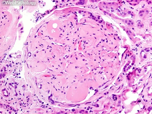
Imaging modality: brightfield histopathology. This slide shows a renal glomerulus from a biopsy specimen of diabetic nephropathy. The mesangial matrix is markedly expanded, producing round to nodular consolidations within the tuft. Several Kimmelstiel‑Wilson nodules are evident as acellular, hyaline nodules that distend the mesangial stalks and encroach on capillary loops. The glomerular basement membranes appear thickened on corresponding PAS‑positive and silver‑stained sections (referenced in adjacent images), consistent with nodular diabetic glomerulosclerosis. The surrounding cortex exhibits mild chronic interstitial changes and arteriolar hyalinosis in keeping with chronic diabetic kidney disease. The glomerulus displays relatively extensive mesangial deposition with focally patent capillary lumina, creating a characteristic nodular pattern. Overall, the morphology is diagnostic for diabetic microangiopathy with progressive nodular sclerosis. Clinically, these findings correlate with long‑standing hyperglycemia, proteinuria, and reduced GFR. Pathological significance: KW nodules indicate advanced disease and higher risk of progression to end‑stage renal disease. Differential considerations include non‑diabetic nodular glomerulosclerosis and other glomerulopathies, but the classic KW nodules and mesangial expansion strongly support diabetic nephropathy. This image is valuable for education, pathology review, and correlating histology with clinical diabetes management. It highlights key features for learners: mesangial expansion, KW nodules, PAS positivity, and altered capillary luminal flow in practice.
Diabetes in Chronic Kidney Disease (Diabetic Kidney Disease)
1. Pathophysiology
A. Hemodynamic Mechanisms
- RAAS activation: Hyperglycemia activates renin-angiotensin-aldosterone system (RAAS), raising angiotensin II, which preferentially constricts the efferent arteriole, raising intraglomerular pressure.
- SGLT1/SGLT2 upregulation: Diabetes upregulates sodium-glucose cotransporters in the proximal tubule, leading to increased glucose and sodium reabsorption. This decreases distal delivery of sodium to the macula densa, blunting tubuloglomerular feedback and sustaining hyperfiltration.
- Insulin-like growth factor (IGF-1) and growth hormone: Elevated in early diabetes; stimulate renal hypertrophy and GFR increase.
- Kallikrein-kinin system (KKS): In early/moderate hyperglycemia, renal kallikrein excretion is elevated, correlating with increased RBF and GFR.
B. Metabolic Mechanisms
- Advanced glycation end products (AGEs): Glucose non-enzymatically glycates proteins, forming AGEs that crosslink collagen in the GBM, increase glomerular permeability, and stimulate mesangial matrix expansion via TGF-β.
- Reactive oxygen species (ROS): Hyperglycemia drives mitochondrial ROS production (via polyol pathway, protein kinase C activation, AGE accumulation), promoting oxidative stress, inflammation, and fibrosis.
- TGF-β: Key profibrotic cytokine, stimulates mesangial cell production of extracellular matrix, collagen IV, and fibronectin.
- Dyslipidemia: Contributes to glomerular injury, especially relevant in type 2 diabetes.
- Tubular metabolic reprogramming: Adaptive and maladaptive shifts in tubular metabolism (fatty acid oxidation, glycolysis) contribute to both diabetic and non-diabetic CKD progression. (Brenner & Rector's The Kidney)
C. Genetic Factors
- ACE gene polymorphism (D allele / DD genotype): Associated with higher ACE activity in renal tissues and increased susceptibility to ESKD.
- IL-10 -1082G/A (AA genotype): ~2.5-fold higher susceptibility to diabetic nephropathy.
- IL-6 gene polymorphisms (rs1800796, rs1524107): Predict nephropathy progression.
- ACACB gene (acetyl coenzyme A carboxylase β): Related to proinflammatory responses in renal tubular cells.
- ICAM1 K469E polymorphism: Associated with DKD in Asian populations. (Brenner & Rector's The Kidney, 2-Volume Set)
D. Structural/Histological Changes (Pathology)
- GBM thickening: Detectable on EM within a few years of diabetes onset, even before functional changes.
- Diffuse mesangial sclerosis: Increase in mesangial matrix with mesangial cell proliferation and BM thickening; found after >10 years of disease; leads to nephrotic syndrome when severe.
- Nodular glomerulosclerosis (Kimmelstiel-Wilson lesion): Ball-like deposits of laminated matrix in the periphery of glomeruli. Present in 15-30% of patients with long-term diabetes. Virtually pathognomonic of diabetes. There is a significant correlation between KW nodules and diabetic retinopathy.
- Hyaline arteriolosclerosis affecting both afferent and efferent arterioles - efferent involvement is virtually unique to diabetes.
- Atherosclerosis; vascular compromise leads to nephrosclerosis (finely granular cortical surface).
2. Clinical Features and Natural History
Stages of Diabetic Nephropathy (Classic Mogensen Classification)
| Stage | Feature | Timing |
|---|---|---|
| I | Glomerular hyperfiltration + renal hypertrophy; GFR elevated | Onset of diabetes |
| II | Mesangial expansion; normal albumin excretion | 2-5 years |
| III | Microalbuminuria (30-300 mg/24h); GFR normal or mildly reduced | 5-10 years |
| IV | Macroproteinuria (>300 mg/24h); GFR declining | 10-20 years |
| V | ESKD; GFR <15 mL/min | >20 years |
- Microalbuminuria appears 5-10 years after diabetes onset; classically progresses over 5-10 years to overt proteinuria and declining GFR.
- Contemporary caveat: Up to 24% of T1DM and 50% of T2DM patients with CKD may be normoalbuminuric - the classic albuminuric progression is not universal.
- Proteinuria in DKD can range from 500 mg to 25 g/24h.
- GFR decline rate: 1.8-14 mL/min/year depending on treatment and risk factors. (Harrison's Principles of Internal Medicine 22E, 2025)
Key Clinical Associations
- Retinopathy: Present in >90% of T1DM with nephropathy (vs. ~60% in T2DM). Absence of retinopathy in a T1DM patient with proteinuria should prompt biopsy to exclude another diagnosis.
- Enlarged kidneys may persist even in advanced DKD (unlike most other causes of CKD where kidneys shrink).
- Hypertension: Nearly universal; accelerates progression.
- Nephrotic syndrome: When glomerulosclerosis is severe - edema, hypoalbuminemia, heavy proteinuria.
- Cardiovascular disease: Albuminuria and reduced GFR are both independent risk factors for CV events; many patients die of CV disease before reaching ESKD.
- Risk factors for CKD in T2DM: Retinopathy/neuropathy, age, BP ≥140/80 mmHg, cardiovascular disease history, longer duration of diabetes. (Fuster and Hurst's The Heart, 15th Edition; Harrison's)
3. Management
A. Glycemic Control
- Target HbA1c: Generally ~7% (individualize - more lenient in advanced CKD/elderly due to hypoglycemia risk).
- DCCT (T1DM) and UKPDS (T2DM) both demonstrated that strict glycemic control prevents development and progression of microalbuminuria.
- Metformin: First-line in T2DM; dose-reduce or stop at eGFR <30-45 mL/min/1.73m² (risk of lactic acidosis).
- Insulin: Dose reduction needed as eGFR falls (reduced renal clearance of insulin - paradoxically, insulin requirements may decrease in advanced CKD).
- Sulfonylureas: Use with caution - gliquidone or glipizide preferred in CKD; avoid glibenclamide (active metabolite accumulates). Hypoglycemia risk increases.
- DPP-4 inhibitors: Dose-adjusted for eGFR; generally safe.
- GLP-1 receptor agonists: See below.
- SGLT2 inhibitors: See below.
B. RAAS Blockade (Cornerstone of Therapy)
- ACE inhibitors (ACEi) or Angiotensin Receptor Blockers (ARBs) are first-line anti-proteinuric and renoprotective agents in DKD regardless of blood pressure.
- Reduce intraglomerular pressure by dilating the efferent arteriole.
- Reduce proteinuria and slow GFR decline.
- The ACE gene D allele (high ACE activity) genotype predicts greater benefit.
- Dual ACEi + ARB: Not recommended due to increased risk of hyperkalemia and AKI without additional benefit (ONTARGET trial data).
- ACEi/ARB are contraindicated in pregnancy (second/third trimester - birth defects including renal tubular dysplasia, hypocalvaria).
C. Mineralocorticoid Receptor Antagonists (MRA)
- Spironolactone / Eplerenone: Reduce albuminuria in DKD; use limited by hyperkalemia, especially with ACEi/ARB.
- Finerenone (novel non-steroidal, selective MRA): Attenuates albuminuria; in FIDELIO-DKD (5734 T2DM patients, eGFR 25-75 mL/min/1.73m², albuminuria):
- 18% lower risk of kidney failure, ≥40% eGFR decline, or kidney death vs. placebo.
- 14% reduction in CV outcomes.
- Lower hyperkalemia rate than steroidal MRAs (2.3% discontinuation vs. 0.9% placebo).
- Target: T2DM patients with moderately increased albuminuria + impaired eGFR, or severely increased albuminuria. (NKF Primer on Kidney Diseases 8e; Comprehensive Clinical Nephrology 7th Ed.)
D. SGLT2 Inhibitors (Paradigm-Shifting Therapy)
- CREDENCE (Canagliflozin, eGFR ≥30 to <90, T2DM + DKD): 33% reduction in risk of kidney replacement therapy, doubling of serum creatinine, and death from kidney insufficiency; also reduced major CV events and heart failure hospitalization.
- DAPA-CKD (Dapagliflozin, eGFR 25-75 mL/min, including non-diabetic CKD): Reduced sustained ≥50% eGFR decline, ESKD, and CV/kidney death. Benefit seen even in non-diabetic CKD.
- EMPA-KIDNEY, CREDENCE, and DAPA-CKD data collectively support SGLT2 inhibitors across eGFR as low as 25 mL/min/1.73m² for initiation, with continuation even if eGFR falls further after starting.
- Recommended for T2DM with CKD + albuminuria in addition to RAAS blockade. (NKF Primer 8e; Comprehensive Clinical Nephrology 7th Ed.)
E. GLP-1 Receptor Agonists
- Reduce HbA1c, body weight, and blood pressure.
- Cardiorenal outcome trials (LEADER - liraglutide; SUSTAIN-6 - semaglutide): Significant reductions in new/worsening nephropathy.
- A 2025 Cochrane systematic review (PMID 39963952) confirmed benefits of GLP-1 RAs for people with both CKD and diabetes.
- Preferred agents in T2DM + established CV disease or CKD, alongside or after SGLT2 inhibitors.
- Dose adjustment generally not required for renal impairment (except exenatide - avoid if eGFR <30).
F. Blood Pressure Control
- Target BP: <130/80 mmHg in DKD with albuminuria (ADA/KDIGO).
- ACEi/ARB as first-line; add amlodipine or diuretics as needed.
- Dihydropyridine CCBs are preferred over non-dihydropyridines when added to RAAS blockade.
- Loop diuretics (furosemide) for volume overload in advanced CKD; thiazides lose efficacy at eGFR <30.
G. Lipid Management
- CKD (like diabetes) is a cardiovascular risk equivalent.
- LDL targets: <55 mg/dL if eGFR <30 mL/min/1.73m²; <70 mg/dL in CKD stage 3.
- SHARP trial: Simvastatin 20mg + ezetimibe 10mg reduced major atherosclerotic events by 16% in advanced CKD.
- Statin benefit is attenuated at very low eGFR and has limited benefit in dialysis patients (4D study, AURORA trial negative).
- Statins should NOT be initiated in dialysis patients but should be continued if already on them. (NKF Primer 8e; Comprehensive Clinical Nephrology 7th Ed.)
H. Anemia Management
- Anemia in CKD-DM: multifactorial (EPO deficiency, reduced EPO sensitivity, inflammation, iron deficiency).
- Erythropoiesis-stimulating agents (ESAs): Correcting severe anemia reduces LVH; however, targeting normal hemoglobin with ESAs showed no mortality benefit in RCTs and may increase cardiovascular risk if Hb is normalized (TREAT trial).
- HIF-PH inhibitors (e.g., roxadustat): Emerging agents for CKD anaemia, stimulate endogenous EPO production.
I. Additional Management
| Area | Approach |
|---|---|
| Protein restriction | 0.8 g/kg/day (avoid very low protein in CKD) |
| Smoking cessation | Independently accelerates GFR decline |
| Weight loss | Beneficial in obese T2DM-CKD patients |
| Hyperphosphatemia | Phosphate binders; dietary phosphate restriction |
| Metabolic acidosis | Sodium bicarbonate supplementation; slows CKD progression |
| Hyperkalemia | Potassium restriction; patiromer/sodium zirconium cyclosilicate to allow RAAS blockade continuation |
| Dialysis/transplant | Kidney transplant preferred (better outcomes in DM-CKD); combined kidney-pancreas transplant option in T1DM |
| Pregnancy in DKD | Postpone pregnancy until after transplant if creatinine >1.5 mg/dL; avoid ACEi/ARB in 2nd-3rd trimester |
Key Evidence Summary
| Trial | Drug | Key Result |
|---|---|---|
| CREDENCE | Canagliflozin | -33% kidney composite endpoint in T2DM+DKD |
| DAPA-CKD | Dapagliflozin | -44% kidney/CV composite, including non-diabetic CKD |
| FIDELIO-DKD | Finerenone | -18% kidney composite, -14% CV composite |
| FIGARO-DKD | Finerenone | Reduced CV primary endpoint in T2DM+CKD |
| LEADER | Liraglutide | -22% new/worsening nephropathy |
| SHARP | Simvastatin+Ezetimibe | -16% major atherosclerotic events in advanced CKD |
Recent update (2025): A Cochrane meta-analysis (PMID 39963952) confirms GLP-1 RAs reduce kidney and cardiovascular outcomes in patients with CKD + diabetes. A 2026 individual patient-level meta-analysis (PMID 41183334) reaffirmed albuminuria and proteinuria as powerful markers of both kidney and cardiovascular disease risk.