
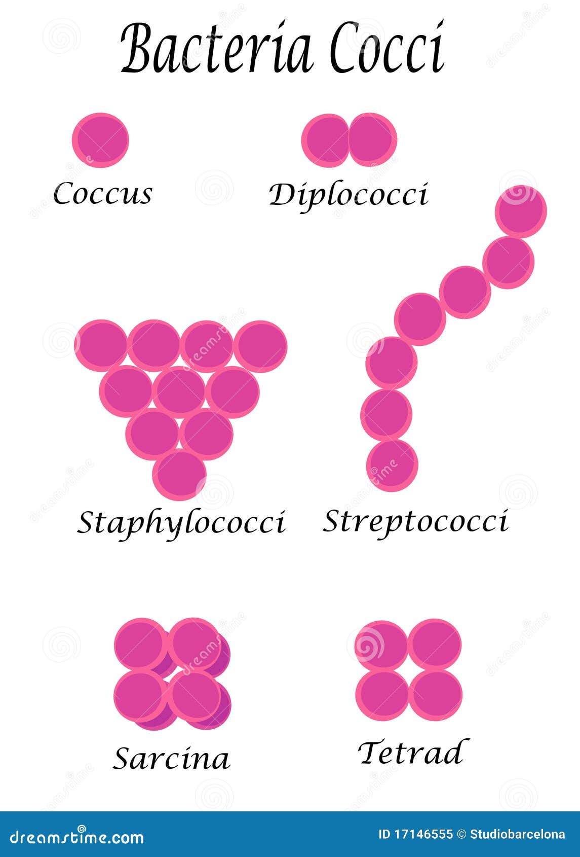
Add individual bacterial images based --- ## 3.2 Morphology and classification of bacteria ...also paying an important role in identifying the bacteria. Staining techniques is... Bacteria are classified into 7 groups depending on their shape: 1. Cocci - (Kokhos means berry) - These are spherical in shape 2. Bacilli - (Baculus means rod) - These are rod shaped cells 3. Vibrios - These are comma shaped, curved rods possessing vibratory motility --- ## Paper -II Microbiology and Pathology (Page 149) 4. Spirilla - These are rigid spiral forms 5. Spirochaetes (Coiled hairs) - These are thin flexuous spiral forms 6. Actinomycetes - These are branching filamentous bacteria. When seen in tissues they appear like radiating rays of the sun (sunray appearance). This is due to the rigid cell wall. 7. Mycoplasma - These are bacteria with defective cell wall. This may be due to spontaneous mutation or due to penicillin administration. Such cells are called as protoplasts, spheroplasts or L forms. ### Arrangement of Bacterial cells 1. Cocci in pairs - Diplococci 2. Cocci in chains - Streptococci 3. Cocci in clusters - Staphylococci 4. Cocci in groups of four - Tetrads 5. Cocci in pockets of eight - Sarcina 6. Bacilli in chains - Streptobacilli 7. Bacilli in chinese letter pattern - Coryne bacteria --- ## Paper -II Microbiology and Pathology (Page 151) ### Difference between Gram positive and Gram negative bacteria | Gram positive bacteria | Gram negative bacteria | |---|---| | Simple cell wall | More complex cell wall | | Thick peptidoglycan cell wall layer | Thin peptidoglycan cell wall layer | | No outer lipopolysaccharide wall layer | Outer lipopolysaccharide wall layer | | Retain crystal violet/iodine | Retain safranin | | Appear (Blue/Purple) | Appear (Pink/Red) | ### Ribosomes These are the centers of protein synthesis.
bacteria morphology shapes cocci bacilli spirilla microscopy images labeled diagram
staphylococci streptococci diplococci sarcina arrangement microscopy images free
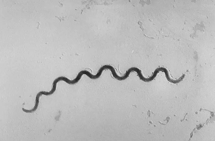
spirochaetes spirochete microscopy image actinomycetes sunray appearance mycoplasma L-form bacteria
3.2 Morphology and Classification of Bacteria
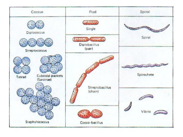
1. Cocci - (Kokhos means berry) - Spherical in shape

2. Bacilli - (Baculus means rod) - Rod-shaped cells
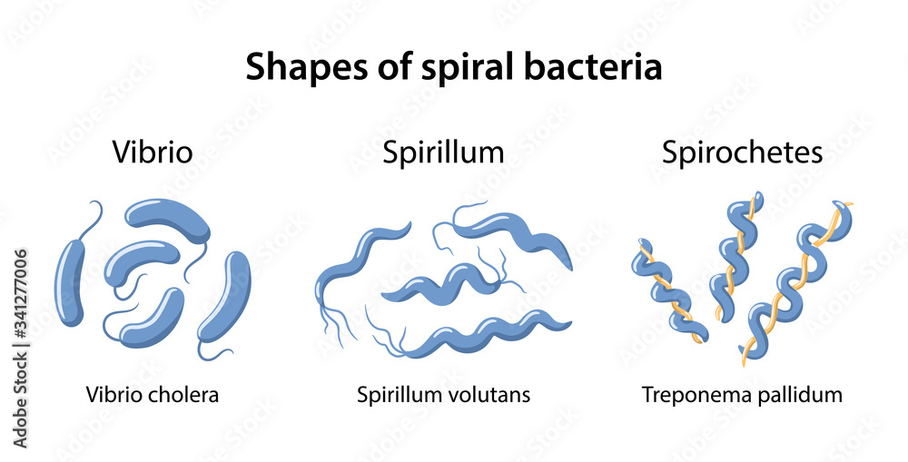
3. Vibrios - Comma-shaped, curved rods possessing vibratory motility
Paper -II Microbiology and Pathology (Page 149)
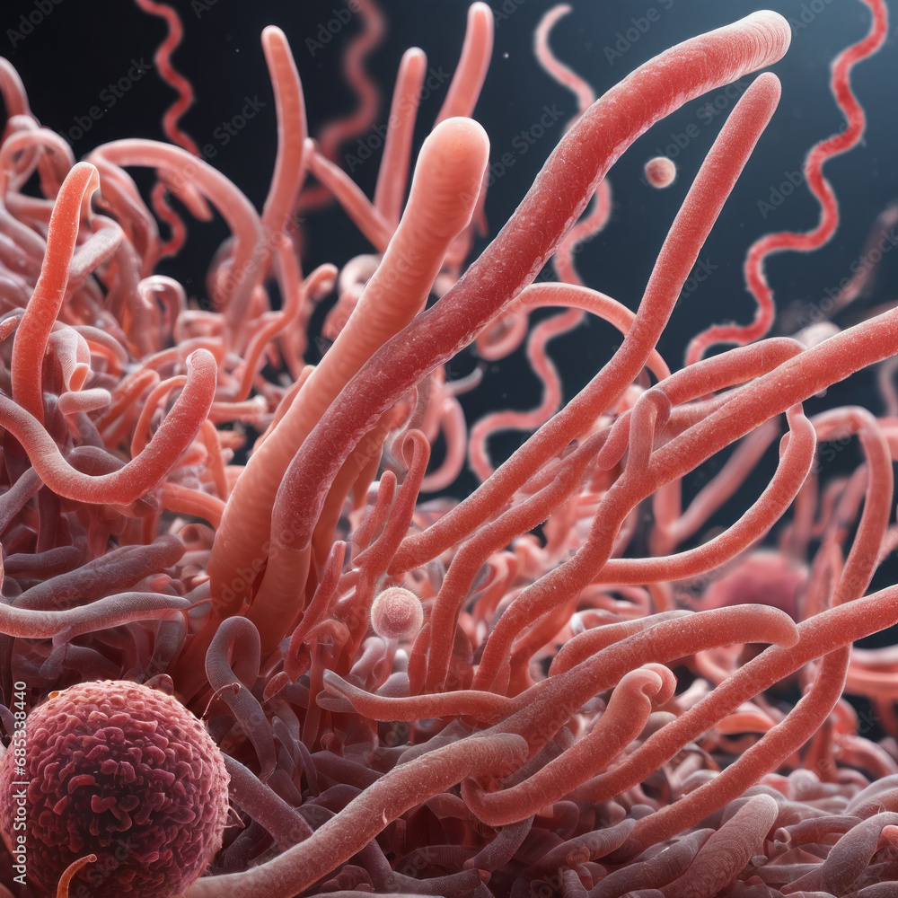
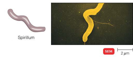
4. Spirilla - Rigid spiral forms
5. Spirochaetes (Coiled hairs) - Thin flexuous spiral forms

Spirilla vs Spirochaetes: Spirilla are rigid spirals (like a corkscrew spring), while spirochaetes are flexible and undulating. Both are visible in the curved shapes column of the overview diagram.
6. Actinomycetes - Branching filamentous bacteria
7. Mycoplasma - Bacteria with defective cell wall
Arrangement of Bacterial Cells
| # | Arrangement | Name |
|---|---|---|
| 1 | Cocci in pairs | Diplococci |
| 2 | Cocci in chains | Streptococci |
| 3 | Cocci in clusters | Staphylococci |
| 4 | Cocci in groups of four | Tetrads |
| 5 | Cocci in pockets of eight | Sarcina |
| 6 | Bacilli in chains | Streptobacilli |
| 7 | Bacilli in Chinese letter pattern | Corynebacteria |
Paper -II Microbiology and Pathology (Page 151)
Difference between Gram Positive and Gram Negative Bacteria
| Gram positive bacteria | Gram negative bacteria |
|---|---|
| Simple cell wall | More complex cell wall |
| Thick peptidoglycan cell wall layer | Thin peptidoglycan cell wall layer |
| No outer lipopolysaccharide wall layer | Outer lipopolysaccharide wall layer |
| Retain crystal violet/iodine | Retain safranin |
| Appear (Blue/Purple) | Appear (Pink/Red) |
Ribosomes
- The overview diagram (SlideShare) covers cocci, rods, spirals, vibrio, spirochetes, sarcinae, and streptobacilli all in one labeled chart - ideal as a reference visual.
- The iStock illustrated diagram shows all cocci arrangements (monococcus through sarcina) plus bacilli and curved forms with clear labels.
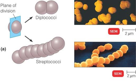
- The SEM/diagram image from MicrobiologyInfo shows diplococci and streptococci with both schematic and actual electron micrograph views.
- The Science Photo Library image shows real spirochaetes under light microscopy.
- The Biology Notes image covers actinomycetes/filamentous forms.
Use individual images not one In chart
cocci bacteria spherical microscopy image wikimedia commons
bacilli rod shaped bacteria microscopy image wikimedia
vibrio cholerae comma shaped bacteria microscopy image
spirilla rigid spiral bacteria microscopy image
3.2 Morphology and Classification of Bacteria
1. Cocci - (Kokhos means berry) - Spherical in shape
2. Bacilli - (Baculus means rod) - Rod-shaped cells
3. Vibrios - Comma-shaped, curved rods possessing vibratory motility
Paper -II Microbiology and Pathology (Page 149)
4. Spirilla - Rigid spiral forms
5. Spirochaetes (Coiled hairs) - Thin flexuous spiral forms

6. Actinomycetes - Branching filamentous bacteria
7. Mycoplasma - Bacteria with defective cell wall
Arrangement of Bacterial Cells
| # | Arrangement | Name |
|---|---|---|
| 1 | Cocci in pairs | Diplococci |
| 2 | Cocci in chains | Streptococci |
| 3 | Cocci in clusters | Staphylococci |
| 4 | Cocci in groups of four | Tetrads |
| 5 | Cocci in pockets of eight | Sarcina |
| 6 | Bacilli in chains | Streptobacilli |
| 7 | Bacilli in Chinese letter pattern | Corynebacteria |
Paper -II Microbiology and Pathology (Page 151)
Difference between Gram Positive and Gram Negative Bacteria
| Gram positive bacteria | Gram negative bacteria |
|---|---|
| Simple cell wall | More complex cell wall |
| Thick peptidoglycan cell wall layer | Thin peptidoglycan cell wall layer |
| No outer lipopolysaccharide wall layer | Outer lipopolysaccharide wall layer |
| Retain crystal violet/iodine | Retain safranin |
| Appear (Blue/Purple) | Appear (Pink/Red) |
Ribosomes
Make presentation
node /home/daytona/skills/shared/scripts/fetch_images.js \ "https://thumbs.dreamstime.com/z/bacteria-cocci-17146555.jpg" \ "https://www.mdpi.com/diagnostics/diagnostics-15-01285/article_deploy/html/images/diagnostics-15-01285-g003-550.jpg" \ "https://as1.ftcdn.net/v2/jpg/06/85/33/84/1000_F_685338440_Ixjtx02vKn39SByDoJresvQFt3UQdwlB.jpg" \ "https://microbiologyinfo.com/wp-content/uploads/2015/05/Spirilla.jpg" \ "https://media.istockphoto.com/id/485943707/photo/spirillum-bacteria.jpg?s=1024x1024&w=is&k=20&c=kQ0YFlO3saRHsuutEKSrVM39IKofMcoHgSWJWx1V19g=" \ "https://biologynotesonline.com/wp-content/uploads/2024/03/image-792.png" \ "https://as1.ftcdn.net/v2/jpg/03/41/27/70/1000_F_341277006_6rFzstpgmDTsK4fJhU5xMUValOqm7924.jpg" 2>&1 | head -5
An error occurred (ValidationException) when calling the ConverseStream operation: The model returned the following errors: prompt is too long: 206500 tokens > 200000 maximum
We encountered an error while processing your request. Please try again or reach out to us for help.