
apoptosis
Reading File
Now I'll search the medical library and PubMed in parallel.
Finding Sources
Searching PubMed
apoptosis mechanisms pathways
Good. The best sources are Robbins Pathology and Cellular and Molecular Immunology. Let me read those sections in parallel.
Reading File
Reading File
Reading File
Excellent content. Let me now read the main Robbins Pathology section and fetch the apoptosis diagram.
Reading File
Loading Image
Now let me fetch the Robbins apoptosis mechanism diagram as well.
Loading Image
I now have comprehensive content from Robbins Pathology and Sleisenger's GI textbook, plus two excellent diagrams. Here is the full answer:
Apoptosis
Apoptosis is a form of programmed cell death in which cells activate intrinsic enzymatic machinery to destroy their own nuclear DNA, proteins, and cytoskeletal components. Unlike necrosis, the plasma membrane remains intact throughout the process; the dying cell is packaged into membrane-bound fragments called apoptotic bodies that are rapidly phagocytosed by macrophages - with no leakage of cell contents and therefore no inflammatory response.
Morphological Features
- Chromatin compaction and condensation (pyknosis)
- Nuclear fragmentation (karyorrhexis)
- Cytoplasmic condensation
- Plasma membrane blebbing and budding off of apoptotic bodies
- Rapid phagocytosis - no inflammation
This is the key distinction from necrosis, where the cell swells, membrane ruptures, and contents spill out, triggering inflammation.
Two Major Pathways
Both pathways converge on activation of caspases - cysteine proteases that cleave after aspartate residues - ultimately activating executioner caspases 3 and 7.
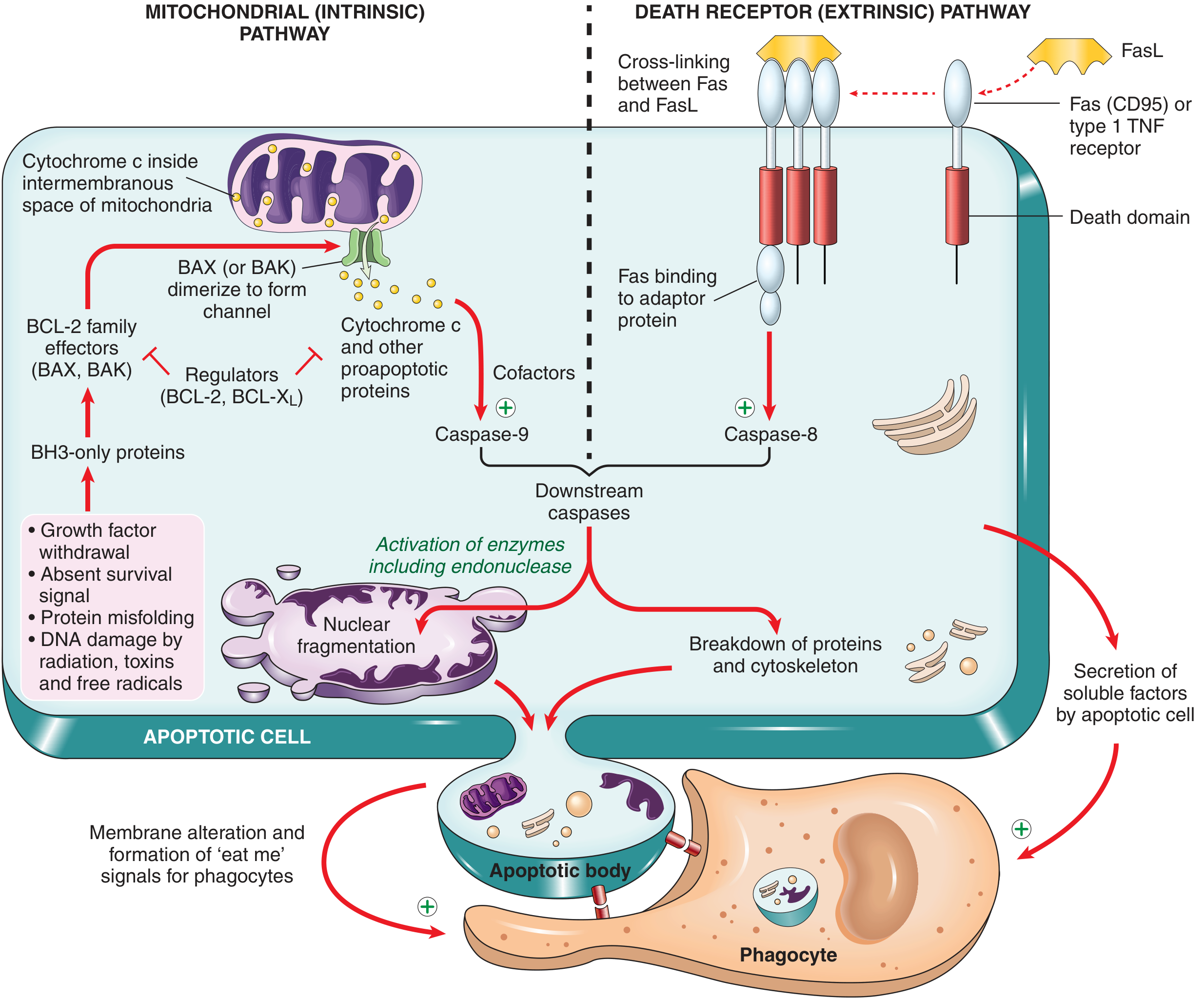
1. Mitochondrial (Intrinsic) Pathway
This is the dominant pathway in most physiologic and pathologic apoptosis.
Triggers:
- Growth factor or survival signal withdrawal
- DNA damage (radiation, toxins, free radicals)
- Protein misfolding / ER stress
- Hypoxia, nutrient deprivation
Mechanism:
- Stress signals activate BH3-only proteins (sensors of the BCL-2 family, e.g., BID, BAD, BIM, PUMA, NOXA)
- BH3-only proteins shift the balance toward proapoptotic effectors BAX and BAK, which dimerize and insert into the outer mitochondrial membrane, forming channels
- Mitochondrial outer membrane permeabilization (MOMP) allows cytochrome c to leak into the cytosol
- Cytochrome c + APAF-1 + procaspase-9 form the apoptosome
- The apoptosome activates caspase-9, which cleaves and activates executioner caspases 3 and 7
Anti-apoptotic checkpoint: BCL-2 and BCL-XL (induced by growth factors) normally hold BAX/BAK in check and maintain mitochondrial membrane integrity. Cancer cells often overexpress BCL-2 (e.g., follicular lymphoma - t(14;18) translocation).
2. Death Receptor (Extrinsic) Pathway
Triggered by ligation of cell surface death receptors of the TNF receptor superfamily, including:
- Fas (CD95) bound by FasL
- TNFR1 bound by TNF
Mechanism:
- Receptor cross-linking induces clustering of death domains in the cytoplasmic tails
- Adaptor proteins (e.g., FADD) are recruited, forming the DISC (Death-Inducing Signaling Complex)
- DISC activates caspase-8 (initiator caspase)
- Caspase-8 directly activates executioner caspases 3 and 7
Note: Caspase-8 can also cleave BID to generate tBID, which amplifies the signal through the mitochondrial pathway - this is the crossover point between the two pathways.
Downstream Execution
Once executioner caspases (3 and 7) are active, they cleave:
- Endonucleases - activate DNase that cleaves DNA at internucleosomal linker regions (produces the classic "DNA ladder" on gel electrophoresis)
- Nuclear lamins - causes nuclear fragmentation
- Cytoskeletal proteins - causes cell shape changes and blebbing
- IAP inhibitors - amplifies caspase activity
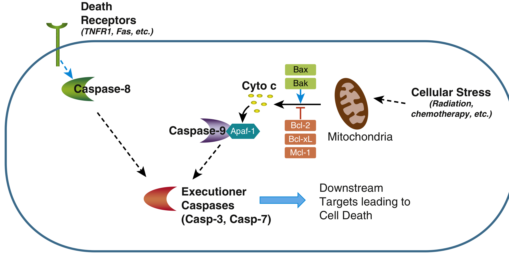
BCL-2 Family - The Master Regulators
| Subfamily | Members | Function |
|---|---|---|
| Anti-apoptotic | BCL-2, BCL-X | Prevent MOMP; inhibit BAX/BAK |
| Proapoptotic effectors | BAX, BAK | Form pores in mitochondrial membrane |
| BH3-only sensors | BIM, BID, BAD, PUMA, NOXA | Activate BAX/BAK; neutralize BCL-2 |
Physiologic Roles of Apoptosis
| Situation | Mechanism |
|---|---|
| Embryonic development (tissue patterning) | Growth factor withdrawal |
| Turnover of intestinal epithelium, lymphocytes | Loss of survival signals |
| Involution of hormone-dependent tissues (endometrium) | Reduced hormone levels |
| Decline of immune cells after an immune response | Loss of survival signals |
| Deletion of self-reactive lymphocytes (central tolerance) | Fas/FasL and mitochondrial pathways |
Pathologic Apoptosis
- Increased apoptosis: neurodegenerative diseases (Alzheimer's, Parkinson's - loss of neurons), viral infections (cytotoxic T lymphocytes kill infected cells via Fas/perforin-granzyme), ischemia-reperfusion injury
- Deficient apoptosis: cancer (BCL-2 overexpression, p53 mutations impair BH3-only protein induction), autoimmune diseases (failure to eliminate self-reactive lymphocytes - mutations in Fas cause autoimmune lymphoproliferative syndrome, ALPS)
Caspase Classification
| Type | Members | Role |
|---|---|---|
| Initiator caspases | Caspase-8, -9 | Activated by DISC or apoptosome; activate executioners |
| Executioner caspases | Caspase-3, -7 | Cleave downstream substrates causing cell death |
Caspases are synthesized as inactive zymogens (procaspases) and activated by proteolytic cleavage - either by another caspase or by focal accumulation of procaspase monomers that self-activate.
Clinical Relevance
- Venetoclax - BCL-2 inhibitor; used in CLL and AML (restores apoptosis in cancer cells)
- TRAIL-based therapies - activate death receptors selectively in tumor cells
- p53 is the chief activator of proapoptotic BH3-only proteins (e.g., PUMA) in response to DNA damage; loss of p53 (the most common mutation in cancer) disables this checkpoint
- Granzyme B from cytotoxic T cells enters target cells via perforin pores and directly activates caspases
Sources:
- Robbins & Kumar Basic Pathology, p. 24-26
- Sleisenger and Fordtran's Gastrointestinal and Liver Disease, p. 23
Recent reviews: Mustafa et al. (2024) provide a comprehensive update on apoptosis signaling in Cells, and Moyer et al. (2025) review apoptosis in cancer biology in Annual Review of Pathology - both consistent with the mechanisms described above.
necrosis
Finding Sources
Searching PubMed
necrosis cell death mechanisms pathology
Reading File
Reading File
Now let me read the morphologic types of necrosis from Robbins (they start around line 220+) and fetch the key images.
Reading File
Excellent - I have all the content I need. Let me now fetch the key histology images for the necrosis types.
Finding Sources
I have all the images and content. Let me fetch the key pathology images to display them.
Loading Image
Loading Image
Loading Image
I have all the material needed. Here is the full, detailed answer on necrosis:
Necrosis
Necrosis is a form of pathologic cell death resulting from severe, irreversible cell injury. It is distinguished from apoptosis by uncontrolled membrane failure, spillage of cellular contents into the extracellular space, and the invariable triggering of an inflammatory response.
Definition and Basic Mechanism
In necrosis, cellular membranes break down, lysosomal enzymes leak out and digest the cell from within, and leaked cellular contents trigger inflammation. The enzymes responsible for digesting dead cells come from two sources:
- Disrupted lysosomes of the dying cells themselves
- Leukocytes recruited as part of the inflammatory response
The released contents include damage-associated molecular patterns (DAMPs) - ATP (from damaged mitochondria), uric acid (DNA breakdown product), and other intracellular molecules normally confined in healthy cells. These bind receptors on macrophages and other cells, triggering phagocytosis and cytokine-driven inflammation.
Causes of Necrosis
- Ischemia (most common) - loss of oxygen supply
- Microbial toxins and infections
- Burns and chemical/physical injury
- Severe hypoxia
- Acute pancreatitis (proteases leak from acinar cells and damage surrounding tissue)
Histologic Features of Necrotic Cells
Cytoplasmic Changes
- Increased eosinophilia (H&E staining) - due to denatured cytoplasmic proteins binding eosin, and loss of RNA (which normally produces basophilia)
- Glassy/homogeneous appearance - loss of glycogen
- Vacuolated, moth-eaten cytoplasm - when organelles are digested
- Myelin figures - whorled phospholipid precipitates from disrupted membranes; can be phagocytosed or calcify
Nuclear Changes (three patterns)
| Pattern | Description |
|---|---|
| Pyknosis | Nuclear shrinkage + increased basophilia; DNA condenses into a dark, shrunken mass |
| Karyorrhexis | Fragmentation of the pyknotic nucleus |
| Karyolysis | Basophilia fades due to DNase digestion of DNA; nucleus dissolves over 1-2 days |
Ultrastructural (EM) Changes
- Discontinuities in plasma and organelle membranes
- Marked mitochondrial dilation with large amorphous intramitochondrial densities
- Disrupted lysosomes
- Intracytoplasmic myelin figures
Morphologic Patterns of Tissue Necrosis
Six distinct patterns are recognized. Most have characteristic gross appearances; fibrinoid necrosis is identified only microscopically.
1. Coagulative Necrosis
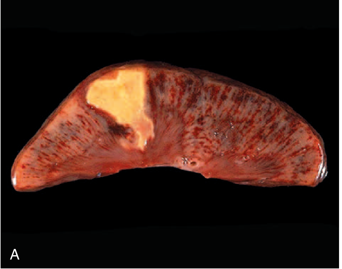
The most common type. The underlying tissue architecture is preserved for several days after injury because the injury denatures both structural proteins and enzymes, halting proteolysis. Affected tissues take on a firm texture. Eosinophilic, anucleate "ghost cells" persist before leukocytes eventually clear the debris.
Cause: Ischemia/infarction in most solid organs
Exception: Brain infarcts cause liquefactive (not coagulative) necrosis
Example: Myocardial infarct, renal infarct
2. Liquefactive Necrosis
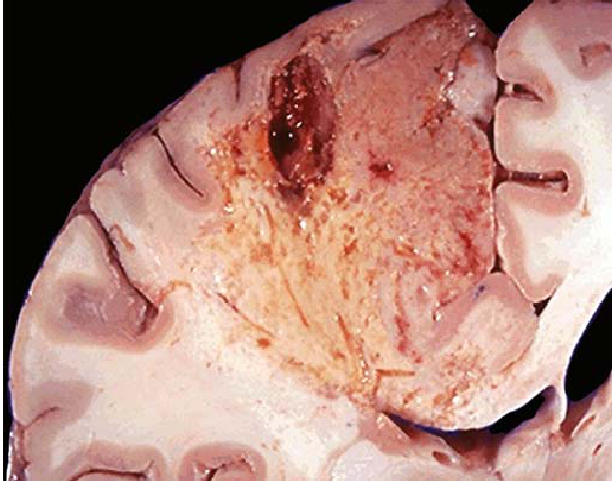
Dead cells are completely digested, converting tissue into a viscous liquid that is removed by phagocytes. When caused by bacterial infection, the material is creamy yellow (pus); a localized collection is an abscess.
Causes:
- Bacterial or fungal infections (leukocyte enzymes digest tissue)
- Brain infarction (for unknown reasons, CNS hypoxia always causes this pattern)
Gross: Soft, fluid-filled cavity
Microscopic: Liquefied tissue, abundant neutrophils
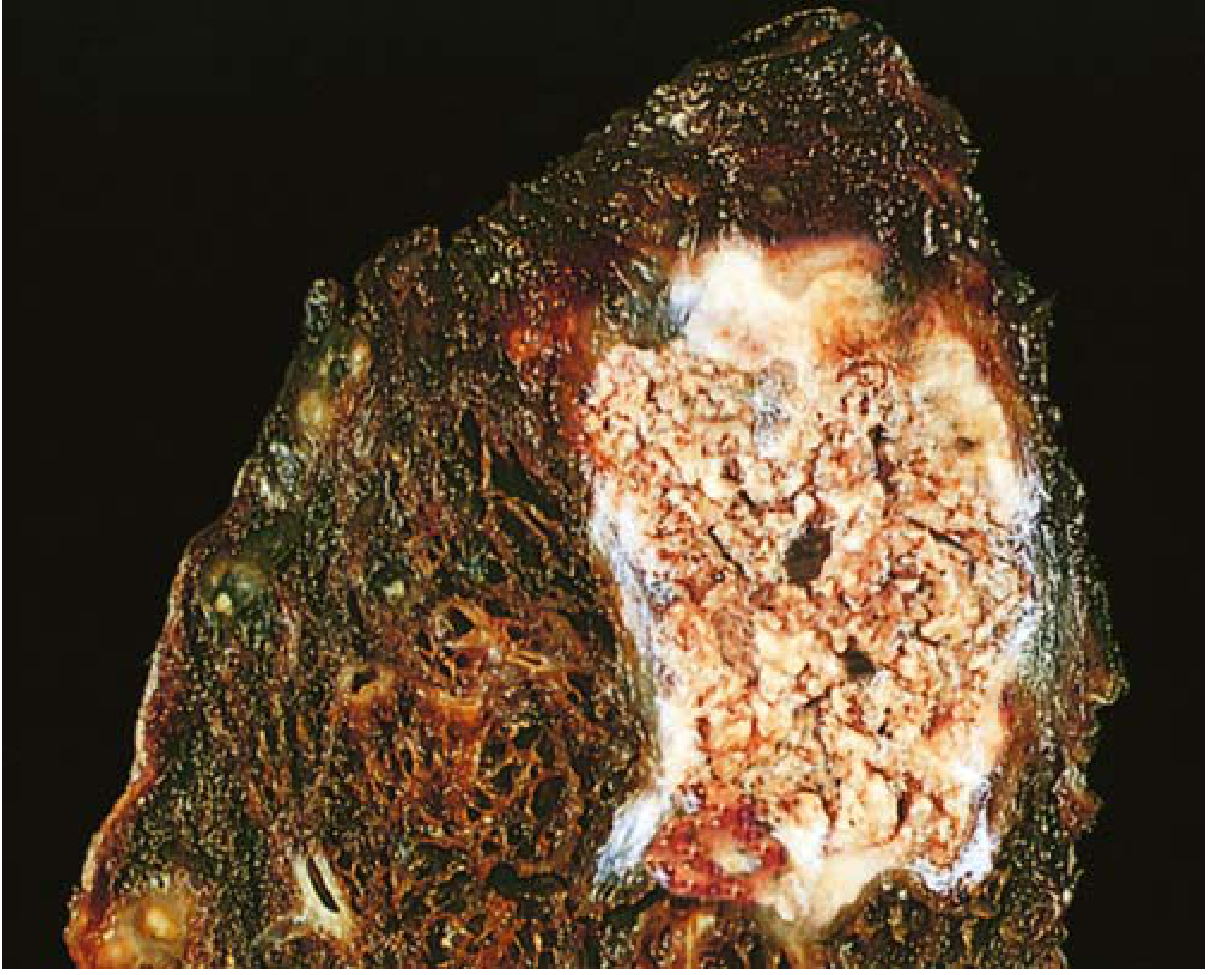
3. Caseous Necrosis
"Cheese-like" - named for its friable, yellow-white gross appearance. On microscopy, necrotic tissue appears as amorphous granular pink material with obliterated tissue architecture (unlike coagulative necrosis, no "ghost" cell outlines remain). The focus is typically surrounded by macrophages and other immune cells forming a granuloma.
Cause: Tuberculosis (classic), other mycobacterial infections, some fungal infections
Gross: Soft, crumbling, yellow-white debris
4. Fat Necrosis
Focal destruction of adipose tissue. Released lipases (from traumatic or pancreatitis-related acinar cell rupture) cleave triglycerides in fat cells. The released fatty acids combine with calcium to form chalky white deposits (saponification).
Causes: Acute pancreatitis, abdominal trauma
Gross: Chalky white opaque spots in mesenteric/peripancreatic fat
Microscopic: Shadowy fat cell outlines + granular basophilic calcium deposits + inflammatory reaction
5. Fibrinoid Necrosis
A special form visible only by light microscopy (no gross correlate). Deposited immune complexes and leaked plasma proteins accumulate in vessel walls, producing a bright pink, amorphous "fibrin-like" appearance on H&E.
Causes:
- Immune complex vasculitis (e.g., polyarteritis nodosa, SLE)
- Malignant hypertension (severe hypertensive arteriopathy) Microscopic: Circumferential bright pink amorphous material in vessel walls
6. Gangrenous Necrosis
Not a distinct histologic pattern - a clinical term for ischemic limb necrosis involving multiple tissue layers. Two subtypes:
| Type | Description |
|---|---|
| Dry gangrene | Coagulative necrosis; no bacterial superinfection; mummified, dry appearance |
| Wet gangrene | Coagulative + liquefactive necrosis from bacterial superinfection; moist, foul-smelling; more dangerous |
Summary Table
| Type | Cause | Location | Gross Appearance | Microscopic |
|---|---|---|---|---|
| Coagulative | Ischemia | All solid organs except brain | Firm, pale, well-demarcated | Ghost cells, preserved architecture |
| Liquefactive | Bacteria, fungi; CNS ischemia | Abscesses; brain | Soft, fluid, pus | Dissolved tissue, neutrophils |
| Caseous | TB, fungi | Lung, lymph nodes | Friable, yellow-white | Amorphous pink debris, granuloma |
| Fat | Pancreatitis, trauma | Mesentery, breast | Chalky white spots | Shadow fat cells, Ca deposits |
| Fibrinoid | Vasculitis, malignant HTN | Vessel walls | None (microscopy only) | Bright pink amorphous deposits |
| Gangrenous | Ischemia ± infection | Limbs | Mummified (dry) or wet/foul | Coagulative ± liquefactive |
Clinical Significance: Serum Biomarkers of Necrosis
The membrane disruption of necrosis releases tissue-specific intracellular proteins into the bloodstream, forming the basis of widely used biomarkers:
| Tissue | Biomarker | Clinical use |
|---|---|---|
| Cardiac muscle | Troponin I/T (cardiac-specific isoforms) | Detected as early as 2 hours post-MI; gold standard for MI diagnosis |
| Hepatocytes | ALT, AST (transaminases) | Hepatocellular necrosis |
| Bile duct epithelium | Alkaline phosphatase (thermostable isoform) | Cholestatic injury |
| Skeletal muscle | CK-MM, myoglobin | Rhabdomyolysis |
Necrosis vs. Apoptosis - Key Differences
| Feature | Necrosis | Apoptosis |
|---|---|---|
| Cause | Severe pathologic injury | Physiologic or controlled pathologic signals |
| Membrane integrity | Lost | Maintained until phagocytosis |
| Cell size | Swells | Shrinks |
| Inflammation | Yes - always | No |
| DNA fragmentation | Random, smeared | Internucleosomal ("DNA ladder") |
| Caspases | No | Yes |
| Fate of contents | Leak into ECM | Packaged in apoptotic bodies |
Sources:
- Robbins & Kumar Basic Pathology, p. 21-27
- Robbins, Cotran & Kumar Pathologic Basis of Disease, p. 1377-1390
okky i want a suggestio ....what if i study a topic from usmle first aid first then watch baunn prakesh youtube lecture with pathoma and the study the topic from robbins
This is a solid instinct, and the sequence you're describing is actually a well-known high-yield approach. Here's an honest breakdown:
Your Proposed Flow
First Aid → Bhanu Prakash (YouTube) + Pathoma → Robbins
What Works Well About This
First Aid first is smart. It gives you the "skeleton" - the testable facts, buzzwords, and high-yield associations. You know what matters before you go deep. This prevents the classic mistake of drowning in Robbins detail without knowing what's actually tested.
Bhanu Prakash + Pathoma is an excellent pairing for the visual/conceptual layer. Bhanu Prakash's drawings make mechanisms stick visually. Pathoma (Husain Sattar) bridges the "why" between basic pathology and clinical disease - written and video together reinforce each other well.
Robbins last is the right place for it. By the time you open Robbins, you already have the framework, so you're not building from zero - you're adding depth, context, and understanding to something you already know. This makes Robbins far less overwhelming.
One Honest Concern
This sequence is time-heavy. Four passes over every topic means you need to be disciplined about:
- Not re-reading everything at equal depth each pass
- Keeping First Aid and Pathoma as your "home base" - Robbins is supplemental, not mandatory for every topic
- Some topics (e.g., basic cell injury, inflammation, neoplasia) reward deep Robbins reading. Others (e.g., many organ system minutiae) do not need it.
A Small Suggested Tweak
Consider this tighter version:
| Step | Resource | Purpose |
|---|---|---|
| 1 | First Aid | High-yield skeleton, buzzwords |
| 2 | Pathoma (book + video) | Mechanism + clinical reasoning |
| 3 | Bhanu Prakash | Visual reinforcement, mnemonics |
| 4 | Robbins (selective) | Only for topics you still don't fully understand |
Swapping Pathoma before Bhanu Prakash makes slightly more sense because Pathoma builds the conceptual framework, and Bhanu Prakash then reinforces it visually. But honestly, the difference is minor - both together work regardless of order.
Bottom Line
Your approach is good and defensible. The sequence makes logical sense. The only trap is treating all four resources as equally mandatory for every single topic - that path leads to burnout. Use Robbins as a reference and reward, not a requirement. Keep First Aid + Pathoma as your core, and you'll be in strong shape.
free radical injury
Finding Sources
Finding Sources
Finding Sources
Good. Best sources are Basic Medical Biochemistry and Robbins. Let me read both in parallel, plus the Robbins section on free radical injury.
Reading File
Finding Sources
Reading File
Reading File
Excellent content. Now let me read the Robbins section on antioxidant defenses and the effects of ROS, plus fetch the key diagram.
Reading File
Reading File
Loading Image
Now let me get the antioxidant defense section and the cellular damage diagram from Basic Medical Biochemistry.
Reading File
Loading Image
I now have all the content and images needed for a comprehensive answer. Here it is:
Free Radical Injury
What is a Free Radical?
A free radical is any molecule or atom that contains one or more unpaired electrons in its outer orbital. This makes it highly unstable and chemically reactive - it aggressively extracts electrons from neighboring molecules, turning them into radicals and triggering a self-perpetuating chain reaction of damage.
Oxygen is itself a biradical (two unpaired electrons), and it readily accepts single electrons to form the most important class of free radicals in biology: Reactive Oxygen Species (ROS).
The Principal ROS in Cell Injury
| Species | Nature | Generation | Removal |
|---|---|---|---|
| Superoxide (O₂⁻) | Radical | Incomplete O₂ reduction in mitochondria (CoQ); NADPH oxidase in leukocytes | SOD → H₂O₂ |
| Hydrogen peroxide (H₂O₂) | Non-radical (but reactive precursor) | From O₂⁻ via SOD; peroxisomal oxidases | Catalase, glutathione peroxidase → H₂O |
| Hydroxyl radical (•OH) | Most reactive and damaging | Fenton reaction: H₂O₂ + Fe²⁺ → •OH + OH⁻ + Fe³⁺; also from ionizing radiation hydrolyzing water | Glutathione, vitamin E |
| Hypochlorite (OCl⁻) | Reactive | H₂O₂ + Cl⁻ via myeloperoxidase (MPO) in neutrophils | - |
| Nitric oxide (NO•) | Radical | Nitric oxide synthase (NOS) from arginine | Reacts with O₂⁻ to form peroxynitrite (ONOO⁻) |
Key reactions to know:
- Fenton reaction: H₂O₂ + Fe²⁺ → •OH + OH⁻ + Fe³⁺ (the hydroxyl radical is generated)
- Haber-Weiss reaction: O₂⁻ + H₂O₂ → •OH + OH⁻ + O₂ (superoxide reduces Fe³⁺ back to Fe²⁺, enabling cycling)
Sources / Generation of Free Radicals
1. Mitochondrial Oxidative Phosphorylation (Normal Metabolism)
~3-5% of all O₂ consumed is converted to ROS. Electrons "leak" from coenzyme Q (CoQH•) in the electron transport chain and reduce dissolved O₂ to superoxide. Normal aerobic metabolism is therefore a constant source of low-level ROS.
2. Phagocytic Leukocytes (Respiratory Burst)
Neutrophils and macrophages deliberately generate ROS to kill microbes:
- NADPH oxidase (phagocyte oxidase) in the phagolysosomal membrane produces large amounts of O₂⁻ → H₂O₂
- Myeloperoxidase (MPO) converts H₂O₂ + Cl⁻ → hypochlorite (bleach) - the most potent antimicrobial ROS
- ROS released from leukocytes can cause collateral tissue injury in inflammation
3. Cytochrome P450 Enzymes
CYP enzymes transfer single electrons to substrates, with risk of accidentally releasing free-radical intermediates. This is amplified by enzyme induction from alcohol, drugs, and toxins.
- Classic example: CCl₄ (carbon tetrachloride) → CCl₃• radical by CYP → hepatocyte necrosis (lipid peroxidation of ER membranes)
4. Exogenous Sources
- Ionizing radiation (X-rays, γ-rays) - hydrolyzes water → •OH + H•
- UV radiation
- Air pollutants, herbicides (e.g., paraquat)
- Ischemia-reperfusion - xanthine oxidase converts hypoxanthine + O₂ → uric acid + superoxide upon reperfusion
Mechanisms of Cell Injury by Free Radicals
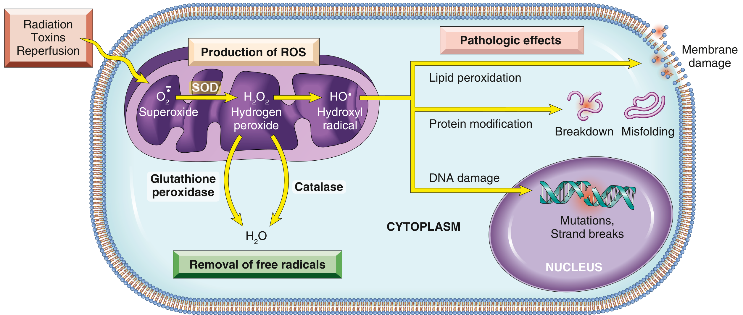
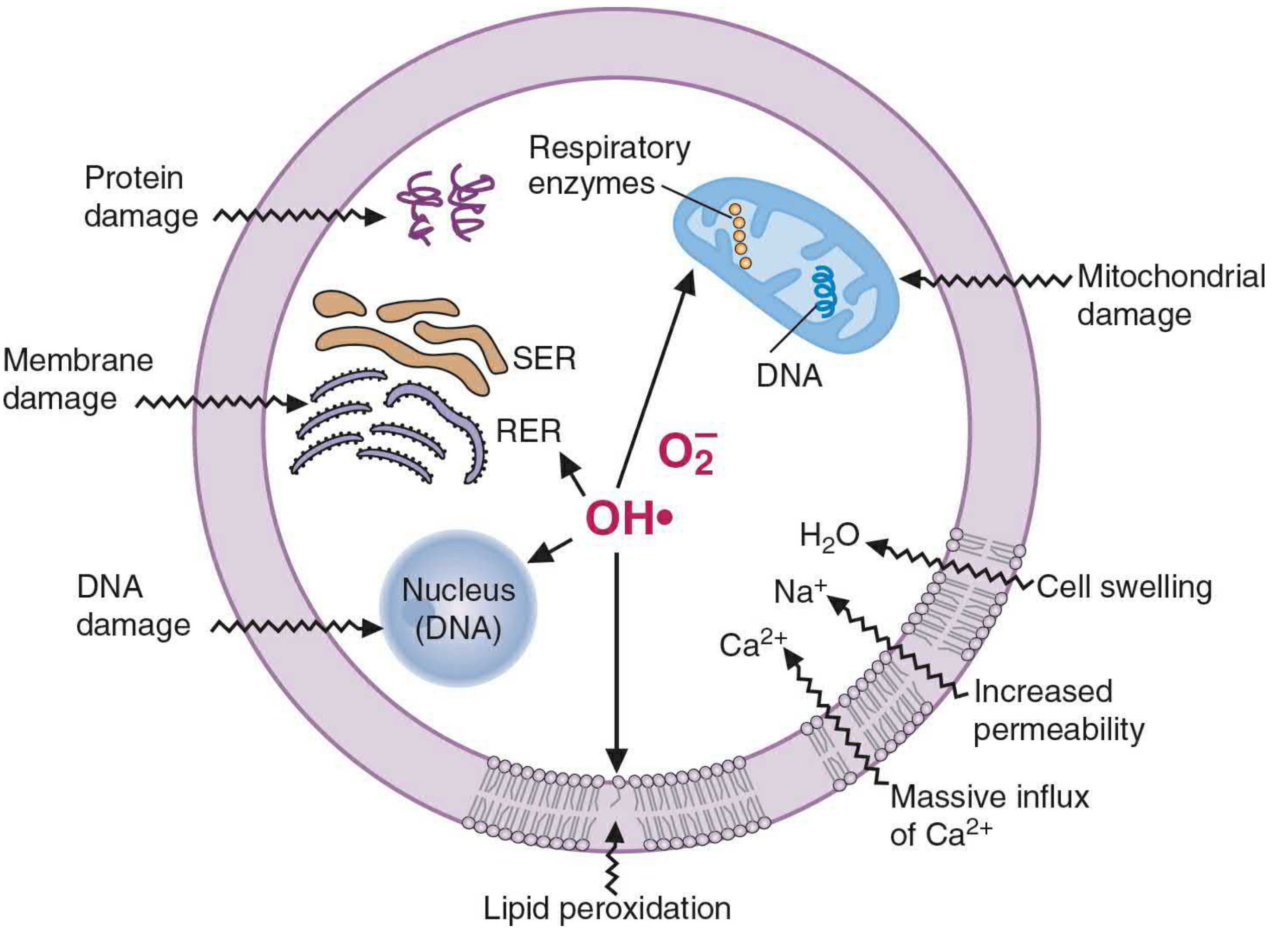
1. Lipid Peroxidation (Most Important)
- •OH attacks polyunsaturated fatty acids (PUFAs) in membrane phospholipids - extracts a hydrogen atom
- Creates a lipid radical (L•) → reacts with O₂ → lipid peroxyl radical (LOO•)
- LOO• attacks adjacent fatty acids → autocatalytic chain reaction propagates
- Products: malondialdehyde (MDA) (urinary/serum biomarker of oxidative damage), ethane, pentane
- Result: damage to plasma membrane, mitochondrial membrane, ER membrane → increased permeability → massive Ca²⁺ influx → cell death
2. Protein Damage
- •OH oxidizes sulfhydryl (-SH) groups on cysteine residues → disulfide bridges → crosslinking → protein aggregation and degradation
- Oxidation of other amino acid residues (methionine → methionine sulfoxide; seen in lens cataracts)
- Damaged proteins may misfold → triggers the unfolded protein response (UPR)
- Direct polypeptide fragmentation can also occur
3. DNA Damage
- •OH attacks the deoxyribose backbone → single- and double-strand breaks
- Base modifications: guanine → 8-hydroxyguanine (commonly measured oxidative DNA lesion)
- Localized by Fe²⁺ binding to DNA (Fenton reaction near DNA = very damaging)
- Consequences: mutations, apoptosis, malignant transformation, aging
4. Calcium Dysregulation (Indirect)
- Lipid peroxidation of plasma and ER membranes → membrane permeability ↑ → Ca²⁺ influx
- Excess cytosolic Ca²⁺ activates proteases and phospholipases → amplifies membrane damage
Antioxidant Defenses
The cell has layered enzymatic and non-enzymatic defenses to neutralize ROS before they cause damage. Oxidative stress = when ROS production overwhelms these defenses.
Enzymatic Antioxidants
| Enzyme | Reaction | Location |
|---|---|---|
| Superoxide dismutase (SOD) | 2 O₂⁻ + 2H⁺ → H₂O₂ + O₂ | Mitochondria (Mn-SOD), cytoplasm (Cu/Zn-SOD) |
| Catalase | 2 H₂O₂ → 2 H₂O + O₂ | Peroxisomes |
| Glutathione peroxidase (GPx) | H₂O₂ + 2 GSH → GSSG + 2 H₂O | Cytoplasm, mitochondria |
Non-Enzymatic (Free-Radical Scavengers)
| Antioxidant | Mechanism | Notes |
|---|---|---|
| Vitamin E (α-tocopherol) | Fat-soluble, donates H to lipid peroxyl radicals; terminates lipid peroxidation chain | In membranes |
| Vitamin C (ascorbate) | Water-soluble; scavenges O₂⁻, •OH; regenerates vitamin E | In cytoplasm |
| Glutathione (GSH) | Donates electron to neutralize H₂O₂ and lipid peroxides; major cellular redox buffer | Kept reduced by glutathione reductase using NADPH |
| β-Carotene / vitamin A | Quenches singlet oxygen and lipid radicals | Lipid soluble |
| Selenium | Cofactor for GPx | Essential micronutrient |
| Ceruloplasmin, transferrin | Bind free iron/copper → prevent Fenton reaction | Sequester transition metals |
Reactive Nitrogen-Oxygen Species (RNOS)
- Nitric oxide (NO•) at low concentrations: physiologic neurotransmitter and vasodilator (produced by eNOS)
- At high concentrations (from iNOS in inflammation): NO + O₂⁻ → peroxynitrite (ONOO⁻) - a potent oxidant
- RNOS cause protein nitration (nitrotyrosine formation), DNA damage, lipid peroxidation
- Implicated in: Parkinson disease, rheumatoid arthritis, septic shock, neurodegeneration
Diseases Associated with Free Radical Injury
| Category | Examples |
|---|---|
| Cardiovascular | Atherogenesis, ischemia-reperfusion injury (MI, stroke) |
| Neurologic | Parkinson disease, Alzheimer disease, ALS, multiple sclerosis, cerebrovascular disease |
| Metabolic | Diabetes, alcohol-induced liver disease (CCl₄-like mechanism via CYP2E1) |
| Pulmonary | COPD, retrolental fibroplasia (O₂ toxicity in premature neonates) |
| Renal | Acute renal failure |
| Genetic/other | Duchenne muscular dystrophy, Down syndrome, OXPHOS (mitochondrial) diseases |
| Aging | Free radical theory of aging - cumulative oxidative damage to DNA, proteins, lipids |
Ischemia-Reperfusion Injury - High-Yield Integration
This is a classic exam scenario combining free radical injury with ischemia:
- During ischemia: ATP depletes → xanthine dehydrogenase converted to xanthine oxidase; hypoxanthine accumulates
- On reperfusion: O₂ rushes in → xanthine oxidase converts hypoxanthine + O₂ → uric acid + superoxide
- Superoxide burst → lipid peroxidation → membrane damage → necrosis
- Simultaneously: reperfusion restores electron transport, but the reduced mitochondrial electron carriers dump electrons onto O₂ → more superoxide
- Neutrophil influx → respiratory burst → further ROS release
This is why post-MI reperfusion injury occurs and is a target for antioxidant therapy research.
Sources:
- Robbins & Kumar Basic Pathology, p. 29-30
- Basic Medical Biochemistry - A Clinical Approach, 6e, p. 898-911 (Chapter 25)
is nuberal forte use for back pain
Let me look this up - "Nuberal Forte" isn't a name I can place with certainty from memory.
Searching sources...
Searching the Web
Nuberal Forte uses ingredients back pain
Yes, Nuberol Forte is used for back pain. Here's everything you need to know:
What is Nuberol Forte?
Nuberol Forte is a combination tablet containing:
- Paracetamol (Acetaminophen) 650 mg - analgesic and antipyretic
- Orphenadrine citrate 50 mg - skeletal muscle relaxant with anticholinergic properties
Manufactured by Searle Company Ltd (Pakistan). Dose: 1 tablet three times daily.
Why It Works for Back Pain
The combination is specifically effective for back pain because back pain is often a mix of two components:
- Pain - addressed by paracetamol
- Muscle spasm - addressed by orphenadrine (muscle relaxant)
The two drugs together provide greater pain relief than either drug alone - clinical studies confirm the combination has enhanced antinociceptive activity and longer duration of action.
Approved Indications (Relevant to Back Pain)
- Chronic low back pain
- Prolapsed intervertebral disc (slipped disc)
- Painful skeletal muscle spasm
- Sprains, strains, whiplash injuries
- Acute torticollis
- Non-articular rheumatism
- Tension and occipital headaches (neck muscle spasm)
A clinical study published on PMC (NFORTE-EFFECT) with 399 patients showed a significant reduction in pain scores (VAS 6.18 → 3.70, p<0.05) in musculoskeletal conditions including backache.
Important Contraindications
- Glaucoma
- Prostatic hypertrophy / bladder neck obstruction
- Myasthenia gravis
- Esophageal spasm, pyloric or duodenal obstruction
- Hypersensitivity to paracetamol or orphenadrine
- Pregnancy and lactation - not recommended
- Children - not recommended
Common Side Effects
These are mainly from the orphenadrine (anticholinergic) component:
| Side Effect | Reason |
|---|---|
| Dry mouth | Anticholinergic |
| Blurred vision | Anticholinergic |
| Urinary retention | Anticholinergic |
| Tachycardia / palpitations | Anticholinergic |
| Dizziness, sedation | CNS effect |
| Nausea, vomiting | GI |
Bottom Line
Yes - Nuberol Forte is well-suited for back pain, particularly when muscle spasm is a contributing factor (which it usually is in most cases of back pain). For severe pain, it is sometimes co-prescribed with an NSAID like diclofenac or aceclofenac for added anti-inflammatory effect.
if person is contionuslt having low heatbeat is it dangerous
A continuously low heartbeat (called bradycardia - heart rate below 60 bpm) can be either completely normal or genuinely dangerous, depending on the cause and whether symptoms are present.
When a Low Heartbeat is NORMAL
Some people naturally have a slow heart rate and it is not dangerous at all:
- Athletes and fit people - trained hearts pump more efficiently, so a resting rate of 40-60 bpm is common and healthy
- During sleep - heart rate naturally drops
- Some medications - beta-blockers (used for blood pressure/heart conditions) intentionally slow the heart
If the person has a slow heart rate but feels completely fine - no symptoms, no dizziness, no fatigue - it is generally not a concern.
When a Low Heartbeat IS Dangerous
It becomes dangerous when the heart is beating too slowly to pump enough blood to the brain and body. Warning signs to watch for:
| Symptom | What It Means |
|---|---|
| Dizziness or lightheadedness | Brain not getting enough blood |
| Fainting (syncope) | Serious - brain blood flow critically low |
| Chest pain or pressure | Heart muscle may be affected |
| Shortness of breath | Poor cardiac output |
| Extreme fatigue / weakness | Organs not getting enough oxygen |
| Confusion or memory problems | Reduced brain perfusion |
Common Causes of Dangerous Bradycardia
- Heart block - electrical signal between upper and lower heart chambers is blocked
- Sick sinus syndrome - the heart's natural pacemaker malfunctions
- Heart attack - especially affecting the right coronary artery (which supplies the SA node)
- Electrolyte imbalances - high potassium (hyperkalemia) can critically slow the heart
- Hypothyroidism - underactive thyroid slows metabolism including heart rate
- Overdose - beta-blockers, calcium channel blockers, digoxin
- Hypothermia - very low body temperature
At What Rate Does It Become an Emergency?
| Heart Rate | Concern Level |
|---|---|
| 50-60 bpm, no symptoms | Usually fine, especially in fit individuals |
| 40-50 bpm, no symptoms | Warrants medical evaluation |
| Below 40 bpm | Concerning even without symptoms |
| Any rate with fainting, chest pain, or breathlessness | Medical emergency - seek care immediately |
Bottom Line
The symptoms matter more than the number alone.
- Slow heart rate + no symptoms = often normal, but worth checking once with a doctor
- Slow heart rate + any symptoms (fainting, dizziness, chest pain, breathlessness) = see a doctor urgently
- Slow heart rate + collapse or loss of consciousness = call emergency services immediately
A doctor will do an ECG (electrocardiogram) to see the electrical pattern of the heart and determine whether the cause is benign or requires treatment (such as a pacemaker for severe cases).
inflamation
Finding Sources
Finding Sources
Finding Sources
Reading File
Finding Sources
Reading File
Reading File
Now let me read the mediators of inflammation, leukocyte recruitment, and chronic inflammation cells sections, and fetch the key diagrams.
Reading File
Reading File
Now let me fetch the key diagrams and read the mediators and granuloma sections.
Finding Sources
Reading File
Now let me fetch the key inflammation diagrams.
Loading Image
Reading File
I now have comprehensive content. Let me read the arachidonic acid mediators and systemic effects sections quickly.
Reading File
Excellent - I have all the content I need. Let me now fetch the arachidonic acid pathway diagram.
Loading Image
I have everything needed for a thorough answer. Here it is:
Inflammation
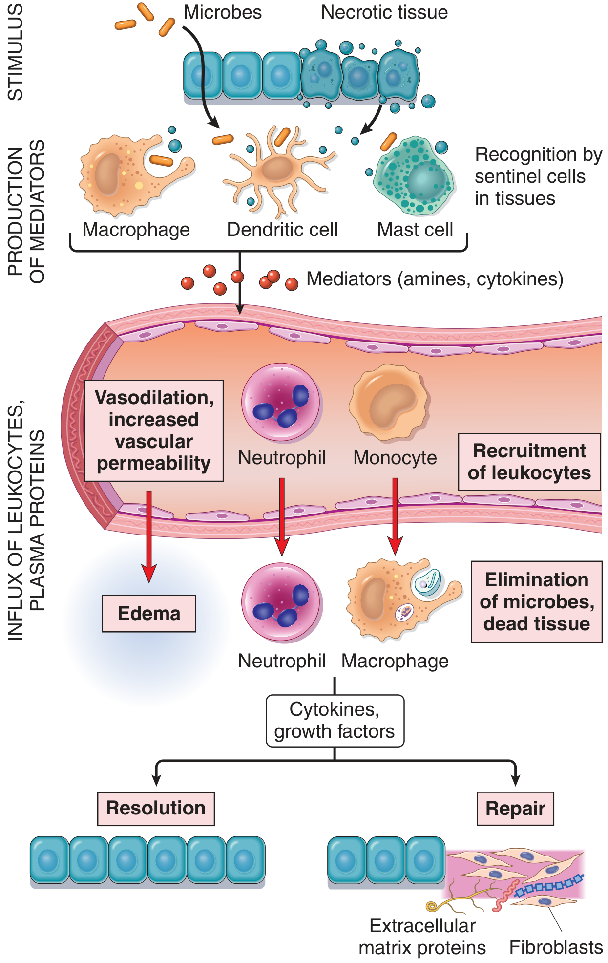
Inflammation is the response of vascularized tissues to infections and tissue damage that brings cells and molecules of host defense from the circulation to the site where they are needed, in order to eliminate the offending agent. It is fundamentally a protective response - without inflammation, infections would go unchecked and wounds would not heal. But inflammation can also cause harm when it is excessive, prolonged, or misdirected.
Cardinal Signs of Inflammation (5 Signs)
Described by Celsius (4 classical) + Virchow (5th):
| Sign | Latin | Mechanism |
|---|---|---|
| Redness (rubor) | Rubor | Vasodilation → increased blood flow |
| Heat (calor) | Calor | Vasodilation → more warm blood to tissue |
| Swelling (tumor) | Tumor | Increased vascular permeability → edema |
| Pain (dolor) | Dolor | Prostaglandins and bradykinin sensitize nerve endings |
| Loss of function (functio laesa) | Functio laesa | Combined effect of above |
Acute vs. Chronic Inflammation - Overview
| Feature | Acute | Chronic |
|---|---|---|
| Onset | Minutes to hours | Days to weeks |
| Duration | Hours to days | Weeks to months |
| Cellular infiltrate | Neutrophils | Macrophages + lymphocytes |
| Tissue injury | Usually mild, self-limited | May be severe and progressive |
| Fibrosis | None | Often present |
| Local/systemic signs | Prominent | Variable, usually modest |
ACUTE INFLAMMATION
Three Major Components
- Vascular dilation - occurs in arterioles; causes redness and warmth; driven by histamine and nitric oxide
- Increased vascular permeability - protein-rich fluid (exudate) leaks into tissues; causes edema; driven by endothelial cell contraction creating interendothelial gaps
- Leukocyte emigration - mainly neutrophils migrate from vessels into tissue
Vascular Changes in Detail
- Vasodilation → slows blood flow → blood becomes more viscous (stasis) → small vessels engorged with red cells (stasis)
- Increased permeability → exudate (high protein, cellular debris) leaks into tissue
- Exudate vs. Transudate: exudate = high protein + cells (inflammatory); transudate = low protein (hydrostatic/osmotic imbalance, not inflammatory)
- Edema = excess fluid; pus = exudate rich in neutrophils + dead cells + microbes
Leukocyte Recruitment - The Steps
This is a multi-step process, each step controlled by specific molecules:
| Step | Process | Molecules Involved |
|---|---|---|
| 1 | Margination | Stasis pushes leukocytes to vessel wall |
| 2 | Rolling | Loose adhesion to endothelium |
| 3 | Adhesion (firm) | Leukocyte sticks firmly to endothelium |
| 4 | Transmigration (diapedesis) | Leukocyte squeezes between endothelial cells |
| 5 | Chemotaxis | Migration through tissue toward injury |
Phagocytosis and Killing
After reaching the site:
- Recognition/attachment - pattern recognition receptors (TLRs) and opsonins (IgG, C3b) enhance binding
- Engulfment - pseudopods surround particle → phagosome formed → fuses with lysosomes → phagolysosome
- Killing - two main mechanisms:
- ROS (respiratory burst): NADPH oxidase generates superoxide → H₂O₂ → myeloperoxidase (MPO) converts H₂O₂ + Cl⁻ → hypochlorite (most potent)
- NO: nitric oxide synthase produces NO → reacts with superoxide → peroxynitrite
- Lysosomal enzymes: elastase, cathepsin G, collagenase, defensins
MEDIATORS OF INFLAMMATION
1. Vasoactive Amines
- Histamine - stored in mast cell granules; released by IgE, C3a/C5a, physical injury; causes vasodilation and increased permeability; acts on H1 receptors
- Serotonin - in platelets; similar to histamine
2. Arachidonic Acid Metabolites (Eicosanoids)
The most pharmacologically important pathway:
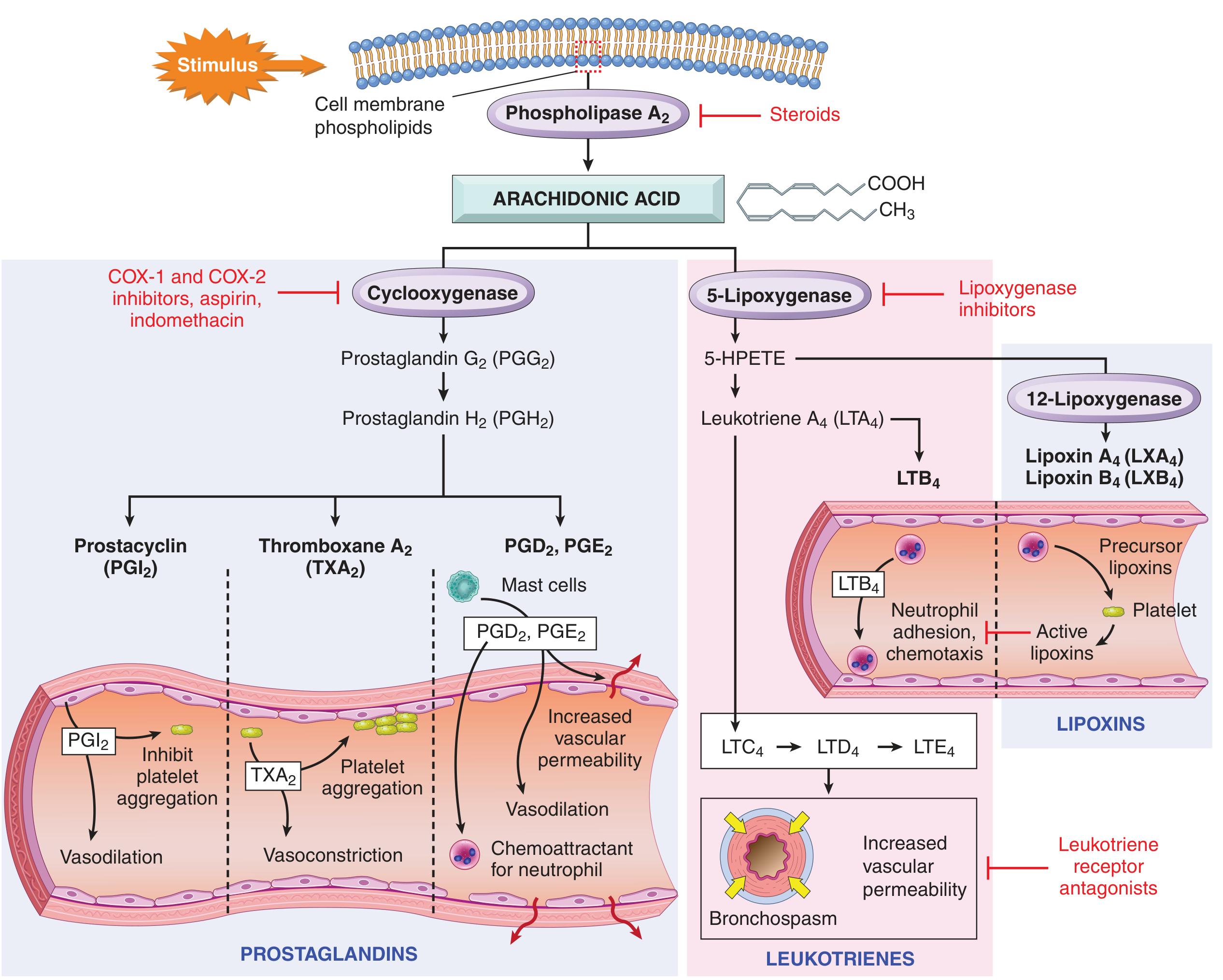
- Stimulus → Phospholipase A₂ releases arachidonic acid from membrane phospholipids
- Two downstream enzymes:
COX pathway → Prostaglandins & Thromboxane:
| Product | Effect |
|---|---|
| PGE₂, PGD₂ | Vasodilation, increased permeability, fever, pain sensitization |
| PGI₂ (prostacyclin) | Vasodilation, inhibits platelet aggregation |
| TXA₂ | Vasoconstriction, platelet aggregation |
5-Lipoxygenase pathway → Leukotrienes:
| Product | Effect |
|---|---|
| LTB₄ | Chemotaxis and neutrophil adhesion |
| LTC₄, LTD₄, LTE₄ | Vasoconstriction, increased permeability, bronchospasm (key in asthma) |
Lipoxins (LXA₄, LXB₄): anti-inflammatory; inhibit neutrophil recruitment; promote resolution
Pharmacologic blockade:
- Aspirin/NSAIDs → inhibit COX-1 and COX-2 → ↓ prostaglandins (pain, fever, inflammation)
- Selective COX-2 inhibitors (celecoxib) → anti-inflammatory, but risk of thrombosis (spare platelet TXA₂ while blocking endothelial PGI₂)
- Corticosteroids → block phospholipase A₂ → block entire AA pathway + suppress cytokine genes (IL-1, TNF, COX-2)
- Zileuton → 5-lipoxygenase inhibitor → ↓ leukotrienes (asthma)
- Montelukast → leukotriene receptor antagonist (asthma)
3. Cytokines and Chemokines
TNF-α and IL-1 - the two master cytokines of acute inflammation, produced mainly by macrophages and mast cells:
- Local effects: endothelial activation (upregulate adhesion molecules), leukocyte recruitment
- Systemic effects (acute phase response): fever (via PGE₂ in hypothalamus), acute phase protein synthesis (CRP, fibrinogen, SAA), leukocytosis, hypotension (shock in sepsis)
IL-6 - stimulates liver to produce acute phase proteins
Chemokines (IL-8/CXCL8, eotaxin, MCP-1) - specialized chemoattractant cytokines that guide leukocytes along concentration gradients to the site of injury
4. Complement System (C3a, C5a, C5b-9)
- C3a and C5a (anaphylatoxins) - stimulate mast cell degranulation → histamine release; vasodilation and increased permeability
- C5a - powerful chemotactic agent for neutrophils and monocytes; also activates leukocytes
- C5b-9 (membrane attack complex) - direct cell lysis
5. Other Mediators
| Mediator | Source | Action |
|---|---|---|
| Bradykinin | Plasma (kinin system) | Vasodilation, increased permeability, pain |
| PAF (platelet-activating factor) | Leukocytes, mast cells | Platelet aggregation, vasodilation, increased permeability |
| Nitric oxide (NO) | Endothelium, macrophages | Vasodilation, microbicidal |
Morphologic Patterns of Acute Inflammation
| Pattern | Appearance | Example |
|---|---|---|
| Serous | Watery, protein-poor fluid; no major cell exudate | Blister (skin burn), pleural effusion (early) |
| Fibrinous | Large exudate, fibrin meshwork; serious injury | Fibrinous pericarditis ("bread and butter"), fibrinous pleuritis |
| Purulent (suppurative) | Pus - neutrophils + necrotic debris; localized = abscess | Bacterial abscesses, empyema |
| Ulcer | Surface defect from necrosis + inflammation | Peptic ulcer, skin ulcer |
Outcomes of Acute Inflammation
Three possible outcomes:
- Complete resolution - offending agent eliminated, tissue returns to normal; debris cleared by macrophages, edema fluid drained by lymphatics (ideal outcome for mild/short-lived injury)
- Healing by fibrosis (scarring) - when tissue destruction is severe, affected tissue cannot regenerate, or abundant fibrin cannot be cleared
- Progression to chronic inflammation - when the injurious agent persists or normal healing is disrupted
CHRONIC INFLAMMATION
Chronic inflammation is a response of prolonged duration (weeks to months) in which inflammation, tissue injury, and repair coexist simultaneously. It may follow unresolved acute inflammation or begin insidiously without a preceding acute phase.
Causes
- Persistent infections - mycobacteria (TB), certain viruses, fungi, parasites (organisms that resist killing)
- Hypersensitivity/autoimmune diseases - self-sustaining immune reactions against self antigens (rheumatoid arthritis, multiple sclerosis, IBD)
- Prolonged exposure to toxic agents - silica dust (silicosis), asbestos, tobacco smoke
- Modern diseases - atherosclerosis, type 2 diabetes, Alzheimer disease (chronic low-grade "sterile" inflammation)
Morphologic Features (3 hallmarks)
- Mononuclear cell infiltrate - macrophages, lymphocytes, plasma cells
- Tissue destruction - caused by persistent offending agent or by inflammatory cells themselves
- Attempts at repair - angiogenesis + fibrosis → scarring
Key Cells
Macrophages - dominant cell type. Two activation states:
- M1 (classically activated) - stimulated by IFN-γ, microbial products → produce TNF, IL-12, NO, ROS → microbicidal and tissue damaging
- M2 (alternatively activated) - stimulated by IL-4, IL-13 → produce TGF-β, VEGF → anti-inflammatory, promote fibrosis and repair
Lymphocytes (T cells):
- Th1 → produce IFN-γ → activate M1 macrophages → amplify inflammation
- Th2 → produce IL-4, IL-5, IL-13 → helminth defense, allergy
- Th17 → produce IL-17 → recruit neutrophils; important in autoimmunity (RA, psoriasis, IBD)
- Bidirectional macrophage-T cell cross-talk perpetuates chronic inflammation
Eosinophils - in IgE-mediated reactions and parasitic infections; granules contain major basic protein (toxic to parasites, also damages epithelium)
Plasma cells - secrete antibodies at sites of chronic inflammation
Granulomatous Inflammation
A special form of chronic inflammation - a nodular collection of activated epithelioid macrophages, often with giant cells (fused macrophages), surrounded by lymphocytes.
Causes:
- Infectious: Tuberculosis (most important), leprosy, syphilis, cat-scratch disease, fungi (Histoplasma, Coccidioides)
- Non-infectious: Sarcoidosis, Crohn disease, silicosis, berylliosis, foreign body reaction
Key histologic features:
- Epithelioid macrophages (abundant pink cytoplasm, low-grade phagolysosomal activity)
- Langhans giant cells (peripheral nuclei in horseshoe pattern) or foreign body giant cells (nuclei randomly scattered)
- Central caseous necrosis (in TB) or no necrosis (in sarcoidosis)
- Surrounding collar of lymphocytes and fibroblasts
Systemic Effects of Inflammation (Acute Phase Response)
Driven mainly by cytokines IL-1, TNF-α, and IL-6:
| Effect | Mediator | Significance |
|---|---|---|
| Fever | PGE₂ (produced in hypothalamus by IL-1/TNF) | Inhibits bacterial growth, enhances immune response |
| Leukocytosis | IL-1, TNF → CSFs from marrow | WBC count elevated; neutrophilia in bacterial infections |
| Acute phase proteins | IL-6 → liver | CRP, fibrinogen, SAA, serum amyloid A; opsonization, complement activation |
| Elevated ESR | Fibrinogen causes RBC aggregation | Useful inflammatory marker |
| Hypotension/shock (severe) | TNF | Septic shock - widespread vasodilation |
| Anemia | Hepcidin (IL-6 → liver) | Sequesters iron, impairs erythropoiesis |
Summary: Pharmacologic Targets in Inflammation
| Drug Class | Target | Blocks |
|---|---|---|
| NSAIDs (aspirin, ibuprofen) | COX-1/COX-2 | Prostaglandins |
| Corticosteroids | Phospholipase A₂, cytokine gene transcription | All AA metabolites + cytokines |
| Antihistamines | H1 receptor | Histamine effects |
| Zileuton | 5-Lipoxygenase | Leukotrienes |
| Montelukast | Leukotriene receptor | LTC₄/D₄/E₄ effects |
| Anti-TNF biologics (infliximab, etanercept) | TNF-α | Chronic inflammation (RA, IBD, psoriasis) |
| Anti-IL-1 (anakinra) | IL-1 receptor | Autoinflammatory diseases |
Source: Robbins & Kumar Basic Pathology, Chapter 2 (Inflammation and Repair), p. 27-47
acute inflamation easy to memorize summary
Acute Inflammation - Quick Memory Summary
The Big Picture (One Sentence)
Infection/injury → tissue sentinels release mediators → vessels dilate + leak → neutrophils rush in → kill the threat → resolve or scar
5 Cardinal Signs
"RRHSP" → Redness, Heat, Swelling, Pain, loss of function
(Rubor, Calor, Tumor, Dolor, Functio laesa)
3 Core Events (Always Remember These 3)
1. VASODILATION → redness + warmth
2. VASCULAR LEAK → edema (exudate leaks out)
3. LEUKOCYTE EMIGRATION → neutrophils flood in
All happen in postcapillary venules.
Leukocyte Recruitment - "RRTAC"
| Step | What Happens | Key Molecule |
|---|---|---|
| Rolling | Loose, tumbling contact | Selectins (P, E, L) |
| Rigid adhesion | Firm sticking | Integrins + ICAM-1 |
| Transmigration | Squeeze between cells | PECAM-1 (CD31) |
| Attraction | Follow chemical trail | Chemotaxis |
| Chemotaxins | What guides them | C5a, LTB₄, IL-8, bacterial N-formyl peptides |
Selectins upregulated by histamine Integrins/ICAM upregulated by TNF + IL-1
Key Mediators - "HAPS CC"
| Mediator | Source | Does What |
|---|---|---|
| Histamine | Mast cells | Vasodilation + leakage (FIRST responder) |
| Arachidonic acid products | Membranes | See below |
| Prostaglandins | COX pathway | Fever, pain, vasodilation |
| Serotonin | Platelets | Vasodilation + leakage |
| Complement (C3a, C5a) | Plasma/liver | Mast cell degranulation, chemotaxis |
| Cytokines (TNF, IL-1) | Macrophages | Fever, leukocytosis, acute phase response |
Arachidonic Acid - The Pathway in 5 Seconds
Membrane phospholipids
↓ (Phospholipase A₂) ← BLOCKED by STEROIDS
Arachidonic acid
↙ ↘
COX 5-LOX
↓ ↓
Prostaglandins Leukotrienes
(fever, pain, (LTB₄ = chemotaxis)
vasodilation) (LTC₄D₄E₄ = bronchospasm)
Drug targets:
- Aspirin/NSAIDs → block COX
- Zileuton → block 5-LOX
- Steroids → block Phospholipase A₂
Exudate vs. Transudate
| Exudate | Transudate | |
|---|---|---|
| Protein | HIGH | Low |
| Cause | Inflammation | Hydrostatic/osmotic pressure |
| Example | Pus, abscess | Heart failure effusion |
Morphologic Patterns - "SFPU"
| Pattern | Key Feature | Classic Example |
|---|---|---|
| Serous | Watery, clear | Skin blister |
| Fibrinous | Fibrin threads | "Bread & butter" pericarditis |
| Purulent | Pus + neutrophils | Abscess, empyema |
| Ulcer | Surface defect | Peptic ulcer |
3 Outcomes of Acute Inflammation
Acute Inflammation
├── 1. RESOLUTION → complete healing (best case)
├── 2. SCARRING/FIBROSIS → tissue can't regenerate
└── 3. CHRONIC INFLAMMATION → agent persists
Systemic Effects (Acute Phase Response)
"FALL"
- Fever (IL-1, TNF → PGE₂ in hypothalamus)
- Acute phase proteins (CRP, fibrinogen → IL-6 → liver)
- Leukocytosis (neutrophilia in bacteria, lymphocytosis in viral)
- Loss of appetite + fatigue (TNF, IL-1)
One-Liner Mnemonics to Lock In
- First mediator released = Histamine (preformed, instant)
- Main cell in acute inflammation = Neutrophil
- Main cell in chronic = Macrophage
- Neutrophil granule enzyme for killing = MPO (makes hypochlorite/bleach)
- Pain mediator = Bradykinin + PGE₂
- Fever mediator = PGE₂ (induced by IL-1 + TNF in hypothalamus)
- Best chemotaxin = C5a (also the most high-yield complement product)
leukocytes extravasation
Finding Sources
Finding Sources
Finding Sources
Reading File
Reading File
Reading File
Loading Image
Now let me read the integrins and chemotaxis sections, and the Cellular Immunology book's integrin table.
Reading File
Reading File
I have everything needed. Here is the comprehensive, well-organized answer:
Leukocyte Extravasation (Emigration)
Leukocyte extravasation is the process by which leukocytes exit the bloodstream and migrate into tissue at the site of infection or injury. It is the central cellular event of acute inflammation and occurs primarily in postcapillary venules.
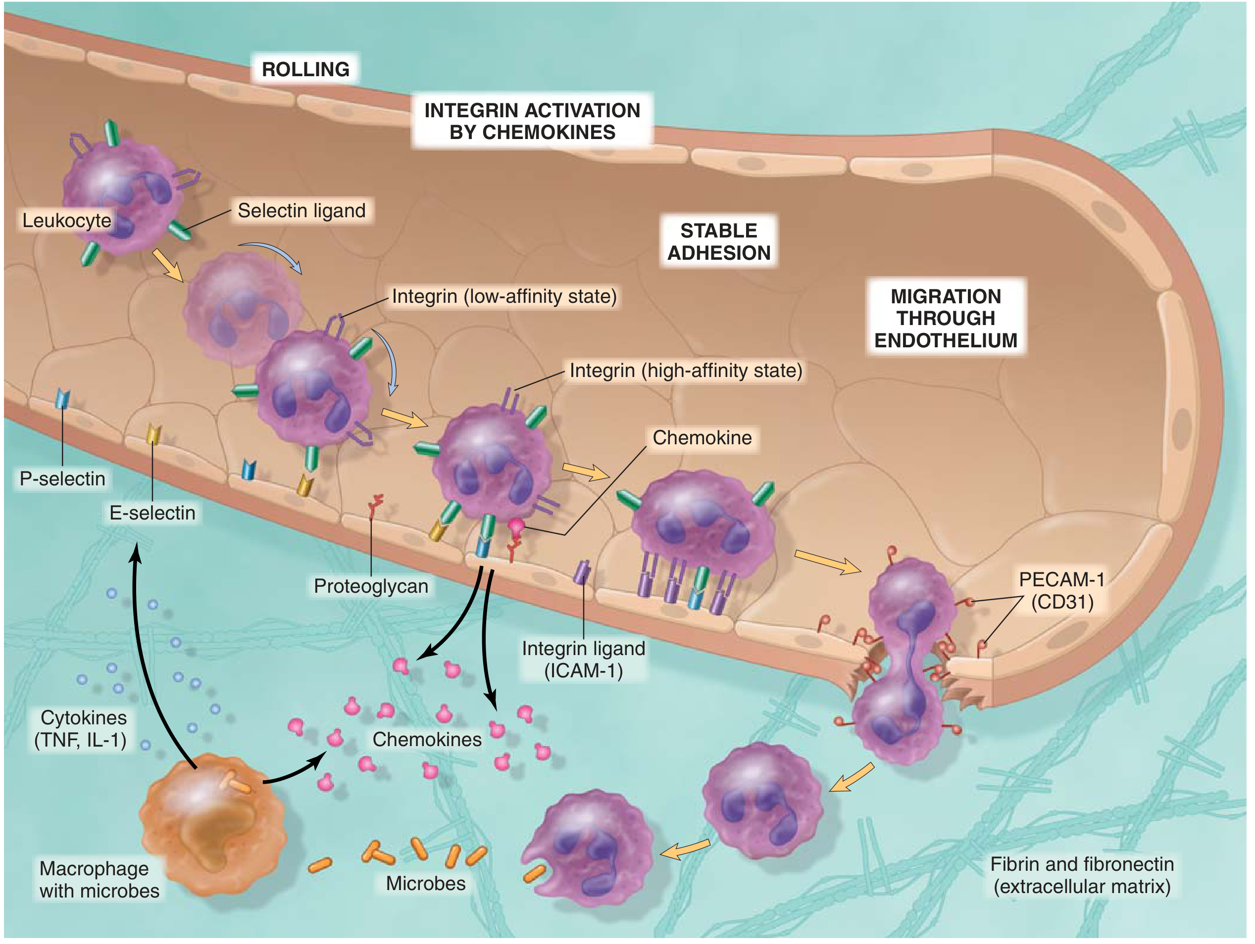
Overview: The Sequence of Events
The process is a sequential, multistep cascade - each step is controlled by different molecules. Think of it as a relay race where different teams hand off to the next:
Margination → Rolling → Activation → Firm Adhesion → Transmigration → Chemotaxis
Step 1: Margination
What happens: Under normal laminar flow, RBCs dominate the central stream and leukocytes are pushed to the periphery. When vasodilation and stasis occur at the site of inflammation, blood flow slows, and leukocytes begin to accumulate along the vessel wall.
Why: Leukocytes are larger and heavier than RBCs - stasis makes them "fall out" toward the endothelium.
Key trigger: Vasodilation → slowed blood flow → stasis
Step 2: Rolling
What happens: Leukocytes make loose, transient contact with endothelial cells and tumble slowly along the vessel surface - they bind, detach, bind again repeatedly.
Molecules involved: SELECTINS
| Selectin | Where Expressed | Upregulated By | Binds To |
|---|---|---|---|
| P-selectin | Endothelium (stored in Weibel-Palade bodies) | Histamine, thrombin - rapid (minutes) | Sialyl-Lewis X on leukocytes (via PSGL-1) |
| E-selectin | Activated endothelium | TNF-α, IL-1 - slower (1-2 hours), via new gene transcription | Sialyl-Lewis X on neutrophils, monocytes, T cells |
| L-selectin | Leukocytes | Constitutive | Sialomucins on endothelium (upregulated by IL-1, TNF) |
Key point: P-selectin acts first and fastest (preformed, just needs to move to surface). E-selectin comes later (requires new protein synthesis). Both bind the same carbohydrate structure on leukocytes: sialyl-Lewis X.
Interactions are low-affinity - quickly broken by shear force of flowing blood → this is why leukocytes roll rather than stick.
Step 3: Chemokine-Mediated Activation (Integrin Upregulation)
What happens: As leukocytes roll, they encounter chemokines displayed on the endothelial surface (bound to proteoglycans). These chemokines bind to G-protein-coupled receptors on the leukocyte surface and trigger intracellular signaling that converts leukocyte integrins from low-affinity → high-affinity conformational state.
Key chemokines:
- IL-8 (CXCL8) - major chemokine activating neutrophil integrins
- MCP-1 - for monocytes
- Eotaxin - for eosinophils
Key concept: The integrins are already present on the leukocyte surface before activation - they just change shape (inside-out signaling) to bind tightly. This is called integrin activation or inside-out signaling.
Step 4: Firm Adhesion (Arrest)
What happens: Activated high-affinity integrins bind their ligands on the endothelial surface firmly. The leukocyte stops rolling completely and becomes anchored.
Molecules involved: INTEGRINS + their LIGANDS
| Integrin (on Leukocyte) | Binds | Ligand (on Endothelium) | Upregulated By |
|---|---|---|---|
| LFA-1 (αLβ2, CD11a/CD18) | → | ICAM-1 | TNF-α, IL-1 |
| MAC-1 (αMβ2, CD11b/CD18) | → | ICAM-1 | TNF-α, IL-1 |
| VLA-4 (α4β1) | → | VCAM-1 | TNF-α, IL-1 |
| α4β7 | → | MAdCAM-1 | Gut-specific endothelium |
Clinical note: Drugs targeting integrins block this step:
- Natalizumab (anti-VLA-4/α4-integrin) - used in multiple sclerosis and Crohn disease
- Vedolizumab (anti-α4β7) - targets gut-specific leukocyte trafficking in IBD
Step 5: Transmigration (Diapedesis)
What happens: The firmly adherent leukocyte squeezes between adjacent endothelial cells at their junctions (paracellular route) and crosses the basement membrane, entering the interstitium.
Key molecule: PECAM-1 (CD31)
- Expressed on both leukocytes and endothelial cells at intercellular junctions
- Homophilic binding (PECAM-1 on leukocyte binds PECAM-1 on endothelium) allows squeezing through
- The process is also called diapedesis
After crossing endothelium:
- Leukocyte must also cross the basement membrane - it secretes collagenases to digest through
- Finally enters extravascular tissue
Site: Predominantly in postcapillary venules - their walls are thin and endothelial cells are most responsive to inflammatory mediators
Step 6: Chemotaxis
What happens: Once in the tissue, leukocytes migrate directionally toward the site of injury/infection by following a chemical concentration gradient (higher concentration = closer to the source).
Major Chemotactic Agents:
| Agent | Source | Type |
|---|---|---|
| C5a | Complement activation | Most potent/high-yield |
| LTB₄ | Arachidonic acid (5-LOX pathway) | Lipid mediator |
| IL-8 (CXCL8) | Macrophages, endothelium | Chemokine |
| N-formyl-Met-Leu-Phe (fMLP) | Bacterial peptides | Bacterial product |
| Fibronectin fragments | ECM breakdown | Extracellular matrix |
Mechanism: Chemotaxins bind GPCRs on leukocytes → activate phospholipases → cytoskeletal rearrangement (actin polymerization at leading edge) → directional amoeboid movement
Timing and Cell Type Specificity
| Time After Injury | Dominant Cell | Reason |
|---|---|---|
| 0-6 hours | Neutrophils | Most numerous in blood; fastest response; bind P/E-selectin quickly |
| 24-48 hours | Monocytes/Macrophages | Longer-lived; replace neutrophils; amplify response |
Exceptions to this pattern:
- Pseudomonas infection → neutrophils persist for days
- Viral infections → lymphocytes predominate early
- Parasites/allergy → eosinophils dominate
- Hypersensitivity reactions → lymphocytes, macrophages, plasma cells
Clinical Significance - Defects in Extravasation
| Disease | Defect | Consequence |
|---|---|---|
| Leukocyte Adhesion Deficiency (LAD) type I | Absent CD18 (β2 integrin) → no LFA-1 or MAC-1 | Recurrent severe bacterial infections, no pus formation, delayed umbilical cord separation |
| LAD type II | Absent sialyl-Lewis X (fucosylation defect) | Absent selectin binding → impaired rolling |
| Chédiak-Higashi syndrome | Defective lysosome-phagosome fusion | Impaired killing after phagocytosis |
Summary Table - All Steps at a Glance
| Step | Process | Key Molecule on Leukocyte | Key Molecule on Endothelium | Triggered By |
|---|---|---|---|---|
| 1 | Margination | - | - | Vasodilation, stasis |
| 2 | Rolling | L-selectin; sialyl-Lewis X (ligand) | P-selectin, E-selectin | Histamine (P), TNF/IL-1 (E) |
| 3 | Activation | Chemokine receptors → integrin activation | Chemokines on proteoglycans | IL-8, MCP-1 |
| 4 | Firm adhesion | LFA-1, MAC-1 (integrins) | ICAM-1, VCAM-1 | TNF-α, IL-1 |
| 5 | Transmigration | PECAM-1 (CD31) | PECAM-1 (CD31) | Physical squeezing |
| 6 | Chemotaxis | Chemokine receptors (GPCRs) | - | C5a, LTB₄, IL-8, fMLP |
Sources:
- Robbins & Kumar Basic Pathology, p. 28-31
- Cellular and Molecular Immunology (Abbas), p. 137-152
name all caspase amd thrier function
Finding Sources
Searching the Web
all caspases 1-14 complete list functions apoptosis inflammation
Finding Sources
Finding Sources
Reading File
Reading File
I have all the content from the textbooks. Here is the complete, organized answer:
All Caspases and Their Functions
Caspases (Cysteine-dependent Aspartate-specific Proteases) are a family of at least 14 caspases (1-14) in mammals. They are synthesized as inactive zymogens (procaspases) and activated by proteolytic cleavage. They fall into three broad functional groups.
The 3 Functional Categories
CASPASES
├── APOPTOTIC
│ ├── Initiator (upstream) → 2, 8, 9, 10
│ └── Executioner (downstream) → 3, 6, 7
└── INFLAMMATORY (non-apoptotic) → 1, 4, 5, 11, 12
GROUP 1: Initiator Caspases (Apoptosis - Upstream)
These are activated FIRST. They sense the death signal and activate executioners downstream.
| Caspase | Pathway | Activation Platform | Key Function |
|---|---|---|---|
| Caspase-2 | Intrinsic | PIDDosome (PIDD + RAIDD) | DNA damage sensor; can trigger mitochondrial permeabilization; also has roles in genomic stability; least understood initiator |
| Caspase-8 | Extrinsic (Death receptor) | DISC (Fas + FADD) | Activated by Fas/FasL, TNFR1 ligation; activates executioner caspases; also cleaves BID → tBID (crossover to intrinsic pathway); inhibited by FLIP |
| Caspase-9 | Intrinsic (Mitochondrial) | Apoptosome (APAF-1 + cytochrome c) | Central initiator of mitochondrial pathway; activated when cytochrome c leaks from mitochondria; directly activates caspase-3 |
| Caspase-10 | Extrinsic (Death receptor) | DISC | Similar to caspase-8; activated at DISC by death receptors (TRAIL-R1/R2, DR3, Fas); activates executioner caspases |
GROUP 2: Executioner Caspases (Apoptosis - Downstream)
These are activated by initiator caspases and carry out the actual demolition of the cell.
| Caspase | Key Substrates Cleaved | Effect |
|---|---|---|
| Caspase-3 | ICAD (inhibitor of CAD/DNase) → DNA laddering; PARP; nuclear lamins; cytoskeletal proteins (actin, fodrin); Bcl-2; FAK; β-catenin | Master executioner - most important; coordinates nuclear fragmentation, cytoskeletal breakdown, membrane blebbing |
| Caspase-6 | Nuclear lamins (lamin A especially) | Nuclear envelope breakdown; nuclear condensation; also activates caspase-8 (feedback amplification) |
| Caspase-7 | Similar to caspase-3; PARP; ER stress targets | Redundant with caspase-3; important in ER stress-induced apoptosis; activates caspase-activated DNase |
What executioner caspases do collectively:
- Cleave ICAD → releases CAD (caspase-activated DNase) → cuts DNA at internucleosomal sites → DNA ladder on gel electrophoresis
- Cleave nuclear lamins → nuclear fragmentation
- Cleave actin, keratin → cell shrinkage and blebbing
- Cleave PARP (DNA repair enzyme) → block DNA repair
- Cleave Bcl-2 and Bcl-XL → destroy anti-apoptotic proteins + release pro-apoptotic fragments
- Cleave beta-catenin, FAK → loss of cell-cell contact
GROUP 3: Inflammatory Caspases (Non-apoptotic)
These do not primarily cause apoptosis. They process pro-inflammatory cytokines and mediate pyroptosis (inflammatory cell death).
| Caspase | Activation Platform | Key Function |
|---|---|---|
| Caspase-1 | Inflammasome (NLRP3, NLRC4, AIM2, etc.) | Cleaves pro-IL-1β → active IL-1β; cleaves pro-IL-18 → active IL-18; cleaves Gasdermin D → forms pores → pyroptosis; master regulator of innate inflammatory response |
| Caspase-4 (human) | Non-canonical inflammasome; binds intracellular LPS directly | Cleaves Gasdermin D → pyroptosis; activated by cytosolic gram-negative bacterial LPS (bypasses NLRP3); equivalent to caspase-11 in mice |
| Caspase-5 (human) | Non-canonical inflammasome; LPS | Similar to caspase-4; cleaves Gasdermin D → pyroptosis |
| Caspase-11 (mouse only) | Non-canonical inflammasome; intracellular LPS | Mouse equivalent of human caspase-4/5; detects cytosolic LPS from gram-negative bacteria; activates Gasdermin D |
| Caspase-12 | ER stress sensor | Activated by ER stress; attenuates caspase-1 activity (anti-inflammatory role in humans); may mediate ER stress-induced apoptosis in rodents; pseudogene in most humans |
The Remaining Caspases (Less High-Yield Clinically)
| Caspase | Category | Key Notes |
|---|---|---|
| Caspase-13 | Inflammatory | Found only in bovines; does not exist in humans |
| Caspase-14 | Non-apoptotic, non-inflammatory | Expressed only in differentiating keratinocytes of the skin; involved in cornification (skin barrier formation, filaggrin processing) and UV protection; NOT involved in classical apoptosis or inflammation |
Caspase Activation - How They Switch ON
All caspases are born inactive (procaspases). Three activation mechanisms:
- Proximity-induced dimerization - initiator caspases; brought together at platforms (DISC, apoptosome, inflammasome) → self-activate by autocleavage
- Proteolytic cleavage by another caspase - executioner caspases are cleaved by initiator caspases
- Conformational change - some caspases activated by allosteric changes
IAPs (Inhibitor of Apoptosis Proteins) - e.g., XIAP - normally inhibit caspases-3, -7, and -9. Smac/DIABLO released from mitochondria neutralizes IAPs, permitting caspase activation.
Master Summary Table
| Caspase | Group | Pathway/Platform | Key Role |
|---|---|---|---|
| 1 | Inflammatory | Inflammasome | IL-1β + IL-18 processing; pyroptosis (Gasdermin D) |
| 2 | Initiator | PIDDosome | DNA damage → apoptosis; genomic stability |
| 3 | Executioner | Activated by 8 and 9 | Master demolisher: DNA laddering, nuclear fragmentation, blebbing |
| 4 | Inflammatory | Non-canonical inflammasome (LPS) | Pyroptosis - human equivalent of caspase-11 |
| 5 | Inflammatory | Non-canonical inflammasome (LPS) | Pyroptosis - human |
| 6 | Executioner | Activated by 3 | Lamin A cleavage; nuclear breakdown |
| 7 | Executioner | Activated by 3/9 | ER stress apoptosis; redundant with caspase-3 |
| 8 | Initiator | DISC (Fas/FADD) | Extrinsic pathway; cleaves BID (crossover) |
| 9 | Initiator | Apoptosome (APAF-1 + cyt c) | Intrinsic/mitochondrial pathway master |
| 10 | Initiator | DISC | Death receptor pathway (TRAIL, Fas) |
| 11 | Inflammatory | Non-canonical (LPS) - mouse | Mouse pyroptosis; gram-neg bacteria sensor |
| 12 | ER stress | ER stress | Anti-inflammatory modulator; pseudogene in most humans |
| 13 | - | - | Bovine only; not in humans |
| 14 | Differentiation | Keratinocytes | Skin cornification; UV protection; NOT apoptotic |
High-Yield One-Liners for Exams
- Most important executioner = Caspase-3 (do not confuse with initiators)
- Intrinsic pathway initiator = Caspase-9 (activated at apoptosome)
- Extrinsic pathway initiator = Caspase-8 (activated at DISC)
- Crossover between pathways = Caspase-8 cleaves BID → tBID → activates intrinsic
- Inflammation caspase = Caspase-1 (inflammasome → IL-1β, IL-18, pyroptosis)
- Pyroptosis = inflammatory caspases cleave Gasdermin D → membrane pores → cell death with cytokine release
- Granzyme B from cytotoxic T cells bypasses the entire cascade and directly cleaves and activates caspase-3
complement system diagram
Finding Sources
Reading File
Finding Sources
Loading Image
Reading File
Loading Image
Here are the two best complement system diagrams from the textbooks, followed by a complete written breakdown:
Complement System
Diagram 1 - All 3 Pathways + Effector Functions (Robbins Pathology)
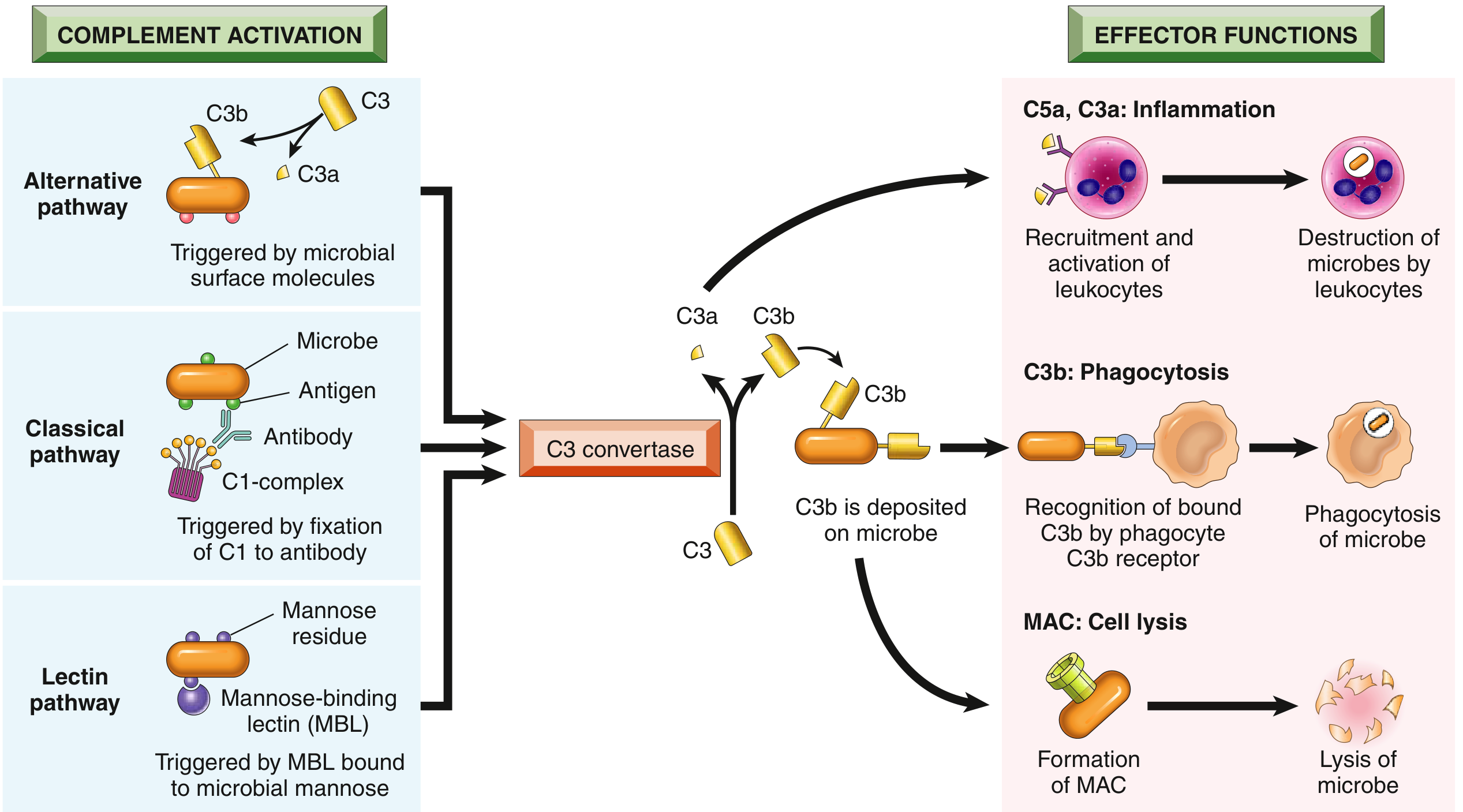
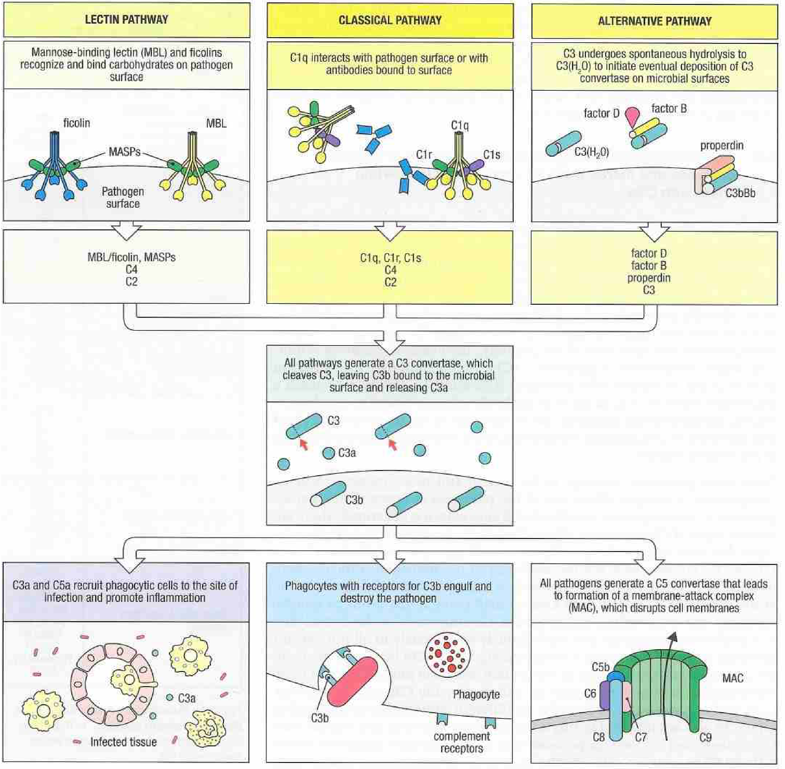
Diagram 2 - Detailed Molecular Components (Janeway's Immunobiology)
The 3 Activation Pathways
All three pathways converge on a single critical step: cleavage of C3 by a C3 convertase.
1. Classical Pathway
Trigger: Antibody (IgM or IgG) bound to antigen on a pathogen surface
Ag-Ab complex
↓
C1q binds Fc region of antibody → C1r + C1s activated
↓
C1s cleaves C4 → C4a (released) + C4b (binds surface)
↓
C4b + C2 → C1s cleaves C2 → C4b2a = C3 CONVERTASE (classical)
↓
C3 convertase cleaves C3 → C3a + C3b
Key proteins: C1q, C1r, C1s, C4, C2
C3 convertase: C4b2a
Memory trick: Classical = C1, C4, C2 (numbers go out of order - 1, 4, 2)
2. Lectin Pathway
Trigger: Mannose-binding lectin (MBL) or ficolins bind to mannose residues on microbial surfaces (antibody-independent)
MBL / Ficolin binds pathogen carbohydrates
↓
MASPs (MBL-associated serine proteases) activated
↓
MASPs cleave C4 and C2 (same as classical from here)
↓
C4b2a = C3 CONVERTASE (lectin) → same as classical
Key proteins: MBL, ficolins, MASP-1, MASP-2, C4, C2
C3 convertase: C4b2a (same as classical)
3. Alternative Pathway
Trigger: Spontaneous C3 hydrolysis; amplified by microbial surfaces (LPS, fungal cell walls, IgA aggregates) - no antibody needed
C3 spontaneously hydrolyses → C3(H₂O) (fluid phase)
↓
C3(H₂O) + Factor B → Factor D cleaves B → C3(H₂O)Bb
↓
This generates more C3b → C3b + Factor B + Factor D → C3bBb = C3 CONVERTASE (alternative)
↓
Properdin (Factor P) stabilizes C3bBb convertase
Key proteins: C3, Factor B, Factor D, Properdin
C3 convertase: C3bBb
Memory trick: Alternative = B, D, Properdin (factors not numbered)
The Convergence Point: C3 Cleavage
C3 convertase (from any pathway)
↓
C3 ──────────────────────→ C3a + C3b
↙ ↘
Released into Covalently binds
plasma microbial surface
(anaphylatoxin) (opsonin + feeds C5 convertase)
C5 Convertase and the MAC
Once C3b accumulates on the surface:
C3b + C3 convertase = C5 CONVERTASE
Classical/Lectin: C4b2a3b
Alternative: C3bBbC3b
↓
C5 → C5a (released) + C5b (stays on surface)
↓
C5b + C6 + C7 + C8 + poly-C9 → MAC (Membrane Attack Complex)
↓
Pore in membrane → lysis of cell
The 3 Effector Functions - "OIL" or "AOP"
| Function | Molecule | Mechanism |
|---|---|---|
| Inflammation (Anaphylatoxins) | C3a, C4a, C5a | Mast cell degranulation → histamine; vasodilation; increased permeability; C5a = most potent chemotaxin for neutrophils |
| Opsonization | C3b, iC3b | Coats microbe surface; phagocytes (neutrophils, macrophages) have CR1 and CR3 complement receptors → enhanced phagocytosis |
| Lysis | MAC (C5b-9) | Poly-C9 forms pores in membrane → water + ion influx → cell swells and lyses; most effective against thin-walled gram-negative bacteria (especially Neisseria) |
Regulatory Proteins (Control of Complement)
The complement system must be tightly regulated to prevent self-destruction:
| Regulator | Controls | Mechanism |
|---|---|---|
| C1 inhibitor (C1-INH) | Classical pathway | Inhibits C1r and C1s; also inhibits kallikrein/bradykinin (deficiency = hereditary angioedema) |
| C4b-binding protein (C4BP) | Classical/Lectin | Binds C4b; displaces C2a; promotes C4b degradation |
| Factor I | All pathways | Cleaves C3b → iC3b (inactive); requires cofactors |
| Factor H | Alternative pathway | Competes with Factor B; cofactor for Factor I; protects self cells |
| DAF (CD55) | Classical + Alternative | Accelerates decay of C3 convertases on self cells |
| CD59 (Protectin) | MAC | Blocks poly-C9 assembly; prevents MAC on self cells |
| Carboxypeptidase N | Plasma | Inactivates C3a and C5a (cleaves arginine from C-terminus) |
Clinical Deficiencies - High Yield
| Deficiency | Consequence |
|---|---|
| C1q, C4, C2 (classical pathway) | Recurrent infections + SLE-like autoimmune disease (defective immune complex clearance) |
| C3 | Most severe - recurrent severe bacterial infections (encapsulated organisms) |
| C5-C9 (terminal components) | Recurrent Neisseria infections (meningococcal, gonococcal) - MAC needed to kill gram-negatives |
| Factor D, Properdin (alternative pathway) | Neisseria infections (especially in males - properdin is X-linked) |
| C1-INH | Hereditary angioedema - bradykinin accumulates → swelling (NOT mast-cell-mediated) |
| CD55/CD59 (DAF/Protectin) | PNH (Paroxysmal Nocturnal Hemoglobinuria) - complement lyses own RBCs |
| MBL | Recurrent infections in early childhood |
Memory Summary
3 PATHWAYS → all make C3 CONVERTASE → cleave C3 → C3a + C3b
C3a/C5a → INFLAMMATION (anaphylatoxins, chemotaxis)
C3b → OPSONIZATION (phagocytosis)
C5b-9 → MAC → LYSIS
Classical: Antibody → C1q → C1r/s → C4 → C2 → C4b2a
Lectin: MBL → MASPs → C4 → C2 → C4b2a
Alternative: Spontaneous C3 → Factor B + D + Properdin → C3bBb
Sources:
- Robbins & Kumar Basic Pathology, p. 37-39
- Janeway's Immunobiology 10e, p. 76-77
just briefly explain whats inflammation
Inflammation - Brief Explanation
Inflammation is the body's protective response to injury or infection.
When your tissues are damaged - by a cut, a burn, bacteria, or a dead cell - the body detects the danger and sends help. This "help" is the inflammatory response.
What it does:
- Increases blood flow to the area (causing redness and warmth)
- Makes blood vessels leaky so defensive proteins flood into the tissue (causing swelling)
- Recruits white blood cells - mainly neutrophils - to kill microbes and clean up debris
The purpose: Eliminate the threat, clear the damage, and start healing.
The cardinal signs you feel: redness, heat, swelling, pain, and loss of function - all of these are side effects of the defensive response, not the infection itself.
Two types:
- Acute - fast (minutes to days), mainly neutrophils, usually resolves. Example: infected cut, appendicitis
- Chronic - slow and prolonged (weeks to months), mainly macrophages and lymphocytes, causes ongoing tissue damage. Example: tuberculosis, rheumatoid arthritis
The key idea: Inflammation is protective by design, but it can cause harm when it's excessive (septic shock), misdirected (autoimmune disease), or persistent (chronic diseases like atherosclerosis, diabetes, Alzheimer's).
mediators of acute inflamation
Finding Sources
Your credits are exhausted. You can purchase a top-up or subscribe to a plan to continue using Orris. If you're already subscribed, your daily credits will reset tomorrow.
Create a study guide on acute inflammation and its mediators
Your credits are exhausted. You can purchase a top-up or subscribe to a plan to continue using Orris. If you're already subscribed, your daily credits will reset tomorrow.
This is a shared conversation. Sign in to Orris to start your own chat.