
Peripheral smear result in. Beta thalassemia
Reading File
In beta thalassemia, the peripheral smear shows microcytic hypochromic red cells, target cells (codocytes), nucleated red blood cells, basophilic stippling, and poikilocytosis. Severity varies by whether it's trait, intermedia, or major.
Searching sources...
Finding Sources
Finding Sources
Good results from multiple textbooks. Let me read the key sections in parallel.
Reading File
Reading File
Reading File
Excellent - rich content from multiple textbooks. Let me fetch the peripheral smear image from Tietz which was referenced, and also the pathophysiology diagram.
Loading Image
Loading Image
Reading File
I now have comprehensive, well-sourced information with actual peripheral smear images. Here is the full answer:
Peripheral Smear Findings in Beta Thalassemia
The peripheral smear findings differ significantly depending on whether the patient has beta thalassemia minor (trait), intermedia, or major.
Beta Thalassemia Major
CBC: Severe anemia with Hb 30-65 g/L, MCV 48-72 fL, MCH 23-32 pg.
Peripheral smear shows:
| Finding | Explanation |
|---|---|
| Microcytes | Reduced beta-globin → small, poorly hemoglobinized cells |
| Hypochromia | Insufficient HbA formation; large central pallor area |
| Macrocytes | Also present (mixed picture) |
| Target cells (codocytes) | May have a bridge joining central and peripheral pigment zones |
| Nucleated red blood cells (NRBCs / normoblasts) | Reflect intense erythropoietic drive - characteristic finding of thalassemia major/intermedia |
| Basophilic stippling (prominent) | Residual ribosomes/RNA in poorly hemoglobinized cells |
| Polychromasia | Reticulocytosis from compensatory erythropoiesis |
| Poikilocytosis | Bizarre cell shapes due to membrane damage by alpha-chain precipitates |
| Occasional spherocytes | From membrane damage |
| Schistocytes (occasional) | Fragmented cells from mechanical hemolysis |
"A characteristic markedly abnormal RBC morphology is noted on the peripheral blood smear; this includes a large number of microcytes and/or macrocytes, prominent basophilic stippling, numerous target cells, which may have a bridge joining the central and peripheral pigment zones, polychromasia, and occasional spherocytes, schistocytes, and nucleated red cells."
- Tietz Textbook of Laboratory Medicine, 7th ed.
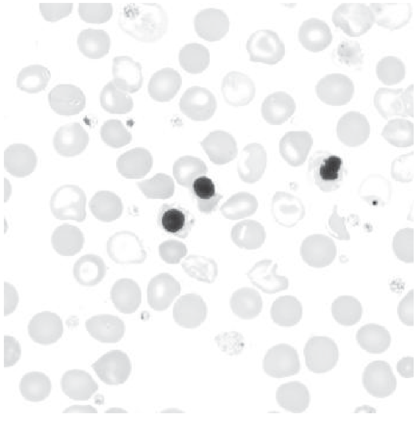
Beta Thalassemia Minor (Trait)
These patients are usually asymptomatic with mild or absent anemia.
Peripheral smear shows:
- Hypochromia
- Microcytosis
- Basophilic stippling
- Target cells
"The peripheral blood smear typically shows hypochromia, microcytosis, basophilic stippling, and target cells."
- Robbins, Cotran & Kumar Pathologic Basis of Disease
Beta Thalassemia Intermedia
Findings lie between those of minor and major - microcytic hypochromic cells, target cells, NRBCs (fewer than in major), basophilic stippling, and polychromasia.
Why These Findings Occur
The pathophysiology diagram below explains the mechanism:
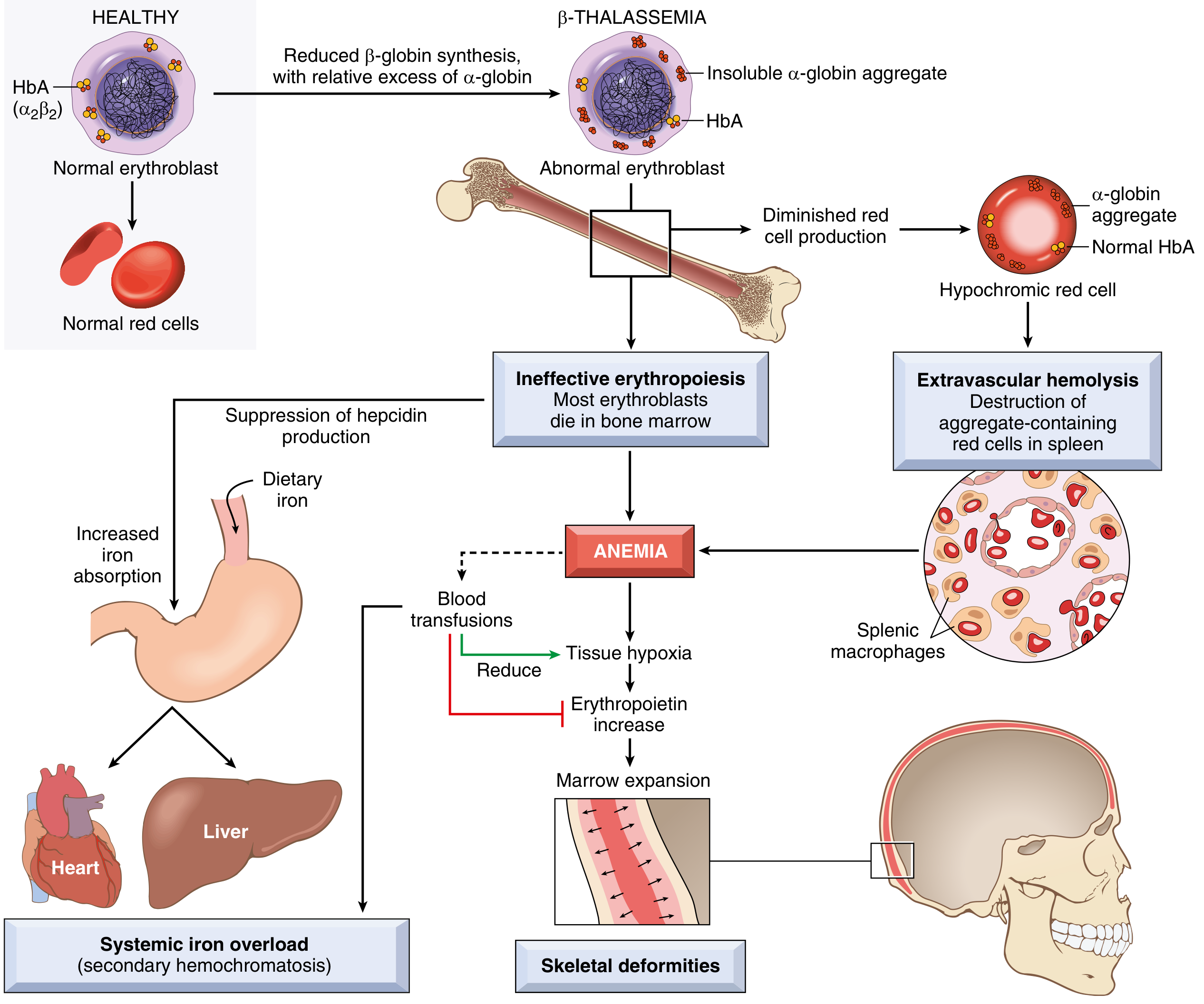
- Reduced beta-globin synthesis → inadequate HbA → microcytic, hypochromic cells
- Excess unpaired alpha-globin chains form toxic precipitates → membrane damage → bizarre shapes (poikilocytosis, schistocytes, spherocytes)
- Massive ineffective erythropoiesis - most erythroblasts die in the marrow → NRBCs released into circulation
- Compensatory erythropoietic drive → polychromasia, reticulocytosis
Summary Table
| Feature | Major | Intermedia | Minor (Trait) |
|---|---|---|---|
| Microcytosis/Hypochromia | +++ | ++ | + |
| Target cells | +++ | ++ | + |
| Basophilic stippling | +++ | ++ | + |
| NRBCs | +++ (characteristic) | + | Absent |
| Poikilocytosis | +++ | ++ | Mild |
| Polychromasia | ++ | + | Minimal |
| Schistocytes/Spherocytes | Occasional | Rare | Absent |
Key distinguishing point: Circulating nucleated red blood cells (NRBCs) that are poorly hemoglobinized are characteristic of beta-thalassemia major and intermedia, and help distinguish them from iron deficiency anemia (which has similar microcytic/hypochromic morphology but no NRBCs).
Sources: Tietz Textbook of Laboratory Medicine (7th ed.); Robbins & Kumar Basic Pathology; Robbins, Cotran & Kumar Pathologic Basis of Disease.
This is a shared conversation. Sign in to Orris to start your own chat.