
Breast carcinoma
"breast cancer"[MeSH Terms] AND "treatment"[MeSH Terms]
breast carcinoma histology invasive ductal
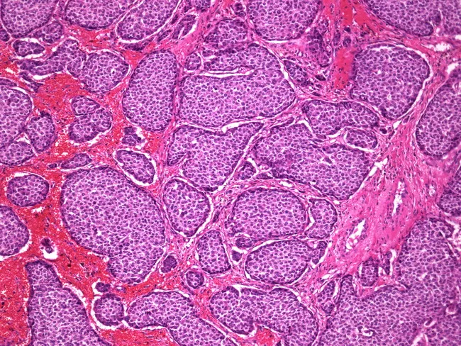
Imaging modality: Light microscopy of Hematoxylin and Eosin stained breast tissue sections. The section shows invasive carcinoma of the breast with neuroendocrine differentiation, characterized by small to large nests of tumor cells embedded in a fibrovascular stroma. Tumor cells are polygonal with uniform cytoplasm and round to oval nuclei; the nests are tightly packed and separated by thick pink (eosinophilic) septa, consistent with desmoplastic fibrous stroma. The overall architecture resembles ductal carcinoma of the breast, with features consistent with an invasive ductal carcinoma with neuroendocrine differentiation (carcinoid-like breast tumor). The histology may overlap with neuroendocrine tumors but in this context, the nest pattern within breast parenchyma supports a primary breast carcinoma rather than a metastatic neuroendocrine tumor. Immunohistochemical confirmation with neuroendocrine markers (synaptophysin, chromogranin) and hormone receptor status (estrogen receptor, progesterone receptor) would aid definitive classification and therapeutic planning. The diagnostic significance lies in recognizing neuroendocrine differentiation as a histologic variant of breast carcinoma, which can influence prognosis and treatment, and underscores the relationship between carcinoid-like breast tumors and conventional ductal carcinomas. Clinically relevant use cases include education on breast cancer histology, differential diagnosis of neuroendocrine breast tumors, and correlation with imaging and molecular profiling for targeted therapy.
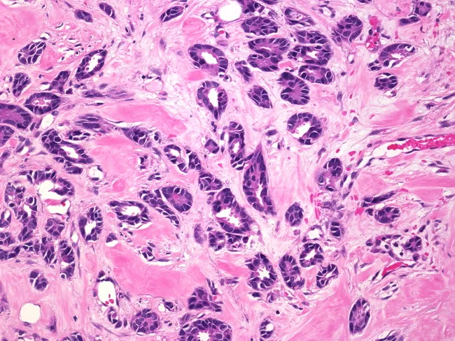
High-magnification hematoxylin and eosin stained histology image of male breast tissue showing invasive tubular carcinoma. The specimen reveals an infiltrating architecture composed of uniformly small, cuboidal to columnar epithelial cells forming well-differentiated, angulated tubular glands with open lumina embedded in desmoplastic fibrous stroma. The myoepithelial layer surrounding lumina is variably diminished, consistent with invasive ductal carcinoma with tubular differentiation. Nuclei are relatively small, round to oval with mild hyperchromasia and minimal pleomorphism; mitotic activity is infrequent. The stroma is pink, densely fibrous, with sparse inflammatory infiltrate. There is no marked necrosis or mucin lakes. The tubular glands demonstrate low-grade cytology and orderly tubular architecture characteristic of tubular carcinoma, a well-differentiated histologic variant of breast cancer. In male patients, this lesion is rare but generally portends a favorable prognosis compared with high-grade ductal carcinomas. The image highlights histopathologic hallmarks of tubulary differentiation and invasion, underscoring the diagnostic value of routine H&E staining in distinguishing tubular carcinoma from other invasive breast cancers such as conventional invasive ductal carcinoma or mucinous variants. Clinically, tubular carcinoma in men is frequently estrogen receptor positive with good response to hormonal therapy after definitive surgical management. Indeed.
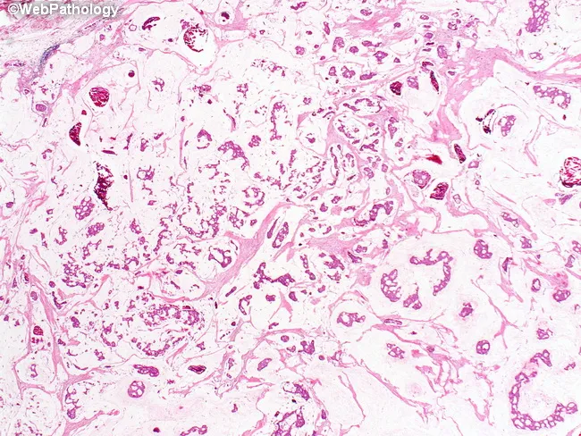
Imaging modality and technique: Light microscopy of formalin-fixed, paraffin-embedded breast tissue section stained with Hematoxylin and Eosin (H&E). Primary subject: mucinous (colloid) carcinoma of the breast. Location: breast parenchyma, mammary gland, invasive component within a mucinous matrix. Visual features: clusters and sheets of malignant epithelial cells dispersed within abundant extracellular mucin pools; mucin lakes with variable density; delicate fibrous septa partially partitioning the mucin pools; cells frequently arranged in small clusters or cords; nuclei show mild to moderate atypia with occasional prominent nucleoli; mitotic activity typically low to moderate. Background mucin appears pale eosinophilic and may conform to the tumor margins. In-situ component, if present, is rare or absent. Pathologic interpretation: mucin-producing invasive ductal carcinoma with abundant extracellular mucin, the histologic subtype. Diagnostic significance: this pattern associates with a distinct biology and a favorable prognosis compared with conventional invasive ductal carcinoma; it influences therapeutic decisions and may correlate with hormone receptor positivity and variable HER2 status. Clinical correlation: correlate histology with immunohistochemistry receptor profile, imaging findings, patient age, and clinical risk to guide surgical planning, adjuvant therapy, and surveillance. Differential considerations include standard invasive ductal carcinoma, signet-ring cell variants, and metastatic mucinous adenocarcinoma to the breast; confirm with ancillary studies.
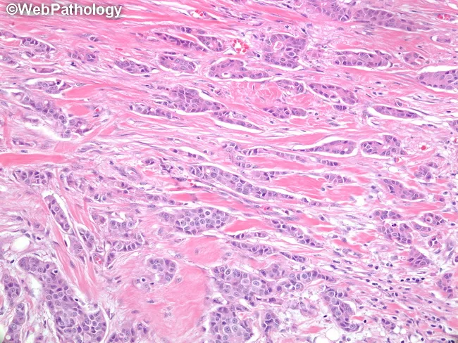
Histology section examined under light microscopy after Hematoxylin and Eosin staining of breast tissue. The specimen shows invasive ductal carcinoma with glandular/tubular differentiation. Tubule formation occupies approximately 30–40% of the tumor, corresponding to a Nottingham score of 2 for tubule formation. Nuclear grade and mitotic rate are not provided in this description. The histology reveals invasive nests of polygonal cells with pleomorphic, hyperchromatic nuclei set in desmoplastic stroma. Central lumina are present within tubulo-glandular structures, supporting ductal morphology. The tumor infiltrates surrounding parenchyma and disrupts normal architecture. This pattern indicates intermediate differentiation, consistent with Nottingham grade II. Morphologic heterogeneity may include focal solid areas coexisting with tubular elements. Clinically, intermediate grade tumors commonly have variable prognosis and treatment implications. Therapy decisions integrate receptor status (ER/PR/HER2), tumor size, nodal involvement, and proliferation index. Nottingham grading thus informs prognosis, guides adjuvant therapy, and supports educational interpretation of breast carcinoma histology. This image serves as a teaching resource for pathologists and trainees evaluating ductal neoplasia. The documented tubule-forming component highlights glandular differentiation and remains a salient correlate of tumor biology in breast cancer. In clinical practice, grading combines with receptor profiling to tailor therapies and estimate recurrence risk. Accurate histologic grade improves staging accuracy.
Breast Carcinoma
Epidemiology
-
The majority (75%) are diagnosed after age 50
-
Incidence is highest in North America, Northern/Western Europe, and Australia/New Zealand
-
Incidence and mortality are rising in low-resource countries, largely due to social/lifestyle changes
-
Robbins, Cotran & Kumar Pathologic Basis of Disease, p. 971
-
Robbins & Kumar Basic Pathology, p. 719
Risk Factors
Modifiable Risk Factors
| Risk Factor | Relative Risk / Details |
|---|---|
| Obesity (BMI >30) | RR 1.29 in postmenopausal women |
| Nulliparity / late first pregnancy (>35 yrs) | Increased estrogenic exposure |
| No / short breastfeeding | >12 months breastfeeding is protective |
| HRT use >10 years | RR 1.2 |
| Alcohol (heavy: >4 drinks/day) | RR 1.46 |
| Tobacco (≥25 cigarettes/day) | RR 1.14 |
| Radiation exposure | RR 6 |
Non-Modifiable Risk Factors
| Risk Factor | Details |
|---|---|
| Age | Median age ~60 yrs in the West; ~48 yrs in South Asia |
| Female sex | Rare in males (<1% of all breast cancers) |
| Early menarche / late menopause | Prolonged estrogenic stimulation |
| Personal history of breast cancer | Increased risk of contralateral cancer |
| BRCA1 / BRCA2 mutations | BRCA1 - 50-85% lifetime risk; BRCA2 - 50-60% lifetime risk |
| Dense breast tissue | Higher mammographic density |
| Prior breast biopsy with atypical hyperplasia | 4-5x increased risk |
- Bailey and Love's Short Practice of Surgery 28th Ed., p. 952
- Robbins, Cotran & Kumar, p. 971
Classification
By In Situ vs. Invasive Status
- Ductal Carcinoma In Situ (DCIS): Confined within ducts; most often found on mammography as calcifications. A direct precursor to invasive ductal carcinoma. When cancer develops after untreated DCIS, it is nearly always invasive ductal carcinoma in the same breast.
- Lobular Carcinoma In Situ (LCIS): A marker of increased risk and a precursor lesion. When subsequent cancer develops, 2/3 are in the same breast and 1/3 in the contralateral breast.
| Subtype | Frequency |
|---|---|
| Invasive carcinoma, no special type (NST) / "ductal" | ~75-80% |
| Invasive lobular carcinoma | 10-15% |
| Mucinous (colloid) carcinoma | ~2% |
| Tubular carcinoma | ~2% |
| Papillary carcinoma | ~2% |
| Medullary pattern | ~4% |
| Paget's disease of the nipple | <2% |
| Metaplastic, adenoid cystic, apocrine | Rare |
- Schwartz's Principles of Surgery 11th Ed., p. 596
- Robbins, Cotran & Kumar, p. 978
Molecular / Biomarker Classification
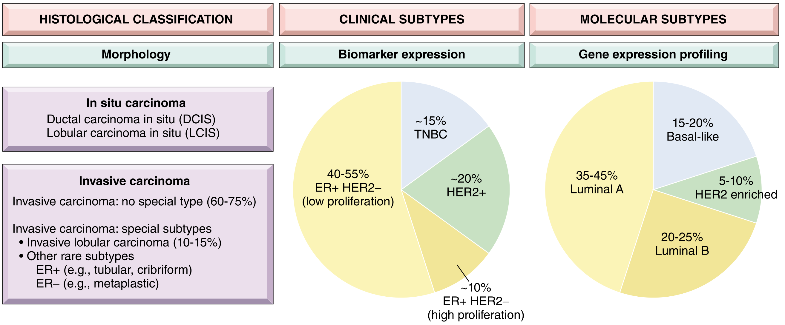
| Clinical Subtype | Molecular Subtype | Frequency | Key Features |
|---|---|---|---|
| ER+/HER2- (Luminal), low proliferation | Luminal A | 35-45% | Best prognosis; endocrine therapy alone often sufficient |
| ER+/HER2-, high proliferation | Luminal B | 20-25% | Higher grade, worse prognosis than A |
| HER2+ | HER2-enriched | ~20% | HER2 gene amplification; respond to anti-HER2 therapy (trastuzumab) |
| Triple Negative (TNBC) | Basal-like | ~15% | ER-, PR-, HER2-; often BRCA1-mutated; worst prognosis; chemotherapy is mainstay |
- Robbins, Cotran & Kumar, p. 972-973
Histologic Grading - Nottingham Score
- Tubule formation (glandular differentiation)
- Nuclear pleomorphism
- Mitotic rate
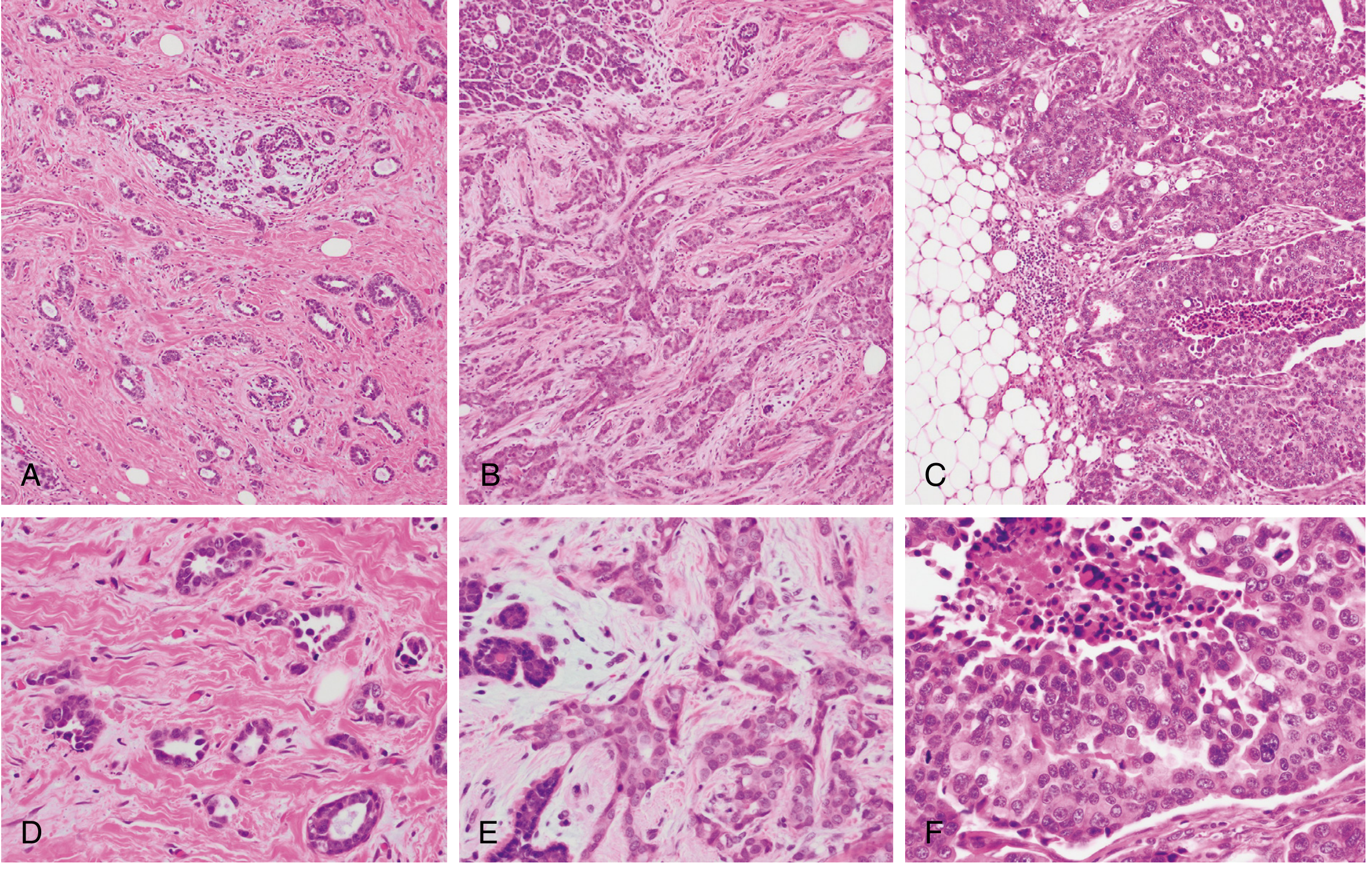
- Grade 1 (3-5): Well differentiated - tubular growth, uniform nuclei, low mitotic rate
- Grade 2 (6-7): Moderately differentiated - mixed solid/tubular, moderate pleomorphism
- Grade 3 (8-9): Poorly differentiated - solid sheets, high pleomorphism, frequent mitoses, necrosis common
Clinical Presentation
- Painless breast lump (most common) - hard, irregular, fixed
- Skin changes: Peau d'orange (dermal lymphatic obstruction), skin dimpling, ulceration
- Nipple changes: Nipple retraction, Paget's disease (eczematous nipple lesion)
- Nipple discharge (especially bloody)
- Axillary lymphadenopathy
- Inflammatory breast carcinoma: Warm, erythematous, edematous breast - mimics mastitis; highly aggressive; dermal lymphatic invasion
Mammographic Features
- Spiculated / stellate mass with irregular margins
- Clustered microcalcifications (fine, stippled)
- Asymmetric density / architectural distortion
- Average size of mammographically detected invasive carcinoma: ~1 cm (vs. ~2-3 cm on palpation)
- Sensitivity increases with age: at age 40, only 10% of mammographic lesions are cancer; rises to >25% after age 50
- About 10% of invasive carcinomas are not visible on standard 2D mammography - digital breast tomosynthesis (DBT), ultrasound, and MRI serve as adjuncts
TNM Staging (AJCC 8th Edition)
| Category | Criteria |
|---|---|
| Tis | DCIS or Paget's disease (without invasive component) |
| T1 | ≤20 mm (T1mi ≤1 mm; T1a 1-5 mm; T1b 5-10 mm; T1c 10-20 mm) |
| T2 | 20-50 mm |
| T3 | >50 mm |
| T4 | Any size + chest wall/skin extension; T4d = inflammatory carcinoma |
| cN0 | No regional lymph node metastases |
| cN1 | Movable ipsilateral Level I/II axillary nodes |
| cN2 | Fixed/matted ipsilateral axillary nodes; internal mammary nodes |
| cN3 | Infraclavicular, supraclavicular, or internal mammary + axillary |
| M0 | No distant metastasis |
| M1 | Distant metastasis (bone, lung, liver, brain most common) |
- Schwartz's Principles of Surgery 11th Ed., p. 605
Prognostic Factors
| Tumor Factors | Host Factors |
|---|---|
| Nodal status | Age / menopausal status |
| Tumor size | Family history |
| Histologic/nuclear grade | Prior breast cancer |
| Lymphovascular invasion | Immunosuppression |
| Hormone receptor status (ER/PR) | Prior chemotherapy/radiation |
| HER2/neu expression | |
| DNA ploidy / S-phase fraction |
- Oncotype DX (21-gene RT-PCR): FDA-approved for node-negative, ER+ breast cancer. Generates a Recurrence Score: low score (0-10) = excellent outcomes with endocrine therapy alone (5-year recurrence-free: 98.7%); high score = benefit from chemotherapy. Validated by the TAILORx trial.
- MammaPrint (70-gene): FDA-approved for Stage I/II, node-negative or 1-3 node positive cancers. Low-risk MammaPrint = 97% distant recurrence-free interval at 5 years. Validated by the MINDACT trial.
Treatment
1. Surgical Management
- Wide local excision + sentinel lymph node biopsy (SLNB)
- Followed by whole-breast radiotherapy
- Equivalent survival to mastectomy in Stage I/II disease
- Contraindicated: multicentric disease, diffuse microcalcifications, inability to achieve clear margins, prior chest RT, pregnancy (relative)
- Preferred for: multicentric disease, large tumor-to-breast ratio, BRCA carriers (prophylactic)
- Skin-sparing and nipple-sparing variants allow reconstruction
- Radical mastectomy is now rarely performed (reserved for chest wall invasion)
- SLNB standard for clinically node-negative disease
- Axillary lymph node dissection (ALND) reserved for positive sentinel nodes (or may be omitted in select cases per Z0011 criteria)
2. Radiation Therapy
- Adjuvant whole-breast radiation after BCS - equivalent local control to mastectomy
- Post-mastectomy radiotherapy (PMRT) for T3/T4, ≥4 positive nodes, or close/positive margins
- Accelerated partial breast irradiation (APBI) is an alternative in low-risk patients
3. Systemic Therapy
- Tamoxifen (SERM) - premenopausal women; 5-10 years
- Aromatase inhibitors (anastrozole, letrozole, exemestane) - postmenopausal women; superior to tamoxifen for most
- CDK4/6 inhibitors (palbociclib, ribociclib, abemaciclib) - added to AI in metastatic/high-risk early-stage ER+ disease
- Ovarian function suppression (OFS) with GnRH agonists in high-risk premenopausal women
- Trastuzumab (Herceptin) - monoclonal antibody; backbone of HER2+ treatment; reduces recurrence ~50%
- Pertuzumab - dual HER2 blockade with trastuzumab (neoadjuvant/metastatic)
- T-DM1 / T-DXd (trastuzumab deruxtecan) - antibody-drug conjugates; used after neoadjuvant failure or in metastatic setting
- Lapatinib, neratinib, tucatinib - tyrosine kinase inhibitors (TKIs); used in resistant/metastatic disease
- Anthracyclines (doxorubicin/epirubicin) + taxanes (paclitaxel/docetaxel) form the backbone
- TNBC and high-grade ER+ tumors most likely to benefit
- Neoadjuvant chemotherapy (NACT): Used to downstage tumors before surgery; pathologic complete response (pCR) is a strong prognostic marker
- Capecitabine: Post-NACT residual disease in TNBC (CREATE-X trial)
- Olaparib, talazoparib - approved for germline BRCA-mutated, HER2-negative metastatic or high-risk early-stage disease
- Pembrolizumab (anti-PD-1) + chemotherapy for PD-L1+ metastatic TNBC (KEYNOTE-522 / 355); also approved as neoadjuvant + adjuvant for high-risk early TNBC
- Atezolizumab + nab-paclitaxel (previously used, now less favored after updated evidence)
4. Metastatic Breast Cancer (Stage IV)
- Goal is palliation, QoL improvement, and life prolongation (rarely curative)
- Bone metastases: Bisphosphonates (zoledronic acid) or denosumab + palliative radiotherapy to weight-bearing lesions
- Visceral crisis: Systemic chemotherapy preferred; oral metronomic chemotherapy for QoL
- CNS metastases: Whole-brain radiotherapy, stereotactic radiosurgery (SRS), or HER2-targeted TKIs (lapatinib/tucatinib cross BBB)
- Surgical resection for solitary visceral metastasis in selected good-performance-status patients
Hereditary Breast Cancer
- BRCA1 (chromosome 17q21): 50-85% lifetime breast cancer risk; 40% ovarian cancer risk; associated cancers are mostly TNBC; DNA repair deficiency (homologous recombination)
- BRCA2 (chromosome 13q12.3): 50-60% lifetime breast cancer risk; 20% ovarian cancer risk; also associated with prostate, pancreatic, and bile duct cancers; most common in male breast cancer
- Women with confirmed BRCA mutations may be offered bilateral risk-reducing mastectomy (reduces risk by >90%) and bilateral salpingo-oophorectomy
- MRI screening (in addition to mammography) from age 25-30 in BRCA carriers
Special Histologic Subtypes - Key Points
| Subtype | Key Features | Prognosis |
|---|---|---|
| Paget's disease of nipple | Eczematous nipple lesion; large pale vacuolated Paget cells in rete pegs; CEA+; associated with DCIS; distinguish from melanoma (S-100+) | Depends on underlying invasive component |
| Inflammatory breast carcinoma | Dermal lymphatic invasion; peau d'orange; T4d; NO discrete mass often; aggressive | Poor - treat with NACT first |
| Invasive lobular carcinoma | Single-file ("Indian-file") cell pattern; loss of E-cadherin; bilateral/multicentric; ER+; hard to detect on mammography | Generally favorable if detected early |
| Mucinous (colloid) carcinoma | Tumor cells floating in mucin lakes; ER+; usually older women | Favorable |
| Tubular carcinoma | Well-formed angulated tubules; low grade; ER+; excellent prognosis | Very favorable |
| Metaplastic carcinoma | Squamous/sarcomatoid differentiation; ER-, PR-, HER2- (TNBC); aggressive | Poor |
Male Breast Cancer
- <1% of all breast cancers; median age 60-70 years
- Most are ER+/PR+ invasive ductal carcinoma
- BRCA2 mutation is the most common genetic risk factor in males
- Treatment mirrors female breast cancer, though mastectomy is usually the surgical approach due to limited breast tissue
- Bailey and Love's Short Practice of Surgery 28th Ed., p. 1000
Key Summary Points (from Robbins & Kumar Basic Pathology):
- Lifetime risk: 1 in 8 American women
- 75% diagnosed after age 50
- ~12% due to germline mutations (BRCA1/2 = half of hereditary cases)
- DCIS is a direct precursor to invasive ductal carcinoma
- Biologic type (luminal/HER2/TNBC) drives prognosis and treatment selection more than histologic type alone