
Im 1st year mbbs student at Hngu university [ Bhagyoday medical college Kadi ] in exam imp questions list i saw one question is 8 mark question is Immunoglobulins [ Classification, structure, function]give a answer of this question easy to understand English language,when I read this question answer I understand easily, give answer this type when I read this question answer first time so much answer I understand easily in first time and when i write this in exam my sir will give me full out of full marks and easy to read and understand,answer give as 1 st year mbbs [ Biochemistry ]and easy to understand and write Thank you
Immunoglobulins - Classification, Structure & Function
(8 Mark Exam Answer - 1st Year MBBS Biochemistry)
INTRODUCTION
Simple memory tip: "Immuno = Immune, Globulin = Protein" - they are immune proteins!
PART 1 - CLASSIFICATION
| Class | Heavy Chain | Form in Body | % in Serum |
|---|---|---|---|
| IgG | Gamma (γ) | Monomer | ~75-80% (Most abundant) |
| IgA | Alpha (α) | Monomer / Dimer | ~10-15% |
| IgM | Mu (μ) | Pentamer | ~5-10% |
| IgD | Delta (δ) | Monomer | <1% |
| IgE | Epsilon (ε) | Monomer | Trace |
Memory trick: "GAMED" = IgG, IgA, IgM, IgE, IgD
PART 2 - STRUCTURE
Basic Structure (Same for ALL Immunoglobulins)
- 2 Heavy (H) chains - large chains (~440 amino acids in IgG)
- 2 Light (L) chains - small chains (~220 amino acids in IgG)
- All 4 chains are joined by disulfide bonds (-S-S-)
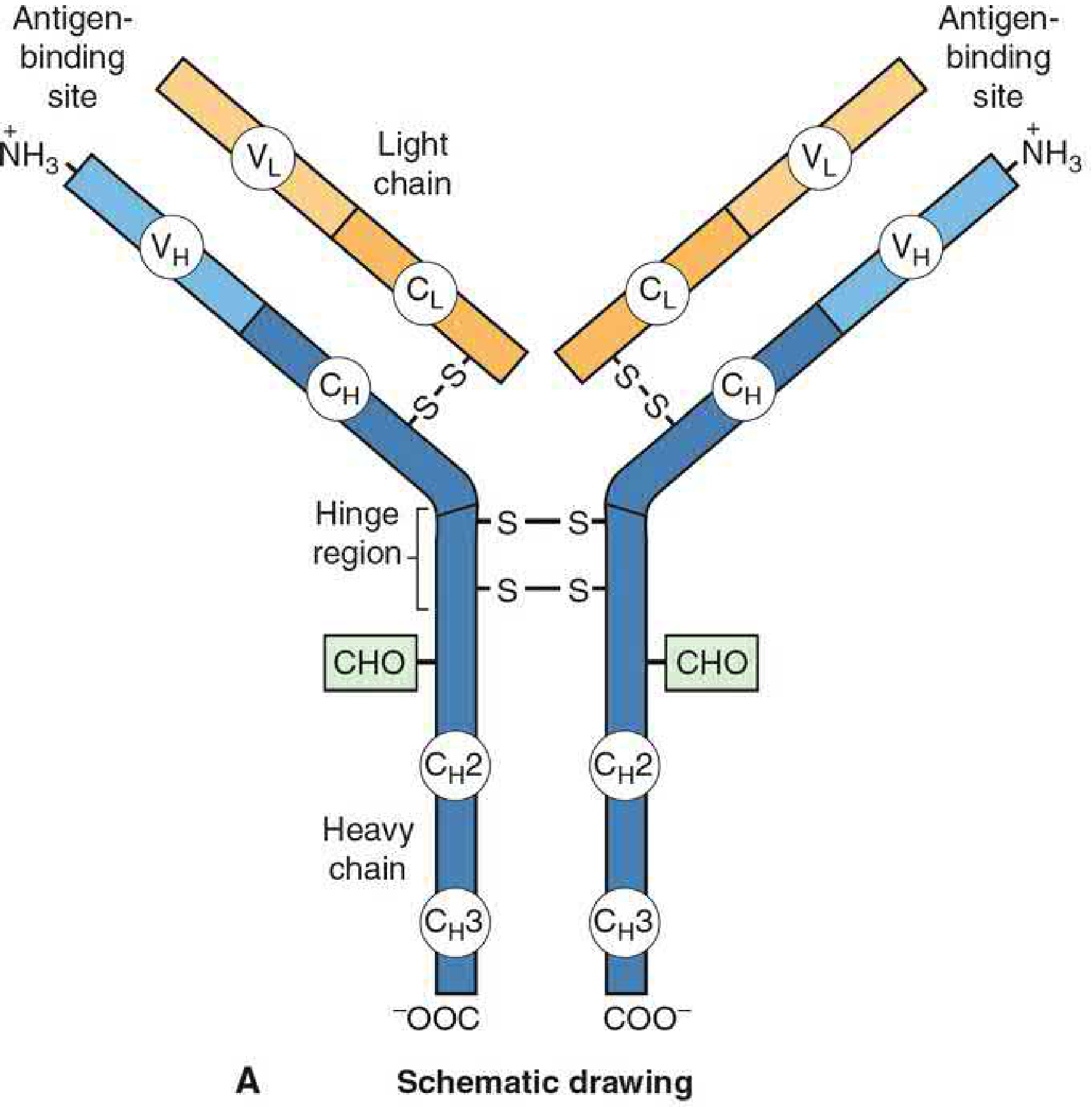
Parts of Each Chain
- Variable (V) region - at the tip of the "Y" - changes from antibody to antibody - this is where antigen binds
- Constant (C) region - at the stem/bottom of the "Y" - same in all antibodies of a given class
Important Fragments (Produced by Enzyme Digestion)
| Fragment | Enzyme | What it contains | Function |
|---|---|---|---|
| Fab (Fragment Antigen Binding) | Papain | V region + part of H chain | Binds antigen |
| Fc (Fragment Crystallizable) | Papain | Constant region of H chain | Binds complement, Fc receptors |
| F(ab')2 | Pepsin | Both Fab arms joined | Binds antigen + precipitates it |
Hinge Region
- Located between Fab and Fc parts
- Gives flexibility to the molecule so it can grab antigens at different angles
- Present in IgG, IgA, IgD
- IgM and IgE do NOT have a hinge region - instead they have an extra pair of constant domains
Light Chain Types
- Kappa (κ) - more common
- Lambda (λ) - less common
- Every immunoglobulin has EITHER two kappa OR two lambda chains (never mixed)
Structure of All 5 Classes
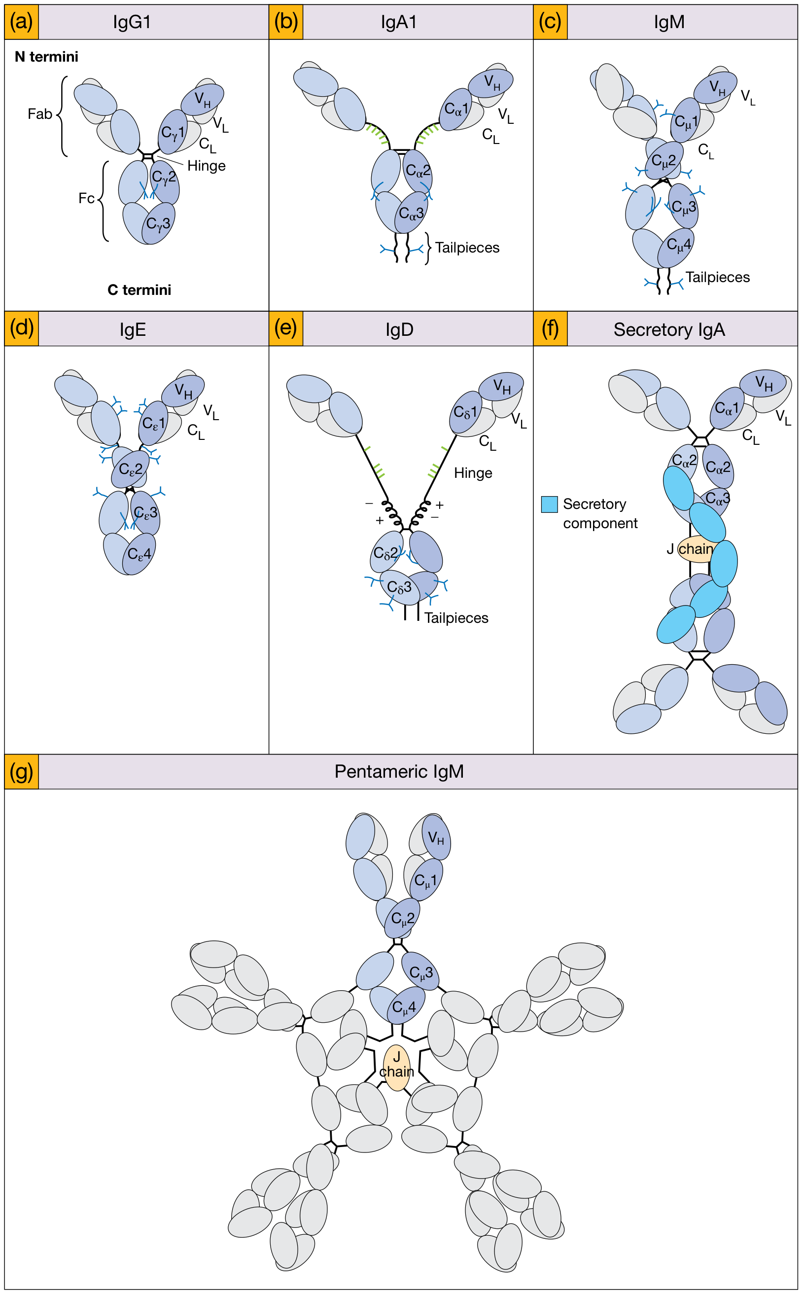
PART 3 - FUNCTIONS OF EACH CLASS
1. IgG - Most Abundant (75-80%)
- Main antibody in blood and tissues
- Only immunoglobulin that crosses the placenta - gives passive immunity to the newborn baby
- Activates complement system
- Binds to phagocytes (opsonization) - helps macrophages eat bacteria
- Main antibody in secondary (memory) immune response
- Has 4 subclasses: IgG1, IgG2, IgG3, IgG4
2. IgA - Guardian of Mucosal Surfaces
- Found in secretions - saliva, tears, breast milk, intestinal juice, respiratory tract
- In secretions it exists as dimeric IgA (2 monomers) linked by a J chain and covered by Secretory Component (SC) - this protects it from digestive enzymes
- Provides first line defense at mucosal surfaces
- Does NOT activate complement (classical pathway)
3. IgM - First Responder
- Pentamer (5 monomers joined by J chain) - largest immunoglobulin
- First antibody produced in primary immune response (appears first after infection)
- Most efficient at activating complement
- Natural blood group antibodies (anti-A, anti-B) are IgM
- Monomeric IgM acts as B-cell receptor (BCR) on B lymphocyte surface
4. IgE - Allergy and Antiparasitic
- Found in very low concentration in serum
- Most IgE is bound to mast cells and basophils (via Fc receptors)
- When antigen (allergen) binds to IgE on mast cells → degranulation → releases histamine → allergic reaction (hay fever, asthma, anaphylaxis)
- Also important against parasitic worm infections (helminths)
5. IgD - B-Cell Receptor
- Found in very low amounts in serum
- Main role is as a receptor on the surface of mature naive B-cells
- Helps in B-cell activation and maturation
- Its exact function is still not fully understood
SUMMARY TABLE (Quick Revision)
| Property | IgG | IgA | IgM | IgD | IgE |
|---|---|---|---|---|---|
| Heavy chain | γ | α | μ | δ | ε |
| Structure | Monomer | Monomer/Dimer | Pentamer | Monomer | Monomer |
| Serum % | 75-80% | 10-15% | 5-10% | <1% | Trace |
| MW (approx.) | 150,000 | 160,000 | 900,000 | 185,000 | 190,000 |
| Crosses placenta? | YES | No | No | No | No |
| Complement activation | Yes | No | Yes (best) | No | No |
| Key function | Main serum Ab | Mucosal defense | Primary response | B-cell receptor | Allergy/parasites |
KEY POINTS TO REMEMBER
- ALL immunoglobulins have the same basic 4-chain Y-shaped structure (2H + 2L chains)
- They differ in their heavy chains (γ, α, μ, δ, ε)
- IgG = most abundant, crosses placenta
- IgA = secretory, protects mucosal surfaces
- IgM = pentamer, first in primary response, best complement activator
- IgE = allergy and antiparasitic
- IgD = mainly B-cell surface receptor
- Variable region = antigen binding | Constant region (Fc) = effector functions
- Draw the Y-shaped IgG structure with labels - examiners love diagrams!
- The GAMED mnemonic will help you remember all 5 classes
- Focus on IgG, IgA, and IgM the most - they have the most marks in questions
- The summary table above is perfect for last-minute revision the night before your exam
Im 1st year mbbs student at Hngu university [ Bhagyoday medical college Kadi ] in exam imp questions list i saw one question is 5 mark question is Immunoglobulins [ Classification, structure, function]give a answer of this question easy to understand English language,when I read this question answer I understand easily, give answer this type when I read this question answer first time so much answer I understand easily in first time and when i write this in exam my sir will give me full out of full marks and easy to read and understand,answer give as 1 st year mbbs [ Biochemistry ]and easy to understand and write Thank you
Immunoglobulins - Classification, Structure & Function
(5 Mark Exam Answer - 1st Year MBBS Biochemistry)
INTRODUCTION
1. CLASSIFICATION
| Class | Heavy Chain | Structure | Serum % |
|---|---|---|---|
| IgG | Gamma (γ) | Monomer | 75-80% |
| IgA | Alpha (α) | Monomer / Dimer | 10-15% |
| IgM | Mu (μ) | Pentamer | 5-10% |
| IgD | Delta (δ) | Monomer | <1% |
| IgE | Epsilon (ε) | Monomer | Trace |
Memory trick: "GAMED" = IgG, IgA, IgM, IgE, IgD
2. STRUCTURE
Basic Structure - Y-Shaped Molecule
- 2 Heavy (H) chains - large (joined at the bottom of the Y)
- 2 Light (L) chains - small (on the arms of the Y)
- All chains held together by disulfide (-S-S-) bonds
Regions on Each Chain:
- Variable (V) region - at the tips of the Y - different in every antibody - binds antigen
- Constant (C) region - at the stem of the Y - same within each class - responsible for effector functions
Two Important Fragments:
- Fab (Fragment Antigen Binding) - the two arms of the Y - binds antigen
- Fc (Fragment Crystallizable) - the stem of the Y - binds complement and Fc receptors on phagocytes
Hinge Region:
- Between Fab and Fc - gives flexibility to grab antigens
- Present in IgG, IgA, IgD
- IgM and IgE have an extra pair of constant domains instead
3. FUNCTIONS OF EACH CLASS
| Class | Key Functions |
|---|---|
| IgG | Most abundant; crosses placenta (neonatal immunity); activates complement; opsonization; major antibody of secondary immune response |
| IgA | Found in secretions (saliva, tears, breast milk); protects mucosal surfaces; exists as secretory IgA (dimer + J chain + secretory component) |
| IgM | First antibody in primary response; pentamer; best complement activator; carries natural blood group antibodies (anti-A, anti-B) |
| IgE | Binds mast cells; triggers allergic reactions (asthma, anaphylaxis); defense against parasitic worms |
| IgD | Surface receptor on naive B-cells; role in B-cell activation |
KEY POINTS
- All Ig have same basic 4-chain structure (2H + 2L chains) shaped like a Y
- Classified by heavy chain type (γ, α, μ, δ, ε)
- IgG = most abundant, only one crossing placenta
- IgA = secretory immunity (mucosal)
- IgM = first + largest (pentamer), best complement activator
- IgE = allergy and antiparasite
- IgD = B-cell receptor
- Write a 2-line intro, then draw the classification table, then the structure with a small Y-diagram sketch, then functions table - this layout looks very organized and scores full marks
- Always draw the Y-shaped structure with labels in the exam - examiners give bonus impression marks for diagrams
- The summary at the end can serve as your conclusion line
Im 1st year mbbs student at Hngu university [ Bhagyoday medical college Kadi ] in exam imp questions list i saw one question is 8 mark question is Vitamin A & Wald visual cycle give a answer of this question easy to understand English language,when I read this question answer I understand easily, give answer this type when I read this question answer first time so much answer I understand easily in first time and when i write this in exam my sir will give me full out of full marks and easy to read and understand,answer give as 1 st year mbbs [ Biochemistry ]and easy to understand and write Thank you
Vitamin A & Wald's Visual Cycle
(8 Mark Exam Answer - 1st Year MBBS Biochemistry)
INTRODUCTION
PART 1 - VITAMIN A
A. Chemistry (Forms of Vitamin A)
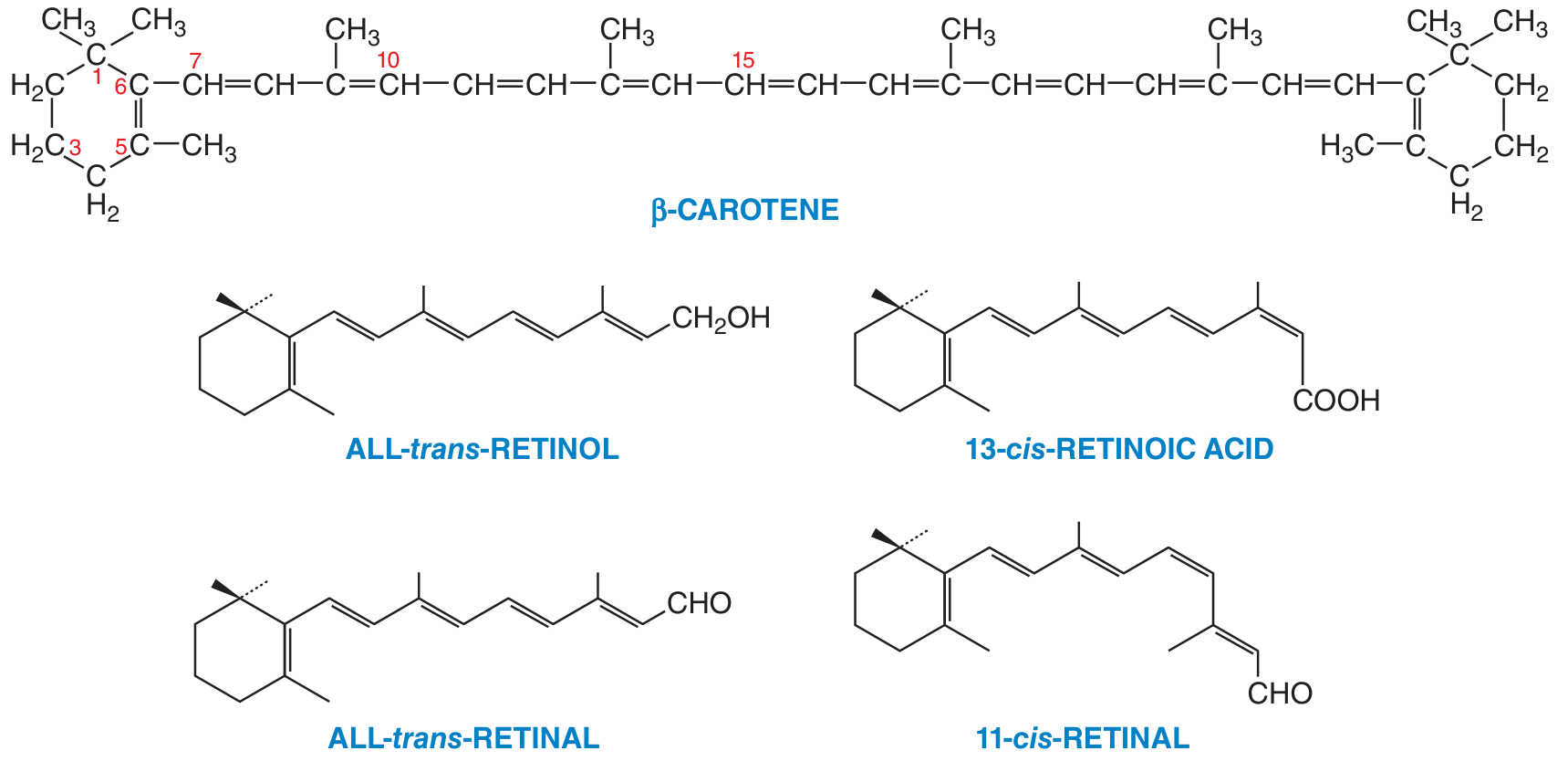
| Form | Also Called | Function |
|---|---|---|
| Retinol | Vitamin A alcohol | Storage + transport form |
| Retinal | Vitamin A aldehyde | Visual cycle (vision) |
| Retinoic acid | Vitamin A acid | Gene expression, growth, epithelium |
| β-Carotene | Provitamin A | Plant source, converted to retinol |
Memory trick: "Retinol → Retinal → Retinoic acid" (these are progressive oxidation steps)
B. Sources of Vitamin A
- Liver, egg yolk, butter, cheese, fish liver oils (cod liver oil), full cream milk
- Dark green leafy vegetables (spinach, fenugreek)
- Orange/yellow fruits and vegetables (carrot, mango, papaya, pumpkin)
Note: β-Carotene is converted to Vitamin A in the intestinal wall. 1 molecule of β-Carotene → 2 molecules of Retinal → 2 molecules of Retinol
C. Absorption, Transport & Storage
- Vitamin A is fat-soluble - requires dietary fat and bile salts for absorption
- Absorbed in the small intestine
- Transported in blood by Retinol Binding Protein (RBP)
- Stored in liver (as retinyl esters) - the liver is the main storage organ
- Daily Requirement: 750 mcg/day (adult)
D. Functions of Vitamin A
| Function | Details |
|---|---|
| 1. Vision | Forms rhodopsin in rod cells - needed for dim light (night) vision |
| 2. Epithelial maintenance | Keeps epithelium moist and healthy; prevents keratinization |
| 3. Growth | Needed for normal bone and tooth development |
| 4. Reproduction | Required for spermatogenesis and fetal development |
| 5. Immunity | Maintains integrity of mucous membranes as a barrier |
| 6. Gene expression | Retinoic acid binds nuclear receptors (RAR, RXR) and controls gene transcription |
| 7. Antioxidant | β-Carotene acts as antioxidant |
E. Deficiency of Vitamin A
| Deficiency Sign | Explanation |
|---|---|
| Night blindness (Nyctalopia) | First and earliest sign - cannot see in dim light |
| Xerophthalmia | Dry eyes due to loss of goblet cells |
| Bitot's spots | Foamy white patches on conjunctiva |
| Keratomalacia | Softening and ulceration of cornea → blindness |
| Follicular hyperkeratosis | Skin becomes rough and scaly (toad skin) |
| Growth retardation | Stunted bone growth in children |
PART 2 - WALD'S VISUAL CYCLE
Background
Key Players (Remember these first!)
| Term | What it is |
|---|---|
| Rod cells | Retinal cells sensitive to dim/low light |
| Cone cells | Retinal cells for bright light and colour vision |
| Opsin | A protein present in rod cells |
| 11-cis-Retinal | The active form of Vitamin A - acts as a "key" |
| Rhodopsin | Opsin + 11-cis-Retinal combined = the light-sensitive pigment |
| Scotopsin | Opsin after separating from retinal (bleached form) |
Steps of the Visual Cycle
IN DARK (Rhodopsin is formed and ready):
WHEN LIGHT FALLS (Vision occurs):
RECOVERY (Dark adaptation - cycle repeats):
- Reduced back to All-trans-Retinol → re-isomerized to 11-cis-Retinal
- OR carried to Retinal Pigment Epithelium (RPE) cells for re-isomerization
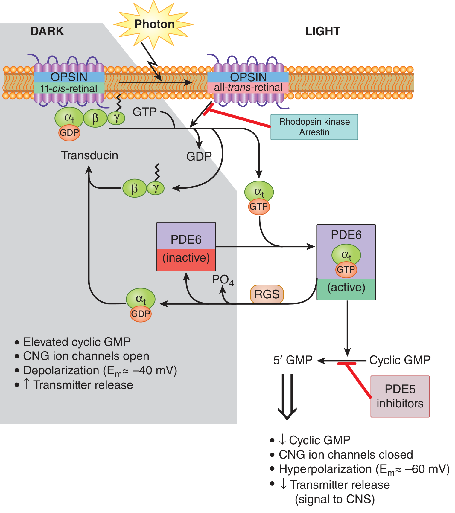
Simple Flow Diagram of the Visual Cycle (Draw this in exam!)
    DARK                              LIGHT
      |                                  |
  11-cis-Retinal + Opsin           Rhodopsin
         ↓                               ↓ (photon hits)
      RHODOPSIN                  All-trans-Retinal + Opsin
      (ready)                         (bleached)
         ↑                               ↓
  Re-isomerization              Transducin activated
  (in RPE cells)                        ↓
         ↑                         PDE6 activated
  All-trans-Retinal ←─────────  cGMP → 5'-GMP
                                        ↓
                               Na+ channels CLOSE
                                        ↓
                               Hyperpolarization
                                        ↓
                             Nerve signal → Brain → VISION
Why Night Blindness Happens in Vitamin A Deficiency
- Without enough Vitamin A, there is not enough Retinal in the body
- So Rhodopsin cannot be regenerated after bleaching
- The rod cells cannot respond to dim light again
- Result: Night blindness (Nyctalopia) - the earliest sign of Vitamin A deficiency
QUICK SUMMARY TABLE
| Topic | Key Point |
|---|---|
| Chemical name | Retinol (Vitamin A) |
| Solubility | Fat-soluble |
| Storage | Liver |
| Transport | Retinol Binding Protein (RBP) |
| Active form in vision | 11-cis-Retinal |
| Visual pigment | Rhodopsin = Opsin + 11-cis-Retinal |
| First deficiency sign | Night blindness |
| Named after | George Wald (Nobel 1967) |
| Cells involved | Rod cells (dim light), Cone cells (colour/bright) |
- Introduction (2-3 lines) - 0.5 mark
- Chemistry/Forms table - 1 mark
- Sources + Absorption - 1 mark
- Functions table - 1.5 marks
- Deficiency table - 1 mark
- Wald's Visual Cycle with diagram - 3 marks (this is the high-value part - always draw the cycle!)
Im 1st year mbbs student at Hngu university [ Bhagyoday medical college Kadi ] in exam imp questions list i saw one question is 8 mark question is Ammonia [ Formation , Transamination , Deamination, Transport, Storage,Detoxification Uses ] give a answer of this question easy to understand English language,when I read this question answer I understand easily, give answer this type when I read this question answer first time so much answer I understand easily in first time and when i write this in exam my sir will give me full out of full marks and easy to read and understand,answer give as 1 st year mbbs [ Biochemistry ]and easy to understand and write Thank you