
Frame a interesting case discussion for a case of myxoid neoplasm
myxoid neoplasm diagnosis pathology
myxoid liposarcoma histology pathology MRI
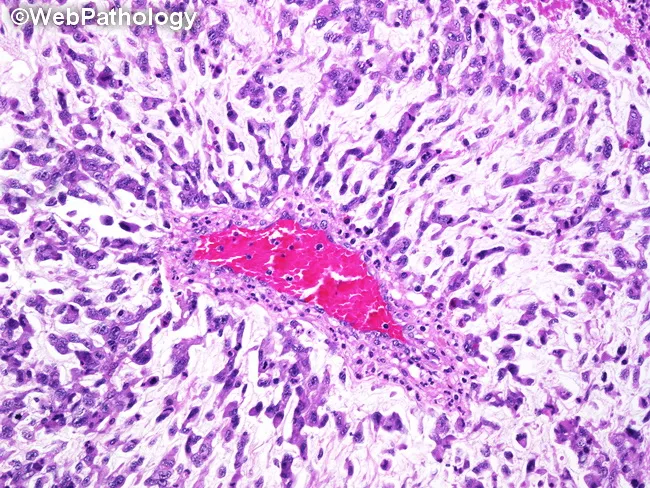
Histology image of a soft tissue mass showing features diagnostic of myxoid liposarcoma, high-grade (round cell) variant. The specimen is a tissue biopsy examined by light microscopy after Hematoxylin and Eosin staining. The architectural pattern reveals a prominent myxoid (mucoid) extracellular matrix with a delicate, arborizing capillary network radiating from vessels, a classic backdrop for myxoid adipocytic tumors. Intermixed are primitive, round to oval tumor cells arranged in cords and short trabeculae extending from the vascular core. The cells have scant cytoplasm, hyperchromatic nuclei, and mitotic activity is variably increased consistent with high-grade transformation. Occasional lipoblasts with scalloped cytoplasm may be present, highlighting adipocytic differentiation amidst the myxoid milieu. The surrounding stroma is relatively cellular in the high-grade areas, with less prominent lipogenic features compared with low-grade components. This image emphasizes the cellularity and architectural disruption associated with the round cell component, which portends a more aggressive clinical behavior and higher metastatic potential. Clinically, identification of high-grade cell populations in a predominantly myxoid liposarcoma influences prognosis and treatment planning, including surgical excision with wide margins and consideration of adjuvant chemotherapy or radiotherapy. Overall, the image exemplifies hallmarks used in pathology reports to classify liposarcoma subtype and grade for diagnostic accuracy.
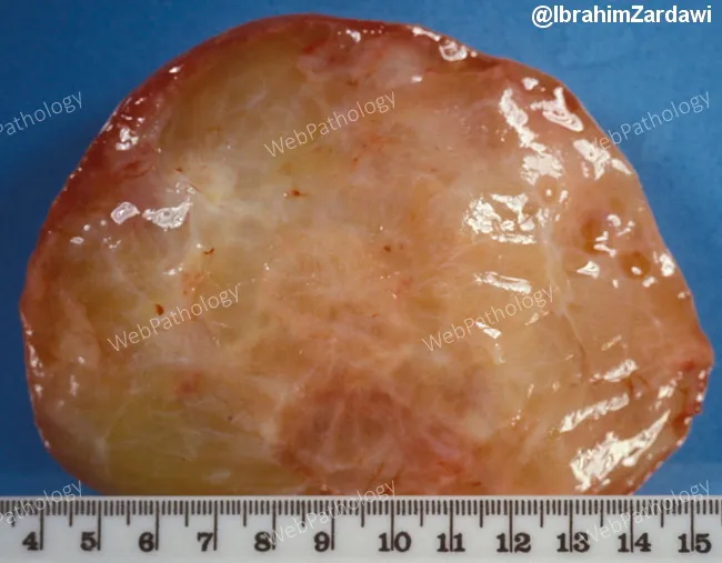
Gross pathology photograph of a myxoid liposarcoma in a soft-tissue tumor specimen. Modality: gross examination with photographic documentation. The specimen is a well-circumscribed soft mass, measuring roughly 8–9 cm in greatest dimension, with a glistening, gelatinous cut surface that reflects abundant myxoid extracellular matrix. The exterior is tan-pink and slightly translucent, with a lobulated contour and a moist, glossy surface. The cut surface reveals a homogeneous, mucinous pale-yellow to pinkish material interspersed with sparse adipose elements; there may be minor hemorrhagic foci at the periphery. The margins appear relatively well defined in the specimen, though in vivo growth can be infiltrative. Overall, the gross morphology is consistent with myxoid liposarcoma, a malignant adipocytic tumor characterized by a plentiful myxoid stroma and delicate capillary networks. This gross pattern correlates with the characteristic histology of myxoid liposarcoma: abundant myxoid stroma containing delicate capillaries and scattered lipoblasts at the microscopic level. Clinically, recognizing this gross pattern aids in prompt pathologic triage, surgical planning, and prognosis. This image is valuable for education in soft tissue sarcomas, tumor grossing technique, and radiopathology correlation, and it supports teaching datasets and searchable image repositories for pathology training, case-based learning, and medical informatics. Educational value assured.
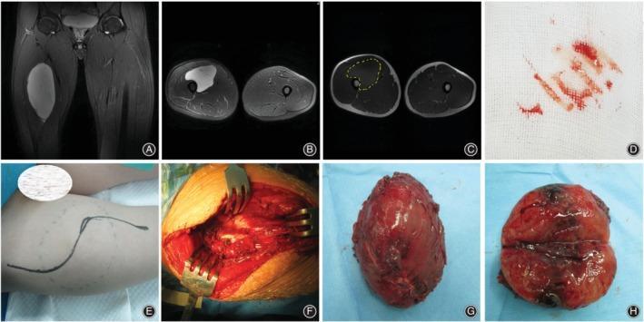
This composite figure illustrates the clinical management and pathology of a primary myxoid liposarcoma in the left thigh of a 20-year-old male. (A-C) Diagnostic MRI in coronal and axial planes shows a large, hyperintense, well-circumscribed ovoid mass within the medial muscle compartment. A yellow dotted line in (C) indicates the planned surgical margins. (D) A biopsy sample on gauze confirms the preoperative diagnosis. (E-F) Surgical progression images depict the planned incision incorporating the biopsy site and the intraoperative field where periosteal resection was performed near the bone. (G-H) Macroscopic pathology of the resected specimen reveals a smooth, glistening, reddish-brown encapsulated mass. Longitudinal sectioning (H) demonstrates a heterogeneous internal architecture with prominent dark regions of central necrosis and cystic degeneration, characteristic of high-grade components or typical evolution of large liposarcomas. The sequence highlights the workflow from diagnostic radiology and biopsy to wide surgical excision and gross pathological assessment in musculoskeletal oncology.
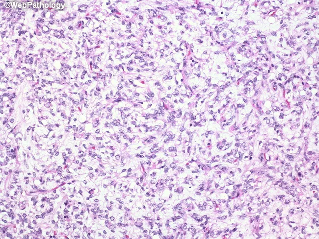
This histopathology image depicts a relatively cellular area within a myxoid liposarcoma on Hematoxylin and Eosin staining using bright-field light microscopy. The tumor is embedded in a prominent myxoid (mucinous) extracellular matrix, rendered in pale bluish-purple, with a delicate, arborizing capillary vascular network that is characteristic of this entity. Scattered lipoblasts are present, identified by cytoplasmic lipid vacuoles that indent (scallop) the nuclei, and by occasional hyperchromatic, rounded to oval nuclei. The neoplastic cells range from small to moderately sized spindle- to stellate-shaped cells with variably eosinophilic, scant cytoplasm and mild nuclear atypia. Cellular areas show increased density compared with the abundant myxoid background. Mitotic figures are uncommon in this field, and there is no conspicuous necrosis. The overall pattern—myxoid stroma with a plexiform capillary network and lipogenic cells—supports a diagnosis of myxoid liposarcoma; higher cellularity regions can be encountered in the cellular variant. This histology distinguishes from other lipomatous neoplasms and from other myxoid sarcomas by the presence of lipoblasts and the characteristic branching vasculature. Clinically, dx is often reinforced by molecular tests for DDIT3 gene rearrangement. Correlate with imaging (MRI/CT) and clinical presentation for staging and treatment planning.
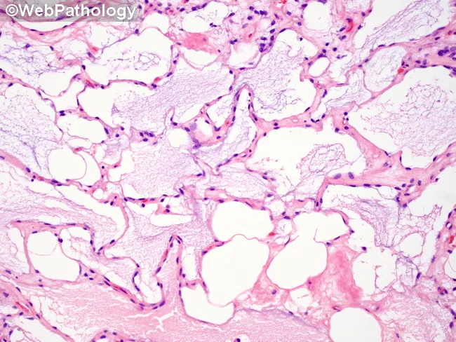
Imaging modality: Light microscopy of hematoxylin-eosin stained histology section from a soft tissue biopsy containing a liposarcoma with prominent myxoid matrix. Anatomical context: soft tissue tumor, unspecified site, intratumoral myxoid stroma with mucin pooling. Visual features: abundant mucin pools create cribriform or lacelike architecture; flat tumor cells line mucin pools with intermittent rounded cells at the pool rims; pseudoacinar patterns arise at the mucin borders; in some areas, a lymphangioma-like appearance is observed; occasional cystic spaces and edema-like basophilic mucin mimic pulmonary edema histology. These features are typical of myxoid liposarcoma; adipocytic differentiation may be seen; scattered delicate capillary networks may accompany the mucin pools. This histology supports diagnosis of myxoid liposarcoma, a soft tissue sarcoma characterized by abundant myxoid stroma and a plexiform capillary network; the mucin-rich milieu contributes to a clear-to-pale appearance on H&E. Diagnostic significance: recognition of myxoid matrix with pooling mucin and cribiform pattern aids distinction from myxofibrosarcoma and other myxoid sarcomas; differential diagnoses include myxoid chondrosarcoma or metastatic mucinous carcinoma depending on location. Clinical correlation: correlate with tumor size, site, and imaging (MRI features of high T2 signal with heterogenous enhancement); prognosis varies with histologic grade and presence of lipoblasts. Potential clinical use cases include diagnostic confirmation and educational illustration for soft-tissue pathology.
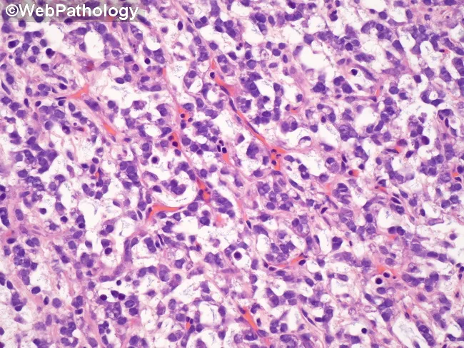
Histopathology image depicts a high-grade myxoid liposarcoma, round cell predominant. Using light microscopy, sections stained with hematoxylin and eosin show primitive-appearing tumor cells arranged in cords, nests, and short trabeculae within a variably myxoid matrix. The tumor displays a delicate, plexiform capillary network typical of myxoid liposarcoma, with prominent branching, thin-walled vessels in a myxoid stroma. Cells are small to medium-sized with hyperchromatic, elongated to round nuclei, scant cytoplasm, and conspicuous mitotic activity in high-grade areas. Occasional lipoblast-like cells may be seen; lipogenic differentiation is variably evident. The background myxoid stroma appears pale pink to blue with abundant mucopolysaccharide matrix. Necrosis is minimal or focal. The lesion lacks overt adipocytic maturation in the high-grade component. Immunohistochemistry is not provided in image, but typical profiles show variable S-100 positivity; molecular hallmark is the FUS-DDIT3 fusion gene due to t(12;16)(q13;p11). Clinically, this histology portends a higher risk of metastasis and recurrence compared to conventional, low-grade myxoid liposarcoma. Potential differential diagnoses include round cell variants of other soft tissue sarcomas such as synovial sarcoma, Ewing/PNET, and malignant peripheral nerve sheath tumor, requiring molecular testing to confirm. The image is educational for pathology education, tumor histology, and diagnostic pathology training; relevant for surgical oncology planning and radiation therapy decisions.
Case Discussion: The Deceptive Thigh Mass - Myxoid Liposarcoma
CASE PRESENTATION
- A 9 x 7 cm ovoid soft tissue mass, medial compartment of the right thigh, deep to the fascia
- Non-tender, poorly compressible, smooth surface, not pulsatile
- No overlying skin changes, no satellite nodules
- No inguinal lymphadenopathy
- Neurovascular status of the limb: intact
QUESTION 1: What is your initial differential diagnosis for a deep, painless soft tissue mass in the thigh of a middle-aged adult?
| Category | Diagnosis | Distinguishing Feature |
|---|---|---|
| Malignant | Myxoid liposarcoma | Myxoid stroma, arborizing capillaries, lipoblasts |
| Malignant | Well-differentiated liposarcoma | Mature adipocytes, MDM2 amplification |
| Malignant | Myxofibrosarcoma | Older patients, more pleomorphic stroma |
| Malignant | Synovial sarcoma | Younger age, calcifications, t(X;18) |
| Benign mimics | Intramuscular lipoma | Mature fat, no lipoblasts, no myxoid stroma |
| Benign mimics | Ganglion / myxoid cyst | Near joints, no deep muscle origin |
| Benign | Nodular fasciitis | Rapidly growing, usually <5 cm, subcutaneous |
Teaching point: Any soft tissue mass >5 cm, deep to fascia, in an adult should be considered malignant until histology proves otherwise. Watchful waiting is NOT appropriate.
QUESTION 2: What imaging would you order, and what would you expect to find?
- T1-weighted: Predominantly isointense to muscle; subtle thin T1-hyperintense fatty septa (the "tell-tale" finding)
- T2-weighted / STIR: Markedly hyperintense - the myxoid component appears like fluid, which can mimic a cyst
- Post-gadolinium: Heterogeneous enhancement, distinguishing it from a simple cyst
- Key diagnostic clue: The combination of fluid-like T2 signal + faint fatty septa is virtually pathognomonic of myxoid liposarcoma
- CT chest: To rule out pulmonary metastases (standard for all STS)
- MRI whole spine / pelvis: Myxoid liposarcoma has a unique predilection for extrapulmonary metastases to fat-rich sites - retroperitoneal fat, bone marrow, spinal epidural fat. Routine spinal MRI staging is recommended for this subtype specifically.
- PET-CT or whole-body MRI: To identify alternative metastatic sites
Clinical pearl: Unlike most other sarcomas, the lungs are NOT the only (or even the most common) site of distant spread in myxoid liposarcoma. The unique tropism for lipomatous sites is explained by the tumor's adipocytic lineage and FUS-DDIT3-driven arrest of adipocyte differentiation. - Schwartz's Principles of Surgery, 11e
QUESTION 3: The biopsy report comes back. What do you expect?
"Sections show a cellular tumor composed of small, primitive mesenchymal cells and lipoblasts at varying stages of differentiation set in a prominent basophilic myxoid matrix. A characteristic branching, thin-walled, arborizing capillary network is present. No significant nuclear pleomorphism. Round cell component estimated at <5%. Consistent with: LOW-GRADE MYXOID LIPOSARCOMA. FISH for DDIT3 (12q13) rearrangement: POSITIVE."
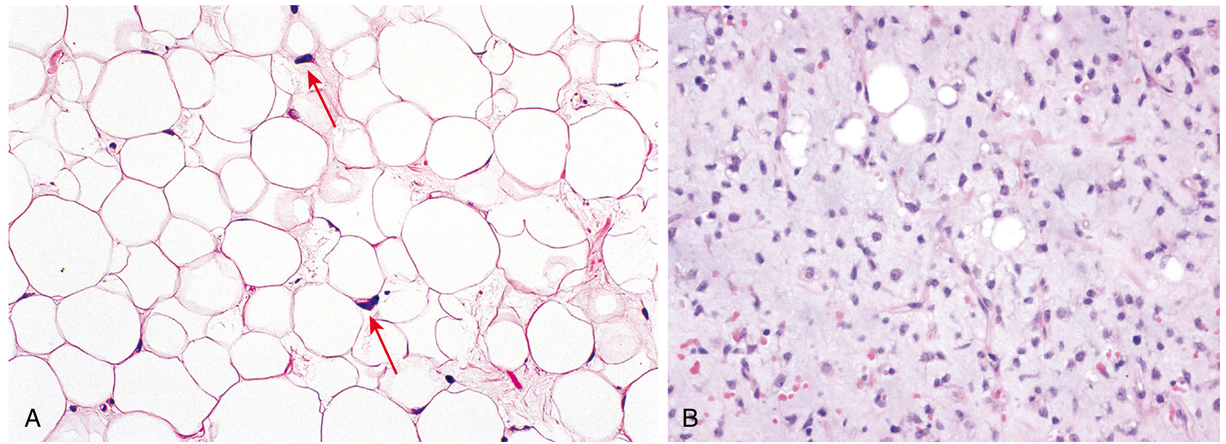
- Myxoid stroma - abundant basophilic extracellular matrix (hyaluronic acid-rich)
- Branching "crow's feet" / arborizing thin-walled capillaries - the most characteristic structural feature
- Primitive mesenchymal cells - small, oval, bland-looking
- Lipoblasts - cells with cytoplasmic vacuoles that scallop ("indent") the nucleus, in varying stages of maturation - resembles fetal fat
- Round cell component - the critical prognostic parameter (discussed below)
"The histologic grade of myxoid liposarcoma is determined not by pleomorphism or histopathologic aggressiveness, but rather by the percentage of round cells within the neoplasm. Tumors with a >5% round cell component are considered high grade." - Current Surgical Therapy 14e
QUESTION 4: What is the molecular basis, and why does it matter clinically?
- t(12;16)(q13;p11) → FUS-DDIT3 fusion: ~90% of cases
- t(12;22)(q13;q12) → EWSR1-DDIT3 fusion: ~10% of cases
"The FUS::DDIT3 fusion gene encodes a chimeric DNA binding protein. Through an uncertain mechanism, this oncoprotein arrests adipocyte differentiation, leading to proliferation of primitive mesenchymal cells." - Robbins & Cotran Pathologic Basis of Disease
- FISH or RT-PCR for DDIT3 rearrangement confirms the diagnosis on biopsy
- Rules out other myxoid tumors (myxofibrosarcoma lacks this translocation)
- Enables diagnosis even when morphology is ambiguous
- Emerging targeted therapies directed at FUS-DDIT3 are under investigation
QUESTION 5: How do you stage and risk-stratify this patient?
- Tumor differentiation score + Mitotic count score + Necrosis score
- Low-grade (G1): <5% round cells, low mitoses, no necrosis
- High-grade (G2/G3): >5% round cells (round cell transformation)
- Stage IA: Low grade, ≤5 cm
- Stage IB: Low grade, >5 cm - our patient
- Stage III: High grade, any size, or any grade with node involvement
- Stage IV: Distant metastasis
- Myxoid liposarcoma has an intermediate prognosis among liposarcoma subtypes
- Local recurrence: ~20%, Metastatic risk: ~50%
- The round cell percentage is the single most important histologic prognostic marker
- Extrapulmonary metastases (bone, retroperitoneum) confer significantly worse prognosis
QUESTION 6: What is the treatment plan?
Surgery (Primary Treatment)
- Wide local excision with clear margins (R0) is the cornerstone of treatment
- Limb-sparing surgery is preferred; amputation reserved for unresectable cases
- Retroperitoneal/intraabdominal locations carry higher recurrence risk due to anatomic difficulty achieving wide margins
Radiation Therapy
- Myxoid liposarcoma is notably radiosensitive - among the most radiation-responsive of all soft tissue sarcomas
- Indications: Tumors >5 cm, close/positive margins, high-grade (round cell) component
- Can be given preoperatively (neoadjuvant - preferred: better tumor downstaging, smaller field) or postoperatively
Chemotherapy
- Myxoid liposarcoma is considered relatively chemosensitive compared to other STS subtypes
- Standard regimens: doxorubicin + ifosfamide (first-line), trabectedin (particularly effective for myxoid liposarcoma - direct activity against FUS-DDIT3)
- Trabectedin (Yondelis) is worth highlighting - it has a specific mechanism of action against FUS-DDIT3 fusion proteins and is approved for advanced/metastatic liposarcoma
- High-grade (round cell) tumors: neoadjuvant chemotherapy followed by radiation, then surgery may be considered
"High-grade myxoid liposarcomas appear to be particularly responsive to radiation therapy as well as systemic therapy, suggesting a potential role for neoadjuvant treatment." - Current Surgical Therapy 14e
Follow-up
- MRI thigh every 3-6 months for 2-3 years, then annually
- CT chest every 6 months
- MRI spine annually (given the extrapulmonary metastatic pattern to bone marrow)
QUESTION 7: Twist - the pathologist calls you back. The round cell component is now re-evaluated at 30%. How does this change management?
- Higher mitotic activity
- Sheets of primitive round cells with scant cytoplasm
- Markedly hyperchromatic nuclei
- Higher metastatic potential - particularly to bone marrow, CNS, and retroperitoneum
- 5-year survival drops significantly compared to pure low-grade myxoid liposarcoma
- Consider neoadjuvant chemotherapy (doxorubicin + ifosfamide) before surgery
- Mandatory whole-body MRI or PET-CT for staging (bone marrow metastases)
- Mandatory spinal MRI (epidural fat metastases)
- More aggressive radiation planning
- Discuss enrollment in clinical trial targeting FUS-DDIT3
SUMMARY TABLE: Myxoid Liposarcoma at a Glance
| Parameter | Details |
|---|---|
| Incidence | 2nd most common liposarcoma; ~5-10% of all adult STS |
| Age | Young to middle-aged adults (2nd-5th decade); most common liposarcoma in children |
| Site | Deep soft tissues, thigh (most common); rarely retroperitoneum |
| Key symptom | Painless, slowly growing deep mass |
| MRI hallmark | Fluid-like T2 signal + subtle T1 fatty septa + heterogeneous enhancement |
| Histology | Myxoid stroma + arborizing capillaries + lipoblasts + primitive cells |
| Molecular | t(12;16) FUS-DDIT3 (~90%) or t(12;22) EWSR1-DDIT3 (~10%) |
| Grading key | % round cell component (>5% = high grade) |
| Unique metastatic pattern | Extrapulmonary: bone marrow, retroperitoneal fat, spinal epidural fat |
| Treatment | Surgery (wide excision) + radiation (highly radiosensitive) ± chemotherapy |
| Chemo-sensitive? | Yes - especially to doxorubicin/ifosfamide and trabectedin |
| Prognosis | Intermediate; local recurrence ~20%, metastatic risk ~50% |
BONUS: Histology Image Gallery

TAKE-HOME MESSAGES
- Any deep thigh mass >5 cm in an adult = sarcoma workup. Do not aspirate, incise, or observe without imaging first.
- MRI before biopsy - the T2-hyperintense "fluid-like" signal with faint fatty septa is virtually diagnostic of myxoid liposarcoma.
- The round cell percentage is everything in grading - >5% transforms it from a low-grade to a high-grade tumor with very different management and prognosis.
- DDIT3 FISH/RT-PCR confirms the diagnosis and distinguishes from other myxoid tumors.
- Stage the spine - the unique extrapulmonary metastatic pattern demands whole-spine MRI as part of staging.
- Trabectedin is the one chemotherapy that directly targets the FUS-DDIT3 fusion protein and has proven activity in this tumor.
- Myxoid liposarcoma is among the most radiosensitive sarcomas - radiation is a key part of multimodal treatment.