
Techniques to identify growth of Viruses in Cell Culture
cytopathic effect virus cell culture
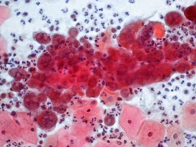
This cervical cytology image from a conventional Pap smear demonstrates the cytopathic effect of herpes simplex virus infection in squamous epithelium. Imaging modality is light microscopy with Pap staining; technique includes fixation, staining, and smear preparation, with magnification around 40x. Anatomical location corresponds to the cervix, specifically the transformation zone where squamous epithelium overlies glandular mucosa. Visual features include numerous large multinucleated squamous cells with nuclear molding, prominent ground-glass nuclei, and perinuclear halos; chromatin marginates and intranuclear eosinophilic inclusions (Cowdry type A) are variably present. A background inflammatory infiltrate contains lymphocytes and neutrophils; adjacent erythrocytes and epithelial cells provide a mixed cytology field. Pathology: characteristic HSV-induced cytopathic effect in cervical epithelium; findings reflect active viral replication and cell-cell fusion, leading to multinucleation and nuclear fragmentation. Diagnostic significance: supports diagnosis of HSV cervicitis in the appropriate clinical context; helps distinguish HSV infection from other cervicitis etiologies such as HPV-related koilocytosis or Chlamydia; may prompt confirmatory testing (HSV PCR, viral culture) and antiviral therapy. Differential considerations: CMV infection with owl’s eye inclusions; koilocytosis from HPV; nonspecific inflammatory changes. Clinical correlation: correlate with genital ulcers, dysuria, sexual history, and lesion distribution; documents viral shedding risk and informs patient counseling.
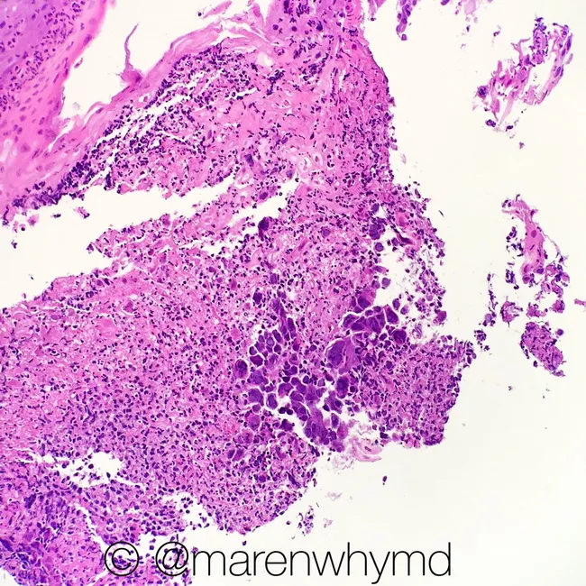
Histopathology image of esophageal mucosa showing herpes simplex virus (HSV) esophagitis. Prepared as a formalin-fixed paraffin-embedded tissue section and stained with Hematoxylin and Eosin (H&E); light microscopy reveals an ulcerated mucosal surface with dense inflammatory infiltrate and focal islands of squamous epithelium. The hallmark cytopathic effect includes multinucleated squamous cells with margination of chromatin and prominent intranuclear ground-glass inclusions (Cowdry type A bodies) within enlarged cells at the ulcer margin. The surrounding stroma exhibits acute and chronic inflammatory cells, edema, and reactive basal cell hyperplasia. These findings distinguish HSV esophagitis from reflux esophagitis and other infectious esophagitides such as cytomegalovirus or candidal disease. The diagnostic significance rests on the classic viral cytopathic changes rather than clinical symptoms alone. Differential considerations include CMV esophagitis with owl’s eye inclusions (larger cells, intranuclear inclusions but different distribution), and candida esophagitis characterized by fungal hyphae and pseudomembrane formation. Clinically, HSV esophagitis supports antiviral therapy (acyclovir/valacyclovir) and prompts evaluation for immunocompromised status or underlying disease. This image is useful for medical education, pathology training, and research on herpesvirus–esophagitis pathology, illustrating the cytopathic effect and ulcer-associated histology in a gastrointestinal biopsy. Correlative immunohistochemistry may confirm HSV infection in ambiguous cases.
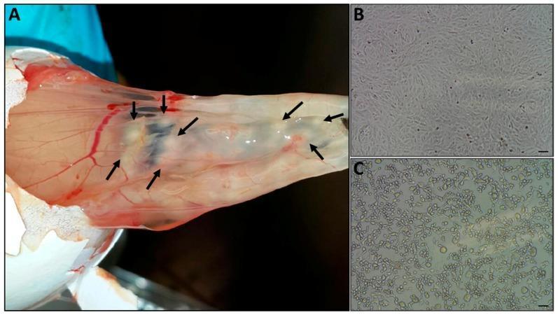
Educational composite image demonstrating virus isolation techniques. Panel A shows a clinical photograph of an embryonated chicken egg with the shell removed to reveal the chorioallantoic membrane (CAM). Black arrows indicate several discrete, elevated, pale-white pock lesions resulting from viral infection. The CAM exhibits visible vascularization with prominent red blood vessels over a translucent membrane. Panels B and C provide a comparison of cell culture morphology (Vero cells) using light microscopy. Panel B displays a control group of uninfected cells, showing a healthy, confluent monolayer of elongated, spindle-shaped cells with clear boundaries. Panel C illustrates significant cytopathic effect (CPE) following inoculation with the pock material. Observed cellular changes in Panel C include rounding of cells, loss of adherence, fragmentation of the monolayer, and increased cellular debris, typical of orthopoxvirus-induced damage. Scale bars (100 μm) are present in the microscopy images for quantitative reference.
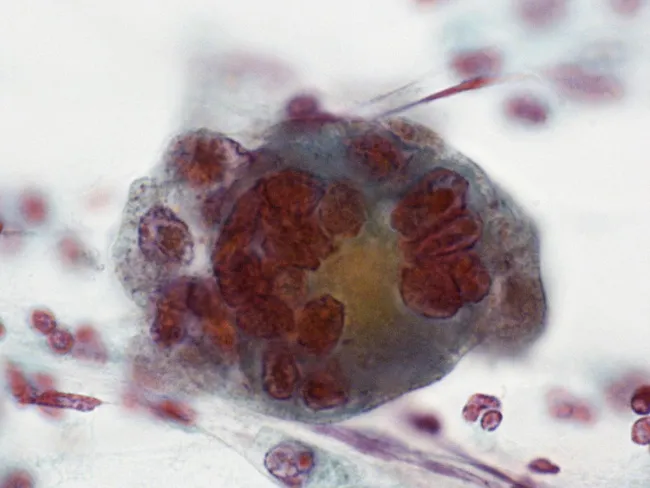
Imaging modality: Conventional cervical cytology (Pap smear) analyzed by light microscopy after Papanicolaou staining. Primary subject: an HSV-induced cytopathic change in cervical squamous epithelium, demonstrated as a multinucleated giant cell with characteristic intranuclear inclusions. Anatomical localization: cervix, transformation zone, squamous epithelium of the lower genital tract. Visual features: a large, rounded cell with abundant cytoplasm, multiple molded nuclei, marginated chromatin, and dense eosinophilic intranuclear inclusions (Cowdry type A). A perinuclear halo and nuclear molding are evident; nuclei tend to crowd together but do not fully overlap. Background typically contains neutrophils or inflammatory cells. Staining accentuates the eosinophilic nuclear inclusions and the halo, enabling recognition of HSV cytopathic effect. Pathological interpretation: cytopathic effects consistent with active herpes simplex virus infection of cervical epithelium (HSV cervicitis). Diagnostic significance: supports clinical suspicion of herpetic cervicitis; guides antiviral therapy and infection control; may prompt confirmatory testing (HSV PCR or viral culture). Differential considerations: cytomegalovirus infection (owl's eye inclusions) can mimic in some respects but shows solitary cells with distinct intranuclear inclusions; koilocytes from HPV are different. Clinical correlation: correlate with symptoms (dysuria, vaginal discharge, genital ulcers) and pregnancy status. Educational relevance: classic cytology example of HSV infection on Pap smear. Reproducible educational reference material.
Techniques to Identify Growth of Viruses in Cell Culture
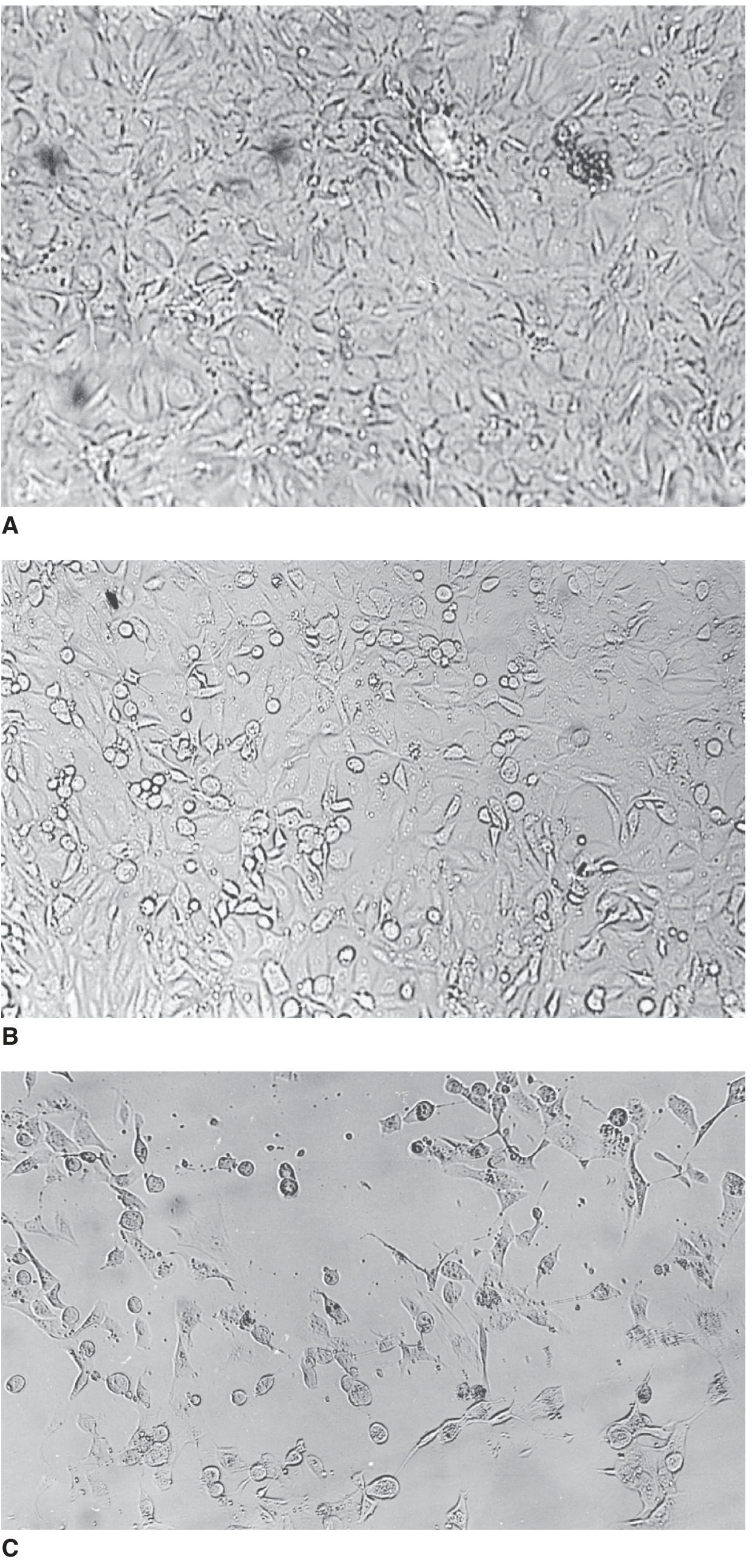
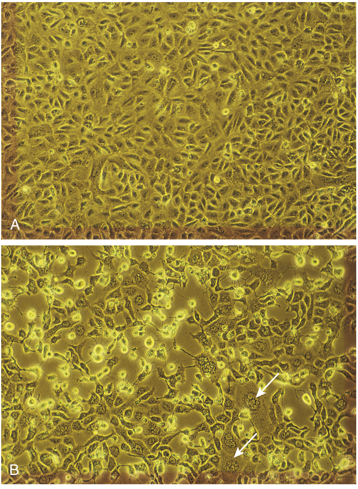
1. Cytopathic Effect (CPE)
- Cell rounding, shrinking, and loss of attachment - common with enteroviruses
- Grapelike clusters of enlarged round cells - adenoviruses
- Syncytia (multinucleated giant cells) formed by virus-induced cell-to-cell fusion - RSV, measles, HSV
- Intranuclear or intracytoplasmic inclusion bodies - HSV (Cowdry type A, intranuclear), CMV ("owl's eye"), rabies (Negri bodies, intracytoplasmic)
- Margination of chromatin - herpesviruses
- Foamy degeneration - some retroviruses
2. Hemadsorption
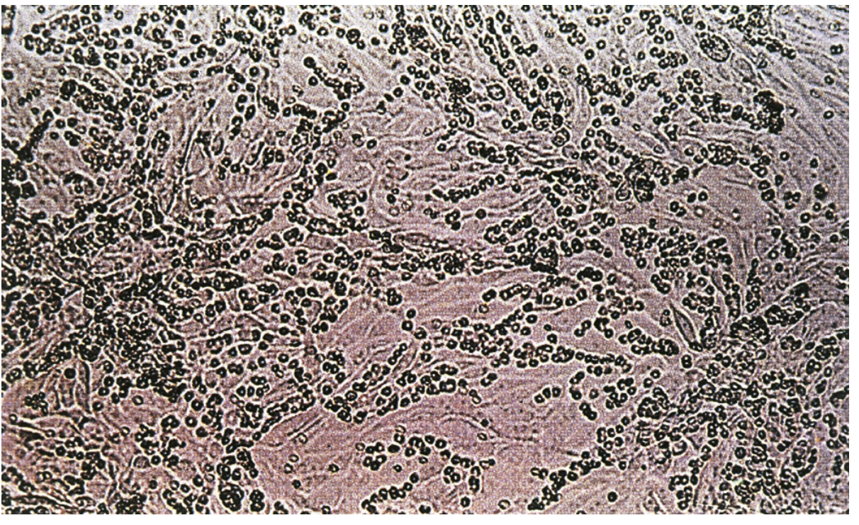
- Principle: Certain viruses (influenza, parainfluenza, mumps, togaviruses) cause the infected cell to express viral hemagglutinin on its membrane. When guinea pig or human red blood cells (RBCs) are added to the culture, they adhere to this glycoprotein on the infected cell surface - a phenomenon called hemadsorption.
- How to test: RBCs are overlaid onto the cell monolayer and, after a short incubation, the monolayer is examined microscopically for clusters of RBCs adhering to cells.
- Detection within: 24-48 hours after infection with influenza or paramyxoviruses
- Confirmation: Inhibition of hemadsorption with a specific antibody identifies the virus type
3. Hemagglutination of Culture Supernatant
- Principle: Viruses released into the culture medium (particularly influenza, paramyxoviruses) can agglutinate RBCs in a test tube or well, even without intact cells.
- The culture supernatant is collected and mixed with RBCs - if hemagglutination occurs, viral replication has taken place.
- Specific identification is done using hemagglutination inhibition (HI): a known antibody that blocks hemagglutination confirms the viral identity.
4. Viral Interference (Heterologous Interference)
- Principle: Some viruses do not produce CPE but suppress the replication of a second "challenge" virus that normally produces CPE in the same cells.
- Method: The cell culture is first inoculated with the test specimen and then challenged with a virus known to produce CPE (e.g., echovirus or ECHO virus for rubella).
- Result: If CPE of the challenge virus is absent or reduced, it indicates the test specimen contained a virus that interfered with it - this reveals replication of the non-cytopathic agent.
- Murray's Medical Microbiology, p. 450: "The rubella virus may not cause a CPE, but it does prevent (interfere with) the replication of picornaviruses in a process known as heterologous interference."
5. Shell Vial Culture (Centrifugation-Enhanced Culture)
- Method: Cell monolayers grown on coverslips inside shell vials are inoculated, then centrifuged at 700 × g for 40 minutes to enhance viral attachment. After 16-24 hours of incubation, cells are stained with specific monoclonal antibodies using direct or indirect immunofluorescence.
- Speed advantage: CMV detected in 18-24 hours versus 2-4 weeks for classic culture.
- Adapted for multiple respiratory viruses using R-Mix cells (a mix of two cell lines), with a pooled IF reagent for initial screening and then virus-specific monoclonals for typing.
- Limitation: Viral isolates are not produced, so this cannot be used for antiviral susceptibility testing.
6. ELVIS (Enzyme-Linked Virus-Inducible System)
- A baby hamster kidney (BHK) cell line is genetically engineered with the HSV UL97 gene promoter linked to the E. coli lacZ gene.
- When HSV infects the cells, it activates the UL97 promoter, which drives β-galactosidase production.
- Addition of a chromogenic substrate produces a blue color, visually indicating HSV infection.
- HSV-1 vs HSV-2 typing is done by adding type-specific monoclonal antibodies to positive cultures.
7. Antigen Detection Assays
- EIA (enzyme immunoassay), DFA (direct fluorescent antibody), IFA (indirect fluorescent antibody), latex agglutination
- Commercial kits available for: HSV 1/2, influenza A/B, RSV, adenoviruses, parainfluenza, rotavirus, CMV
- Useful for viruses that grow poorly or slowly in culture (e.g., rotavirus, hepatitis A virus, CMV)
- Generally less sensitive than viral culture or NAATs
8. Quantitation of Viral Growth
| Method | Principle |
|---|---|
| Plaque assay (PFU) | Serial dilutions are plated; each plaque = one infectious virus. Result expressed as plaque-forming units (PFU/mL) |
| TCD₅₀ | Highest dilution causing CPE in 50% of cultures |
| LD₅₀ | Dilution killing 50% of test animals |
| ID₅₀ | Dilution initiating infection in 50% of test animals |
9. Molecular Detection (NAAT - Nucleic Acid Amplification Tests)
- PCR for DNA viruses, RT-PCR for RNA viruses, real-time PCR for quantitation
- Detect viral genetic material in culture supernatant or directly in specimens
- Faster, more sensitive, and safer than handling live virus
Summary Table
| Technique | Mechanism | Example Viruses |
|---|---|---|
| CPE (Cytopathic Effect) | Visible cell degeneration | Enteroviruses, HSV, adenovirus, RSV |
| Hemadsorption | RBCs adhere to hemagglutinin-expressing cells | Influenza, parainfluenza, mumps |
| Hemagglutination of supernatant | Free virus agglutinates RBCs in medium | Influenza, paramyxovirus |
| Viral Interference | Test virus suppresses CPE of challenge virus | Rubella |
| Shell Vial Culture | Centrifugation + immunofluorescence after 18-24h | CMV, VZV, respiratory viruses |
| ELVIS | β-galactosidase color change in genetically modified cells | HSV 1 & 2 |
| Antigen Detection | EIA / DFA / IFA on infected cells or supernatant | RSV, influenza, CMV |
| Plaque Assay | Count infectious units | Many cytopathic viruses |
| NAAT (PCR/RT-PCR) | Amplify viral nucleic acid | Universal |