
Syndesmophytes detailed
syndesmophytes ankylosing spondylitis spine X-ray
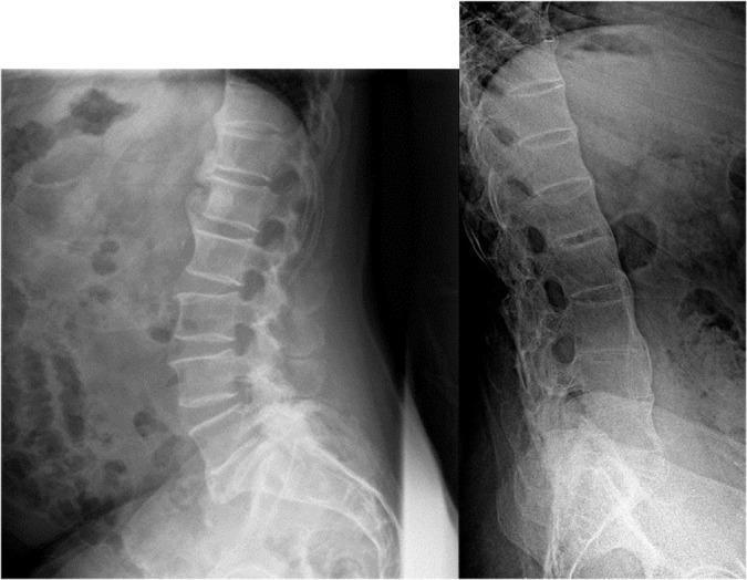
This side-by-side comparison chart consists of two lateral X-ray radiographs of the lumbar spine, demonstrating different patterns of syndesmophyte formation in axial spondyloarthritis. Image A shows Psoriatic Arthritis with axial involvement, characterized by coarse, 'chunky', and asymmetric syndesmophytes. These bony outgrowths appear irregular and do not uniformly bridge the vertebral segments. Image B demonstrates a classic 'bamboo spine' appearance typical of advanced Ankylosing Spondylitis. This is characterized by thin, symmetric, and marginal syndesmophytes that create a continuous vertical bridge of ossification between the vertebral bodies, leading to complete spinal fusion and a rigid, straightened alignment. The educational focus is on the diagnostic differentiation between the irregular, non-marginal ossification of psoriatic spondylitis and the uniform, marginal ossification of axial spondyloarthritis. These visual signs are critical for clinicians in rheumatology and radiology for identifying specific seronegative spondyloarthropathies.
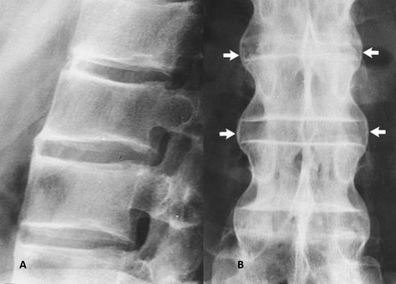
This diagnostic image consists of two radiographic views of the human spine, labeled A and B, demonstrating classic features of ankylosing spondylitis. View A is a lateral X-ray of the lumbar spine showing 'squaring' of the vertebral bodies, where the normal anterior concavity is lost, leading to a rectangular morphology. The intervertebral disc spaces are preserved but show early signs of marginal ossification. View B is an anteroposterior (AP) X-ray illustrating the 'bamboo spine' appearance. White arrows point to syndesmophytes, which are thin, vertical bony bridges formed by the ossification of the outer fibers of the annulus fibrosus. These syndesmophytes connect adjacent vertebral bodies across the intervertebral disc spaces. The fusion of the vertebral column results in a rigid, undulating lateral contour characteristic of advanced axial spondyloarthritis. The image serves as a definitive educational example of progressive spinal fusion and ligamentous calcification in seronegative spondyloarthropathies.
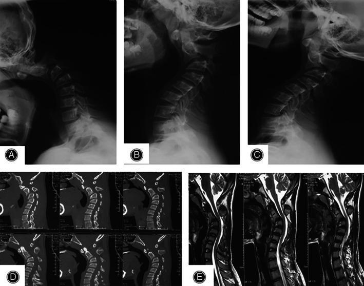
Multimodal imaging of the cervical spine demonstrating severe cervical kyphotic deformity. Panels A, B, and C show lateral x-ray radiography in hyperflexion, neutral, and hyperextension positions, respectively. These views reveal a fixed or progressive kyphosis with a Cobb angle exceeding 70 degrees in the neutral position, with minimal correction on extension. Panel D presents a sagittal CT scan highlighting the bony anatomy; the vertebral bodies maintain relatively normal rectangular morphology and bone density without significant osteophyte formation or syndesmophytes, suggesting a non-degenerative etiology like Ankylosing Spondylitis. Panel E consists of sagittal T2-weighted MRI slices, which show the severe sagittal malalignment but indicate no evidence of significant spinal cord compression or myelomalacia at the apex of the deformity. This imaging series is characteristic for evaluating Ankylosing Spondylitis-induced cervical kyphosis (AICK) and planning surgical correction such as anterior cervical discectomy and fusion (ACDF) or osteotomy.
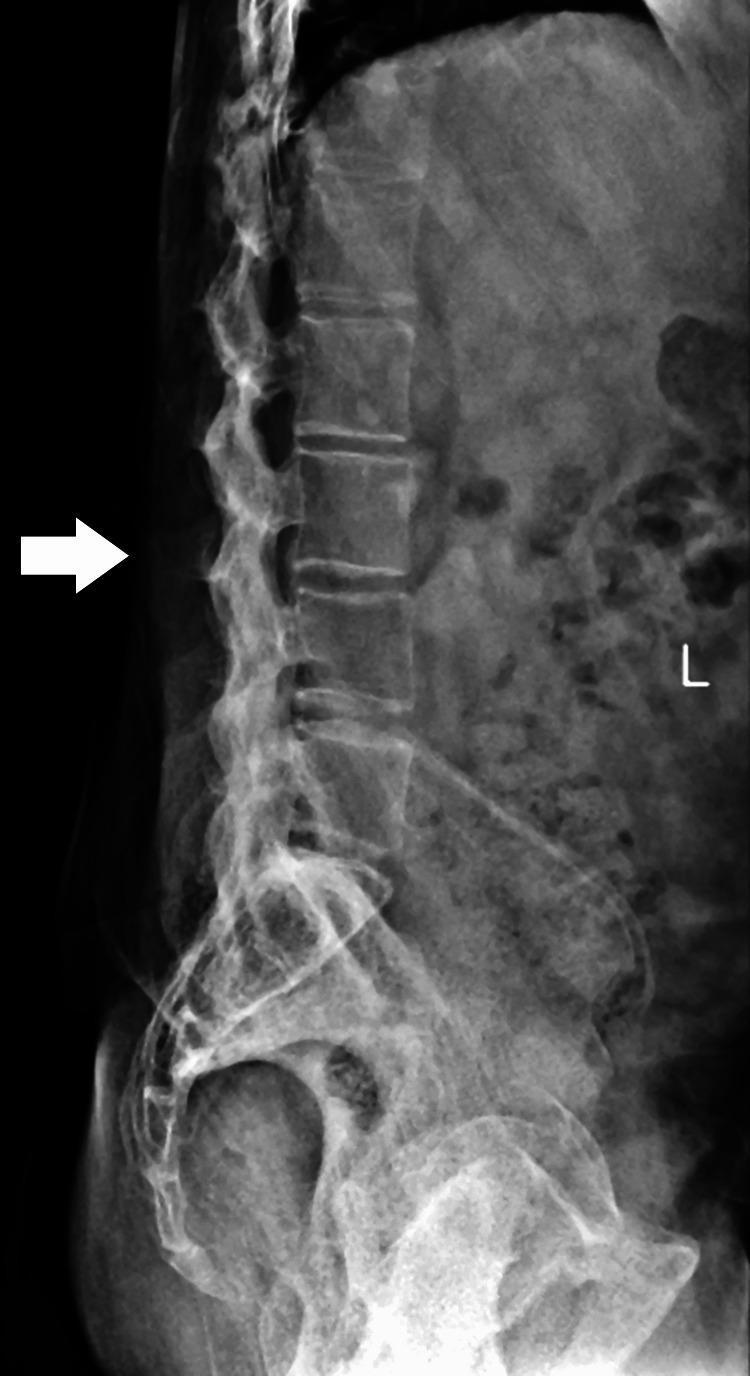
A lateral view X-ray of the lumbosacral spine demonstrating advanced radiographic features of Ankylosing Spondylitis. The image shows the classic 'bamboo spine' appearance, characterized by the formation of thin, vertical marginal syndesmophytes that bridge the intervertebral spaces. There is significant bony ankylosis (fusion) of the vertebral bodies, with blurring and near-complete loss of distinct intervertebral disc spaces. The vertebral endplates appear indistinct and merged. Additional findings include 'squaring' of the lumbar vertebrae and ossification of the spinal ligaments, specifically the interspinous and supraspinous ligaments (indicated by a white arrow pointing to the fused posterior elements). There is a notable loss of the normal lumbar lordosis, suggesting severe spinal stiffness and reduced mobility. The image serves as a definitive educational example of end-stage axial spondyloarthritis involving the lumbar region and sacrum.
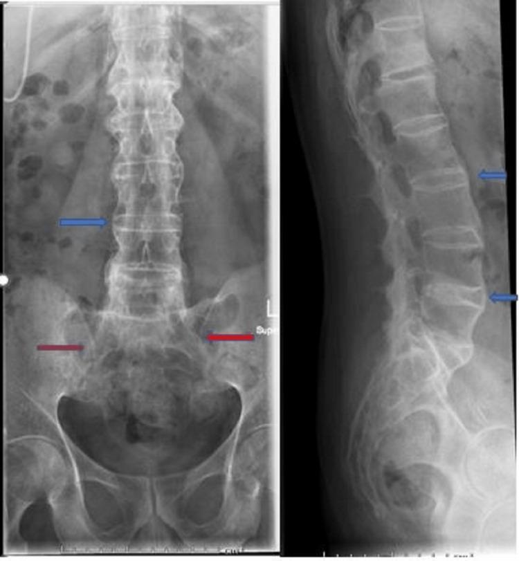
This diagnostic imaging shows anteroposterior (AP) and lateral X-ray views of the lumbar spine and pelvis, demonstrating classic radiographic hallmarks of advanced ankylosing spondylitis. The lumbar spine exhibits 'bamboo spine' morphology, characterized by the ossification of the annulus fibrosus and the formation of syndesmophytes. These vertical bony bridges connect adjacent vertebral bodies (highlighted by blue arrows), resulting in a continuous, straightened, and fused vertebral column with significant loss of normal intervertebral disc space. In the pelvic region, the sacroiliac (SI) joints show evidence of bilateral ankylosis (indicated by red arrows), with complete obliteration of the joint spaces, subchondral sclerosis, and bony fusion. These findings are clinically significant as they represent the end-stage structural damage typical of axial spondyloarthritis, leading to spinal rigidity and decreased mobility. The image serves as a definitive educational example of progressive inflammatory arthritis affecting the axial skeleton.
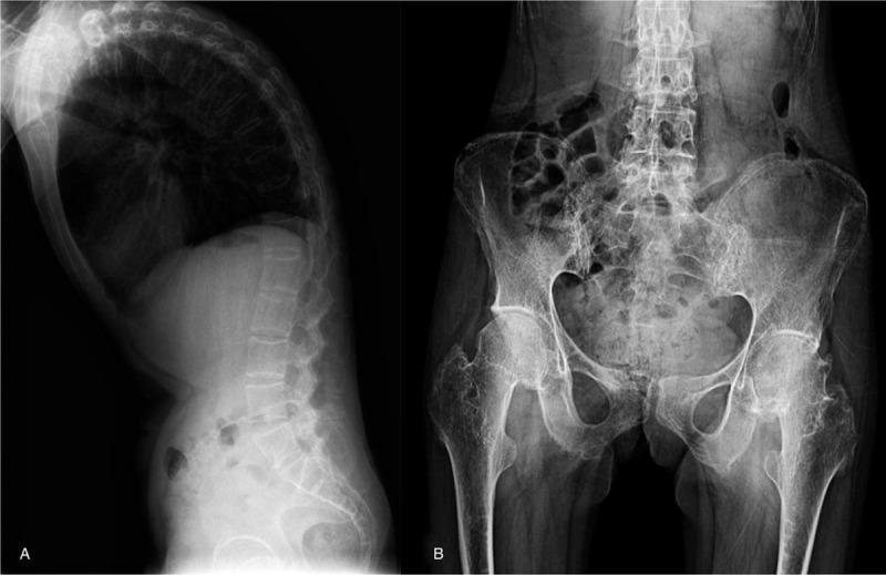
This composite diagnostic image consists of two X-ray radiographs illustrating radiographic manifestations of Ankylosing Spondylitis (AS). Panel A is a lateral view of the spine showing a significant thoracic kyphosis. Key findings include 'squaring' of the vertebral bodies, sclerosis along the vertebral margins, and calcification of the anterior longitudinal ligament (syndesmophytes). The facet joints appear blurred with narrowing or disappearance of the joint spaces. Panel B is an anteroposterior (AP) pelvis radiograph showing bilateral sacroiliitis, characterized by blurred articular surfaces of the sacroiliac joints. The image demonstrates the skeletal progression of a spondyloarthropathy, emphasizing the structural changes in the axial skeleton and pelvic girdle including vertebral fusion and joint erosion. These features are clinically significant for diagnosing advanced autoimmune inflammatory arthritis in the rheumatology domain.
syndesmophytes ankylosing spondylitis formation
Syndesmophytes - Detailed Overview
Definition and Origin
- Firestein & Kelley's Textbook of Rheumatology, p. 1122
- Harrison's Principles of Internal Medicine 22E, p. 2927
Pathogenesis and Molecular Mechanisms
Step 1 - Preceding Inflammation
- Romanus lesion - erosion at the anterosuperior/anteroinferior vertebral corner
- "Shiny corner" sign - focal sclerosis at the same sites
- Vertebral "squaring" - periosteal new bone fills the normal lumbar anterior concavity
Step 2 - Molecular Pathways Driving Bone Formation
| Pathway | Mechanism |
|---|---|
| Wnt signaling | Induces differentiation of mesenchymal stem cells to osteoblasts. DKK-1 (Wnt inhibitor) is inversely related to syndesmophyte formation - low DKK-1 = more bone |
| Sclerostin | Osteocyte-derived Wnt inhibitor; its expression is markedly reduced in axSpA, allowing unchecked osteoblast activity |
| BMP (Bone Morphogenetic Proteins) | BMP-2 was found at entheseal sites in SpA patients; drives endochondral bone formation and ankylosis |
- Rheumatology, 2-Volume Set (2022, Elsevier), p. 300-315
Radiographic Features
- Thin, vertical bony bridges arising from vertebral corners (anterior and lateral aspects)
- Best seen on lateral views of the lumbar and cervical spine
- Progressive bridging across disc spaces causes ankylosis
- Extensive syndesmophyte formation creates the classic "bamboo spine" - a smooth, undulating spinal contour on AP X-ray
- The disc space height is typically preserved (unlike degenerative disc disease)
Differentiating Syndesmophytes from Other Spinal Bony Outgrowths
| Feature | Syndesmophytes (AS) | Osteophytes (Spondylosis) | DISH Ossification | Psoriatic/Reactive Arthritis Syndesmophytes |
|---|---|---|---|---|
| Direction | Vertical | Horizontal/oblique | Flowing, thick | Vertical but coarser |
| Origin | Annulus fibrosus margin (vertebral corner) | Several mm from disc junction | Anterior longitudinal ligament | Annulus fibrosus (paravertebral) |
| Shape | Thin, delicate | Triangular, "parrot-beak" | Thick, flowing candle wax | Bulky, "chunky" |
| Distribution | Symmetric, marginal | Asymmetric, any level | Right-sided thoracic predominance | Asymmetric, non-marginal |
| Disc space | Preserved | Narrowed | Preserved | Preserved |
| SI joints | Bilateral sacroiliitis | Normal | Normal (or minor) | Asymmetric or normal |
- Goldman-Cecil Medicine, p. 2802-2803
- Firestein & Kelley's, p. 1122
Diseases Associated with Syndesmophytes
1. Ankylosing Spondylitis (AS) - Prototype
- Thin, symmetric, marginal syndesmophytes
- Ascend from lumbar to cervical spine
- Bilateral sacroiliitis always present
- Associated with HLA-B27 in >90%
2. Psoriatic Spondyloarthritis
- Coarse, "chunky," asymmetric, non-marginal syndesmophytes
- May skip vertebral levels
- Associated with psoriasis; HLA-B27 in ~50%
3. Reactive Arthritis (formerly Reiter's Syndrome)
- Bulky, nonmarginal, asymmetric syndesmophytes
- Follows genitourinary or GI infection
- Often involves lower thoracic/upper lumbar spine asymmetrically
4. Enteropathic Arthropathy (IBD-associated)
- Resembles AS pattern; often indistinguishable
- Correlates with HLA-B27 positivity
Clinical Implications
- Restricted spinal mobility in all planes
- Increased occiput-to-wall distance (cervical spine)
- Reduced chest expansion (<5 cm)
- Abnormal Schober test (<15 cm on forward flexion)
- Loss of normal lumbar lordosis
- Male sex
- Persistent inflammation (elevated CRP, active MRI lesions)
- Pre-existing syndesmophytes at baseline (strongest predictor)
- Presence of HLA-B27
- Smoking
- Harrison's 22E, p. 2927
Scoring Systems
modified Stoke Ankylosing Spondylitis Spine Score (mSASSS)
- Scores the anterior corners of lumbar and cervical vertebrae on lateral X-ray
- Each corner scored: 0 = normal, 1 = erosion/sclerosis/squaring, 2 = syndesmophyte, 3 = bridging syndesmophyte
- Range: 0-72
- Syndesmophyte formation/growth drives most of the progression score
CT Syndesmophyte Score (CTSS)
- Uses low-dose CT (ldCT) for superior sensitivity
- Detects syndesmophytes earlier than plain radiography
- Higher rate of new bone detection vs. X-ray in prospective studies
- Rheumatology, 2-Volume Set (2022, Elsevier), p. 2967-2981
MRI - Predicting Syndesmophyte Formation
- Corner inflammatory lesions (CIL) - bone marrow edema at vertebral corners; relative risk of 3-5 for new syndesmophyte at that level
- Corner fatty lesions (CFL) - fatty metaplasia following resolved inflammation; stronger predictor than CIL alone
- Sequential: inflammation → fatty metaplasia → new bone (though some syndesmophytes bypass this sequence)
Treatment and Effect on Syndesmophytes
- NSAIDs (continuous use): Celecoxib reduced radiographic progression in one study; non-selective NSAIDs did not replicate this
- Anti-TNF agents: Early data suggested NO inhibition of syndesmophyte formation, but longer-term studies (≥4 years, especially when started early) showed reduced progression
- Anti-IL-17 agents (secukinumab, ixekizumab): Later evidence supports structural inhibition
- No radiographic progression was observed in nr-axSpA patients treated with adalimumab or certolizumab over 2-4 years