
Create a power point presentation of neuro muscular junction anatomy and drug suchynilcholine comprehensive but easy understanding with pictures from standard Anesthesia textbooks like millers and steoltings pharma for Anesthesia resident
neuromuscular junction anatomy diagram motor end plate
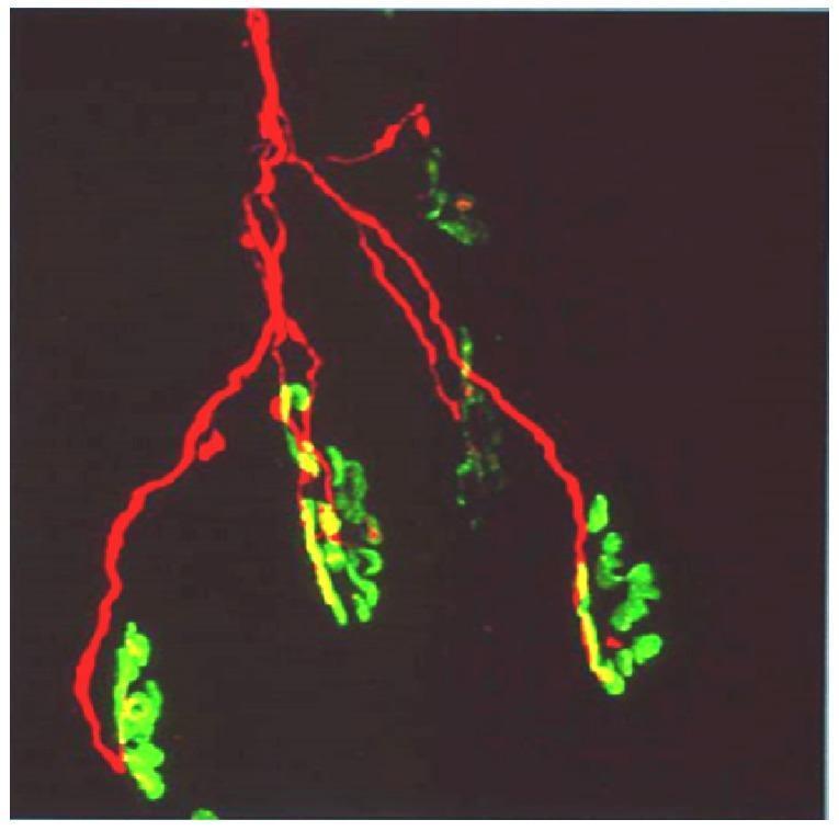
This diagnostic fluorescence microscopy image illustrates the neuromuscular junction (NMJ) architecture. The image features a single motor axon labeled with red fluorescence (TRITC-conjugated anti-neurofilament antibody), showing a characteristic distal branching pattern. These red axonal branches terminate at multiple motor end-plates, which are visualized in green (FITC-conjugated α-bungarotoxin) to reveal the high density of postsynaptic acetylcholine receptors (AChRs). The spatial relationship demonstrates a single axon innervating a cluster of six individual end-plates. Areas of colocalization between the presynaptic nerve terminal and the postsynaptic membrane appear yellow, indicating functional synaptic contact. This visual serves as an educational model for neuroanatomy and toxicology, particularly in the study of neurotoxic snake venoms (like those from the elapid family) that target presynaptic proteins or postsynaptic receptors, leading to neuromuscular paralysis and axonal degeneration.
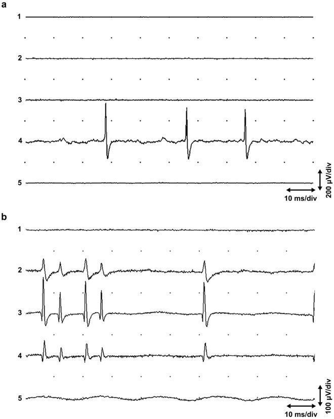
This diagnostic image displays two sets of five-channel needle electromyography (EMG) recordings (a and b) from the human extensor carpi radialis muscle. The electrodes were placed 2 mm apart in parallel within a single muscle fasciculus to study spontaneous neuromuscular junction activity. Panel (a) illustrates local end plate activity. Channel 4 shows non-propagating 'end plate spikes' characterized by high-frequency biphasic waveforms superimposed on low-amplitude, irregular 'end plate noise.' Channels 1, 2, 3, and 5 remain quiescent, indicating the highly localized nature of these potentials. Panel (b) demonstrates propagating end plate spikes. Unlike the local activity in (a), the potentials here are visible across channels 2, 3, and 4. These spikes show variations in amplitude and morphology, reflecting the spatial propagation of action potentials for a short distance along the muscle fiber. Calibrations are provided as 10 ms/div horizontally, with vertical sensitivity at 200 µV/div for (a) and 100 µV/div for (b). This comparison is educationally significant for distinguishing between stationary end plate noise and propagated intrafusal or extrafusal potentials in clinical neurophysiology.
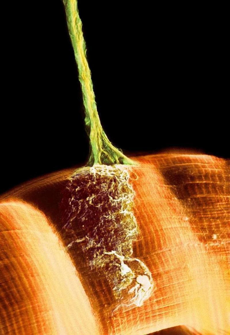
This false-color scanning electron micrograph (SEM) illustrates the morphology of a neuromuscular junction. A single motor axon, depicted in green, descends from the top of the frame to contact a skeletal muscle fiber, shown in orange. The axon exhibits a long, cylindrical structure that tapers and subsequently branches as it approaches the muscle surface. At the terminal end, the axon expands into a complex, granular network of synaptic terminals or motor endplates, which spread across a localized region of the muscle sarcolemma. The muscle fiber displays prominent transverse striations, characteristic of sarcomere organization in skeletal muscle. This visual demonstrates the physical connection between the somatic nervous system and the muscular system, highlighting the site of chemical neurotransmission and the structural adaptation of synapses to maximize contact area with the muscle cell for effective motor signaling.
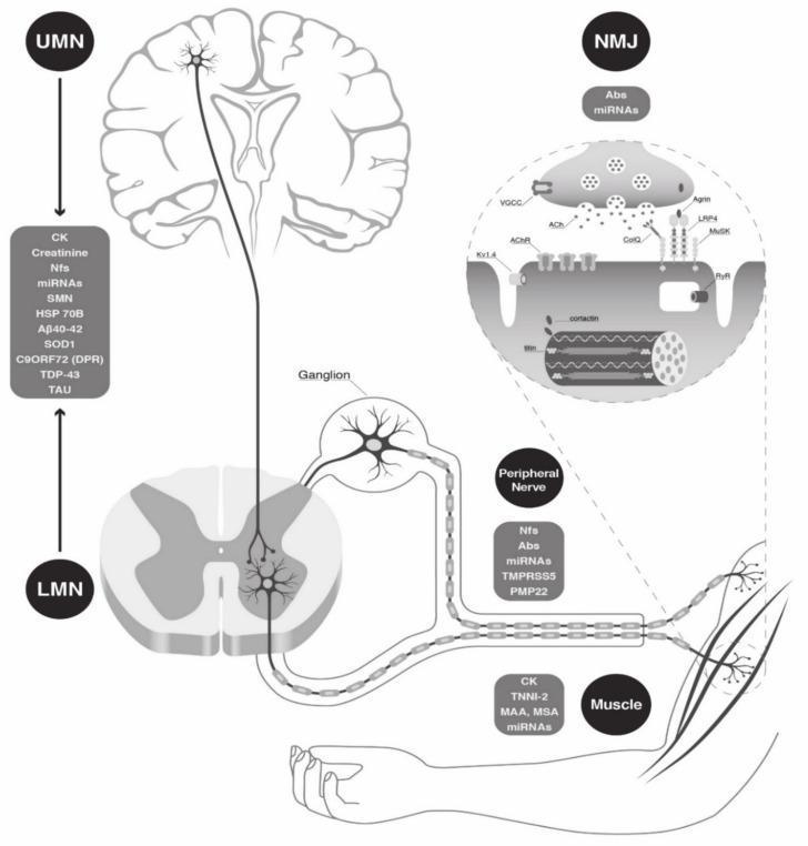
This medical illustration is a pathophysiology diagram detailing the anatomical components of the human neuromuscular unit and their associated circulating biomarkers. The schematic follows a top-down approach: 1) Upper Motor Neuron (UMN), shown within a coronal section of the brain with listed biomarkers including CK, Nfs, miRNAs, and neurodegenerative markers like TDP-43 and A̠40-42. 2) Lower Motor Neuron (LMN), depicted within a cross-section of the spinal cord, showing axons exiting via a ganglion into the peripheral nerve (associated with PMP22 and TMPRSS5). 3) Neuromuscular Junction (NMJ), magnified to show synaptic vesicles, ACh receptors, and muscle endplates with related antibodies (Abs) and miRNAs. 4) Skeletal Muscle, illustrated at the periphery with biomarkers such as CK, TNNI-2, and myositis-specific antibodies (MSA). The diagram serves as an educational tool for neurology and neuromuscular diseases, highlighting the potential for diagnostic and prognostic monitoring across the motor pathway from the central nervous system to the peripheral effector organ.
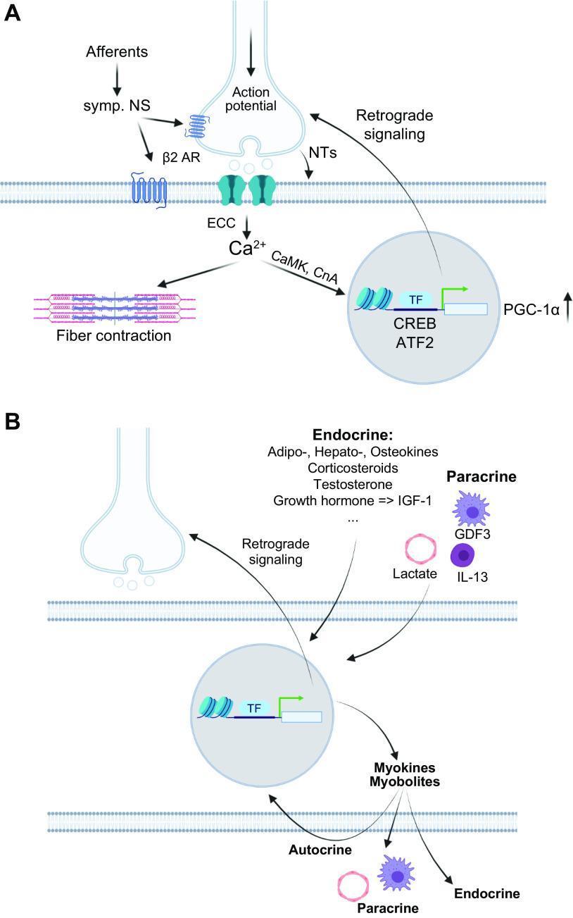
A two-panel (A and B) pathophysiology diagram illustrating motor neuron signaling, excitation-contraction coupling (ECC), and (neuro)endocrine skeletal muscle responses. Panel A depicts the neuromuscular junction where an action potential triggers ECC and the release of intramyocellular calcium (Ca2+), leading to myofiber contraction. It also shows Ca2+ activating signaling pathways (CaMK, CnA) that modulate transcriptional factors (CREB, ATF2) and the coactivator PGC-1̑ in the nucleus, alongside retrograde feedback to the motor neuron via neurotrophic factors (NTs). Adrenergic signaling through ̒2-adrenoreceptors (̒2 AR) from the sympathetic nervous system is also represented. Panel B focuses on 'exerkines,' showing the secretion of myokines and myobolites from the muscle cell. It classifies signaling effects as autocrine, paracrine (e.g., GDF3, IL-13, Lactate), and endocrine. Endocrine factors listed include adipokines, hepatokines, osteokines, corticosteroids, testosterone, and growth hormone-regulated IGF-1, highlighting the systemic coordination between muscle, liver, adipose, and bone tissues during contractile activity.
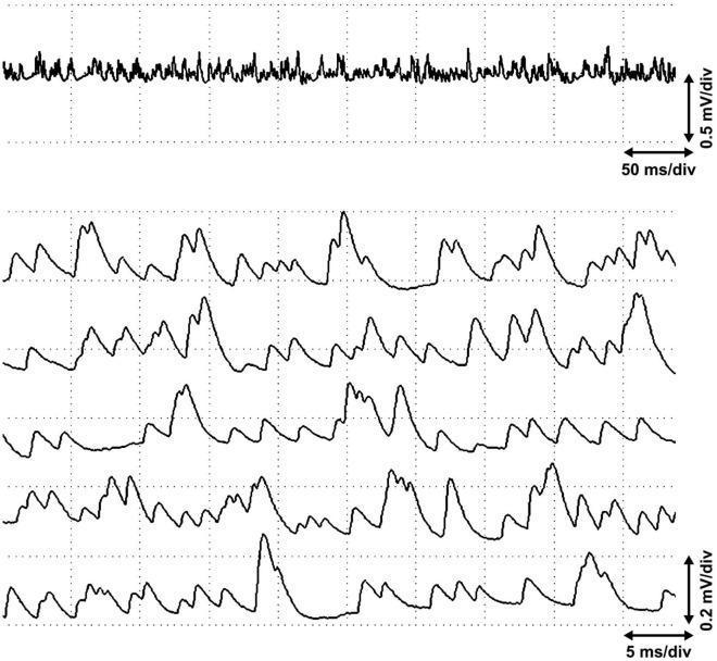
This diagnostic image displays a multi-channel electrophysiological recording of miniature end plate potentials (MEPPs) captured from the end plate zone of the biceps brachii muscle. The upper panel shows a continuous single-channel trace at a scale of 0.5 mV/div and 50 ms/div, illustrating high-frequency, low-amplitude baseline activity known as end plate noise. The lower panel provides a high-resolution, five-channel synchronized view at 0.2 mV/div and 5 ms/div, specifically detailing numerous large and partially stratified MEPPs. These spontaneous postsynaptic potentials are characterized by rapid upward deflections followed by a slower, non-linear decay to baseline. Notable features include the absence of elicited postsynaptic action potentials or 'end plate spikes' despite the high-frequency sequence of potentials. This recording illustrates the physiological spontaneous release of acetylcholine at the neuromuscular junction in an active site, useful for differentiating between normal end plate activity and pathological spontaneous discharges in clinical electromyography (EMG).
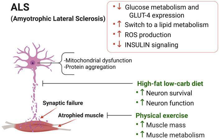
A medical illustration depicting the pathophysiology and proposed therapeutic interventions for Amyotrophic Lateral Sclerosis (ALS). The left side of the diagram shows a degenerating motor neuron and a skeletal muscle unit. Cellular pathology in the neuron is characterized by mitochondrial dysfunction and protein aggregation. The neuromuscular junction is marked by 'Synaptic failure,' leading to 'Atrophied muscle.' A red box highlights metabolic reprogramming in the muscle, showing decreased glucose metabolism, GLUT-4 expression, and insulin signaling, alongside increased lipid metabolism and reactive oxygen species (ROS) production. The right side presents two counteracting therapeutic strategies: a 'High-fat low-carb diet,' which aims to increase neuron survival and function, and 'Physical exercise,' which aims to increase muscle mass and metabolism. The diagram uses specialized arrows to indicate the direction of metabolic changes (red) and the intended positive effects of interventions (green). This educational infographic is designed to illustrate the neuro-muscular metabolic axis in neurodegenerative disease.
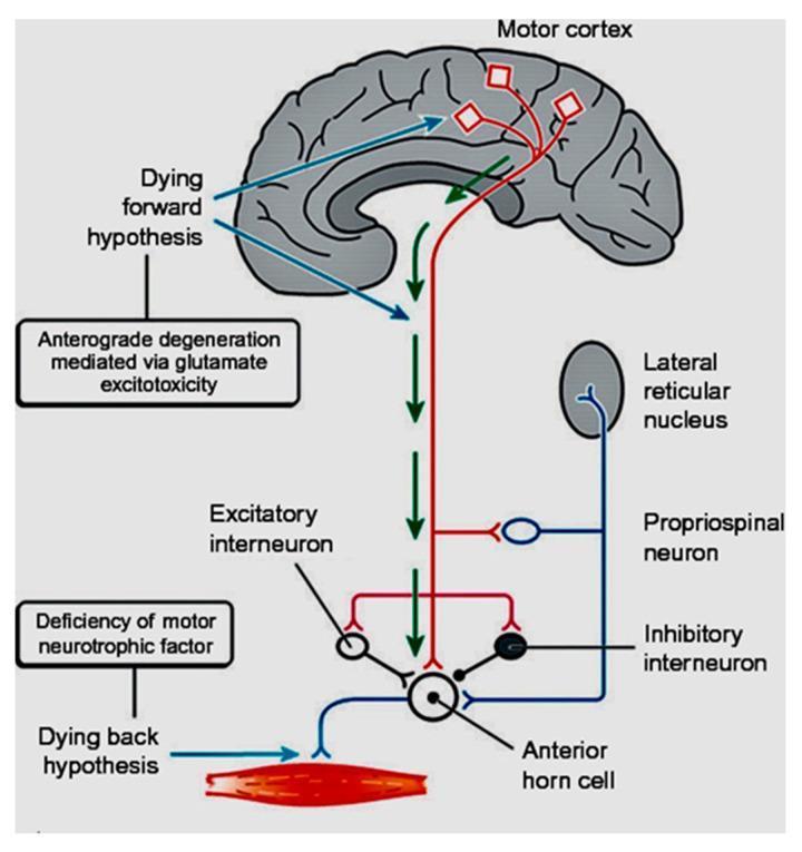
A pathophysiology diagram illustrating the competing hypotheses for neurodegeneration in Amyotrophic Lateral Sclerosis (ALS). The illustration includes a sagittal view of the human brain and the spinal motor circuit. The 'Dying forward hypothesis' is depicted as originating in the motor cortex, showing anterograde degeneration mediated via glutamate excitotoxicity (indicated by green downward arrows). This pathway highlights the involvement of upper motor neurons and their corticofugal projections. In contrast, the 'Dying back hypothesis' is shown starting at the muscle level, suggesting retrograde degeneration caused by a deficiency of motor neurotrophic factors at the neuromuscular junction. The diagram details the lower motor neuron environment, including the anterior horn cell, excitatory and inhibitory interneurons, propriospinal neurons, and the lateral reticular nucleus. This educational graphic serves to differentiate between top-down (cortical) and bottom-up (peripheral) mechanisms of motor neuron disease progression.
succinylcholine mechanism depolarizing neuromuscular blockade acetylcholine receptor
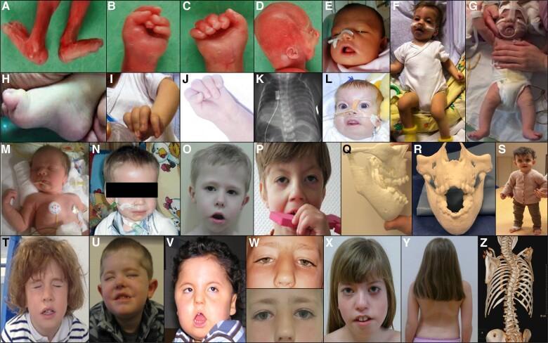
This composite clinical figure illustrates the multifaceted phenotypic spectrum of Fetal Acetylcholine Receptor Antibody-related Disorders (FARAD) across various developmental stages. Panels A–D show severe, lethal manifestations including fetal specimen arthrogryposis multiplex congenita (AMC) with rigid joint contractures of the lower limbs and characteristic clenched fists. Neonatal panels (E–G, L–N) demonstrate congenital hypotonia, bulbofacial weakness requiring nasogastric tubes, and respiratory distress. Musculoskeletal findings include clubfoot (H), limb contractures (I, J), and scoliosis (Y, Z). Advanced diagnostic imaging includes a chest radiograph (K) showing diaphragmatic paresis and 3D CT reconstructions (Q, R, Z) highlighting significant jaw contractures and spinal deformity. Longitudinal clinical photographs (O–X) track the evolution into a persistent myopathic facies in older children, characterized by ptosis, incomplete eye closure (lagophthalmos), and an inverted V-shaped mouth. The collection emphasizes the progression from neonatal bulbar and respiratory impairment to long-term neuromuscular sequelae, illustrating the educational hallmarks of transplacental maternal antibody-mediated fetal neuromuscular blockade.
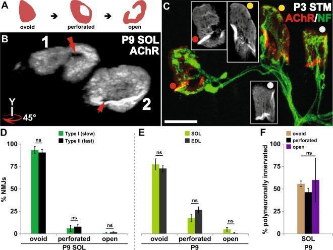
This composite educational image illustrates the maturation of postsynaptic acetylcholine receptor (AChR) aggregates at the neuromuscular junction (NMJ) and its relationship to synapse elimination. Panel A provides a schematic of morphological progression from ovoid plaque to perforated plaque to an open configuration. Panel B displays grayscale confocal images of P9 mouse soleus AChR aggregates, rotated 45 degrees to highlight central perforations (red arrows). Panel C shows a fluorescence micrograph of P3 sternomastoid NMJs labeled for AChR (red) and Neurofilament (green), demonstrating varied maturation stages: open (red dot), perforated (yellow dot), and ovoid (grey dot) within polyneuronally innervated synapses. Panels D and E are bar graphs comparing AChR maturation across muscle fiber types (Type I vs. Type II) and muscles (Soleus vs. EDL), showing no significant difference ('ns'). Panel F quantifies that the percentage of polyneuronally innervated NMJs does not significantly differ based on postsynaptic morphology. The data indicates that postsynaptic AChR maturation occurs independently of the timing of axonal input removal during development.
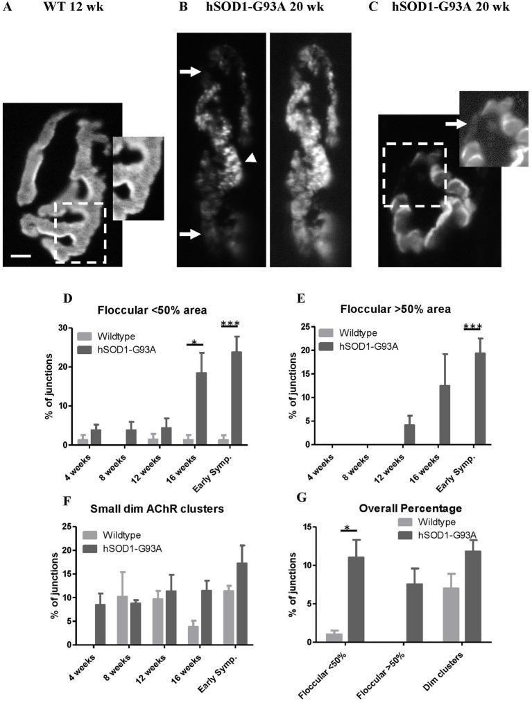
This composite educational graphic illustrates the structural degradation of neuromuscular junctions (NMJs) in an hSOD1-G93A mouse model of Amyotrophic Lateral Sclerosis (ALS). (A) Fluorescence microscopy of a 12-week wild-type NMJ shows a normal 'pretzel-like' acetylcholine receptor (AChR) distribution with crisp borders and a uniform fingerprint-like pattern. (B-C) Corresponding images from 20-week symptomatic hSOD1-G93A mice reveal pathological remodeling, characterized by a granular/floccular AChR appearance (arrowheads), overall labeling dimness (arrows), and fragmented, dim extrajunctional clusters indicating receptor loss or disorganized insertion. Panels D-G present longitudinal quantitative data via bar charts. They demonstrate that while wild-type mice maintain stable NMJ morphology, hSOD1-G93A mice exhibit a significant, age-dependent increase in disrupted AChR patterns. Key findings show that floccular distributions exceeding 50% of the junctional area emerge by 12 weeks and increase sharply by early symptomatic stages (16-20 weeks), correlating structural NMJ decay with disease progression and motor symptom onset.
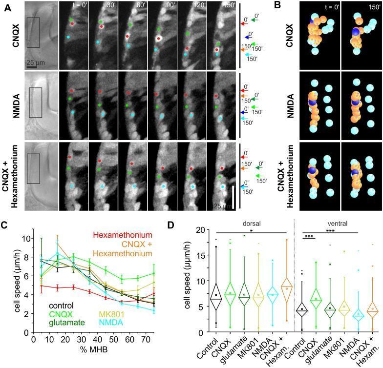
This composite educational graphic details the neurodevelopmental effects of glutamate and acetylcholine (ACh) on tegmental hindbrain nuclei (THN) migration along the midbrain-hindbrain boundary (MHB). Panel A displays time-lapse fluorescent microscopy stills (0–150 min) of zebrafish embryos under three experimental conditions: AMPA receptor blockade (CNQX), NMDA receptor activation (NMDA), and dual blockade (CNQX + hexamethonium). Colored dots track individual neuron positions, illustrating that CNQX increases migration distance, while NMDA markedly inhibits it. Panel B provides digital reconstructions of individual THN tracks (orange and dark blue spheres) after tissue-shift correction (cyan markers), highlighting the linear versus dispersed movement patterns. Panel C is a line graph plotting cell speed (µm/h) against the percentage of MHB progression, demonstrating that blocking AMPA receptors (green) prevents the natural slowing observed at the ventral MHB. Panel D presents statistical analysis via box and whisker plots, showing significant cell speed variances between dorsal and ventral regions under pharmacological treatments, including NMDA and MK801. The data suggests that glutamate acting via AMPA receptors serves as a localized 'slowdown' signal for differentiating neurons.
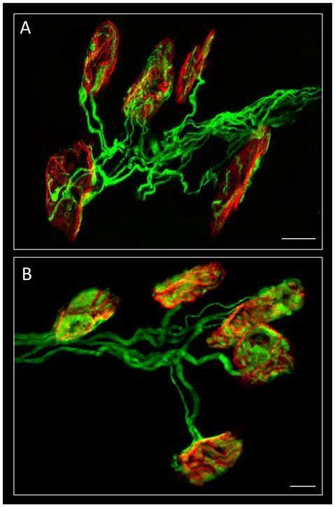
This dual-panel diagnostic image displays confocal immunofluorescence of mouse neuromuscular junctions (NMJs) in the Levator auris longus muscle. Panels A and B demonstrate different innervation states: polyinnervation (A) and monoinnervation (B). The presynaptic axons are stained green using a 200-kD neurofilament antibody, revealing thread-like structures and branching patterns. The postsynaptic nicotinic acetylcholine receptors (nAChR) are labeled red with TRITC-α-bungarotoxin (TRITC-α-BTX), appearing as compact or elongated clusters. Panel A illustrates high-density axonal branching typical of early development, where multiple axons innervate single motor endplates. Panel B showcases a more refined organization with fewer axonal branches per motor unit, representing the results of Hebbian competition and synapse elimination. Areas of signal overlap appear yellow/merged, indicating direct contact between axons and receptor clusters. This image is used to study neurodevelopmental processes, synapse maturation, and the role of signaling pathways (like PKA/PKC) in axonal loss. Scale bar: 10 μm.
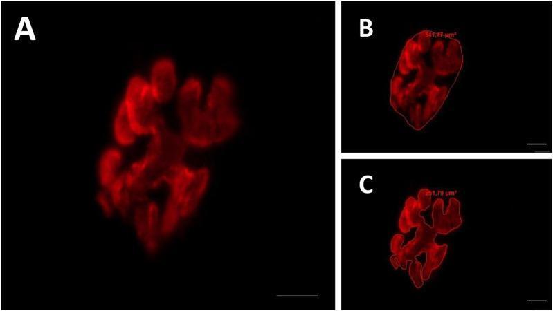
This composite diagnostic image features three panels (A, B, C) showcasing fluorescence microscopy of a mammalian motor endplate at 800x magnification. The primary staining utilizes rhodamine-conjugated alpha-bungarotoxin to visualize the distribution of acetylcholine receptors (AChR) in the postsynaptic compartment. Panel A shows the raw fluorescence signal, revealing a characteristic irregular, clustered morphology of the receptor groups with interspersed dark, non-stained areas. Panel B demonstrates the 'total area' and 'total perimeter' measurement technique, where a red border encompasses the entire synaptic territory, including both receptor clusters and internal gaps (measuring 541.47 µm²). Panel C illustrates the 'stained area' measurement, where the perimeter strictly traces only the fluorescently labeled regions (measuring 251.78 µm²). These images serve as a methodological example for quantifying synaptic plasticity, endplate dispersion, and neuromuscular junction health in neurobiology and clinical physiology research.
phase 1 phase 2 block succinylcholine depolarizing blockade train of four
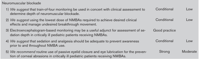
<table> <tr> <td>Neuromuscular blockade</td> <td></td> <td></td> </tr> <tr> <td>1) <i>We suggest</i> that train-of-four monitoring be used in concert with clinical assessment to determine depth of neuromuscular blockade.</td> <td>Conditional</td> <td>Low</td> </tr> <tr> <td>2) <i>We suggest</i> using the lowest dose of NMBAs required to achieve desired clinical effects and manage undesired breakthrough movement.</td> <td>Conditional</td> <td>Low</td> </tr> <tr> <td>3) Electroencephalogram-based monitoring may be a useful adjunct for assessment of sedation depth in critically ill pediatric patients receiving NMBAs.</td> <td>Good practice</td> <td></td> </tr> <tr> <td>4) <i>We suggest</i> that sedation and analgesia should be adequate to prevent awareness prior to and throughout NMBA use.</td> <td>Conditional</td> <td>Low</td> </tr> <tr> <td>5) <i>We recommend</i> routine use of passive eyelid closure and eye lubrication for the prevention of corneal abrasions in critically ill pediatric patients receiving NMBAs.</td> <td>Strong</td> <td>Moderate</td> </tr> </table>
![This Comparison Chart illustrates ionic current time traces and corresponding histograms used for the discrimination of DNA homopolymers and block copolymers via nanopore sensing. The visual displays four distinct 1-second time traces (a-d) recorded at 100 mV, showing ionic current (nA) versus time (sec). In panel (a), poly(dA)5.3k exhibits a single stable blockade level, resulting in one histogram peak. Panels (b) and (c) represent block copolymers [(dT)25-(dC)25]m and [(dA)50-(dC)50]m, both showing two distinct current levels and two corresponding histogram peaks (shaded blue, green, and red). Panel (d) shows [(dT)25-(dC)25-(dA)50]m, which exhibits three current levels reflected as three peaks in the histogram. The current blockade values (approximately 72 pA to 222 pA) demonstrate the educational concept that specific DNA bases (adenine, thymine, and cytosine) cause unique levels of ionic resistance. This diagnostic diagram illustrates the biophysical principles of DNA sequencing and molecular identification in biomedical research.](https://cdn.orris.care/cdss_images/pmc_clinical_VQA_9e57dccb4d96eaa7f5c0254b6bc96e28b18af90dbe274f34044b6e35660d3902.jpg)
This Comparison Chart illustrates ionic current time traces and corresponding histograms used for the discrimination of DNA homopolymers and block copolymers via nanopore sensing. The visual displays four distinct 1-second time traces (a-d) recorded at 100 mV, showing ionic current (nA) versus time (sec). In panel (a), poly(dA)5.3k exhibits a single stable blockade level, resulting in one histogram peak. Panels (b) and (c) represent block copolymers [(dT)25-(dC)25]m and [(dA)50-(dC)50]m, both showing two distinct current levels and two corresponding histogram peaks (shaded blue, green, and red). Panel (d) shows [(dT)25-(dC)25-(dA)50]m, which exhibits three current levels reflected as three peaks in the histogram. The current blockade values (approximately 72 pA to 222 pA) demonstrate the educational concept that specific DNA bases (adenine, thymine, and cytosine) cause unique levels of ionic resistance. This diagnostic diagram illustrates the biophysical principles of DNA sequencing and molecular identification in biomedical research.
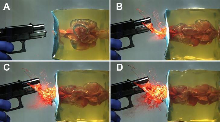
A series of four high-speed photographic frames (A-D) demonstrating the terminal ballistics of a 9 mm Luger shot into a gelatin block at a 1 cm distance, used as a human tissue surrogate in forensic pathology. Panel A (7.2 ms) shows the re-expansion phase of the temporary cavity (TC) following its initial collapse; the cavity appears irregular and multi-lobulated within the transparent gelatin. Panels B (12.5 ms), C (13.3 ms), and D (14.2 ms) illustrate the progression of backspatter. In B, a distinct linear jet of orange-colored fluid (simulated blood/tissue) is ejected from the entry wound toward the firearm muzzle. By C and D, escaping muzzle gases transform this linear jet into a wider, more chaotic spray and aerosol-like pattern with increasing dispersion of droplets. This sequence illustrates the pathophysiology of backspatter formation and the influence of muzzle gases on retrograde material ejection in near-contact gunshot wounds, which is critical for forensic reconstruction and clinical assessment of firearm injuries.
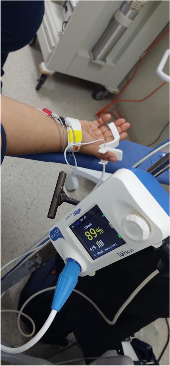
This clinical photograph illustrates the setup for Train-of-Four Ratio (TOFR) monitoring using an acceleromyographic (AMG) device in an anesthetic setting. The image shows the non-dominant hand and forearm of a patient with two peripheral nerve stimulator electrodes positioned over the ulnar nerve at the wrist and distal forearm. A white acceleromyographic sensor is attached to the second or third finger to detect muscular contractions of the adductor pollicis or flexor muscles. Adjacent to the patient, a Dräger TOFScan® monitor displays real-time neuromuscular blockade data, including a TOFR of '89%', a '4/4' twitch response indicated by four vertical bars, and a stimulation current of 40mA. This procedural image demonstrates the clinical application of quantitative neuromuscular monitoring to assess the depth of blockade and ensure safe recovery before extubation. Key educational concepts include the anatomical placement of electrodes for ulnar nerve stimulation and the interpretation of objective TOF values in anesthesiology.
mkdir -p /tmp/workspace/nmj-pptx && ls /tmp/workspace/
~/nmj-pptx/build_pptx.py
import json, subprocess, base64, sys
from io import BytesIO
from pptx import Presentation
from pptx.util import Inches, Pt, Emu
from pptx.dml.color import RGBColor
from pptx.enum.text import PP_ALIGN, MSO_ANCHOR
from pptx.enum.shapes import MSO_SHAPE

# ── Colour palette ──────────────────────────────────────────────
DARK_BLUE   = RGBColor(0x0D, 0x2B, 0x5B)   # deep navy
MED_BLUE    = RGBColor(0x1A, 0x5F, 0x9E)   # brand blue
LIGHT_BLUE  = RGBColor(0xE8, 0xF4, 0xFF)   # very light blue tint
ACCENT_GOLD = RGBColor(0xF5, 0xA6, 0x23)   # amber / gold
WHITE       = RGBColor(0xFF, 0xFF, 0xFF)
DARK_GRAY   = RGBColor(0x22, 0x22, 0x22)
MID_GRAY    = RGBColor(0x55, 0x55, 0x55)
LIGHT_GRAY  = RGBColor(0xF2, 0xF5, 0xF9)
RED_WARN    = RGBColor(0xC0, 0x20, 0x20)
GREEN_OK    = RGBColor(0x1A, 0x7A, 0x40)

# ── Helper – download image from URL ────────────────────────────
def fetch_img(url):
    try:
        result = json.loads(subprocess.check_output(
            ["python", "/tmp/skills/shared/scripts/fetch_images.py", url],
            timeout=30
        ))
        if result and result[0]["base64"]:
            raw = base64.b64decode(result[0]["base64"].split(",", 1)[1])
            return BytesIO(raw)
    except Exception as e:
        print(f"  [WARN] Could not fetch {url}: {e}", file=sys.stderr)
    return None

# ── Presentation setup ───────────────────────────────────────────
prs = Presentation()
prs.slide_width  = Inches(13.333)
prs.slide_height = Inches(7.5)
BLANK = prs.slide_layouts[6]

# ── Utility functions ────────────────────────────────────────────
def add_rect(slide, x, y, w, h, fill_rgb, line_rgb=None, line_pt=0):
    shp = slide.shapes.add_shape(MSO_SHAPE.RECTANGLE, Inches(x), Inches(y), Inches(w), Inches(h))
    shp.fill.solid(); shp.fill.fore_color.rgb = fill_rgb
    if line_rgb:
        shp.line.color.rgb = line_rgb; shp.line.width = Pt(line_pt)
    else:
        shp.line.fill.background()
    return shp

def add_text(slide, text, x, y, w, h, size=18, bold=False, color=None,
             align=PP_ALIGN.LEFT, wrap=True, italic=False, anchor=MSO_ANCHOR.TOP):
    color = color or DARK_GRAY
    tb = slide.shapes.add_textbox(Inches(x), Inches(y), Inches(w), Inches(h))
    tf = tb.text_frame; tf.word_wrap = wrap; tf.vertical_anchor = anchor
    tf.margin_left = tf.margin_right = tf.margin_top = tf.margin_bottom = 0
    p = tf.paragraphs[0]; p.alignment = align
    r = p.add_run(); r.text = text
    r.font.size = Pt(size); r.font.bold = bold
    r.font.italic = italic; r.font.color.rgb = color
    return tb

def add_bullet_list(slide, items, x, y, w, h, size=16, color=None, level1_size=14):
    color = color or DARK_GRAY
    tb = slide.shapes.add_textbox(Inches(x), Inches(y), Inches(w), Inches(h))
    tf = tb.text_frame; tf.word_wrap = True
    tf.margin_left = tf.margin_right = tf.margin_top = tf.margin_bottom = 0
    first = True
    for item in items:
        if isinstance(item, tuple):
            bullet_char, text = item
        else:
            bullet_char, text = "●", item
        if first:
            p = tf.paragraphs[0]; first = False
        else:
            p = tf.add_paragraph()
        r = p.add_run()
        r.text = f"{bullet_char}  {text}"
        r.font.size = Pt(size); r.font.color.rgb = color
    return tb

def add_header_bar(slide, title, subtitle=None):
    """Standard dark-blue header bar at top of slide."""
    add_rect(slide, 0, 0, 13.333, 1.15, DARK_BLUE)
    add_rect(slide, 0, 1.15, 13.333, 0.06, ACCENT_GOLD)
    add_text(slide, title, 0.35, 0.05, 12.5, 0.7,
             size=30, bold=True, color=WHITE, align=PP_ALIGN.LEFT)
    if subtitle:
        add_text(slide, subtitle, 0.35, 0.72, 12.5, 0.42,
                 size=16, bold=False, color=ACCENT_GOLD, align=PP_ALIGN.LEFT)

def add_footer(slide, text="NMJ & Succinylcholine  |  Anesthesia Resident Education"):
    add_rect(slide, 0, 7.25, 13.333, 0.25, DARK_BLUE)
    add_text(slide, text, 0.3, 7.27, 12.5, 0.22,
             size=10, color=WHITE, align=PP_ALIGN.CENTER)

def add_section_label(slide, label, color=MED_BLUE):
    """Small coloured tag in top-right corner."""
    add_rect(slide, 11.3, 1.25, 1.85, 0.32, color)
    add_text(slide, label, 11.3, 1.25, 1.85, 0.32,
             size=11, bold=True, color=WHITE, align=PP_ALIGN.CENTER,
             anchor=MSO_ANCHOR.MIDDLE)

# ════════════════════════════════════════════════════════════════
#  SLIDE 1 – Title
# ════════════════════════════════════════════════════════════════
s = prs.slides.add_slide(BLANK)
add_rect(s, 0, 0, 13.333, 7.5, DARK_BLUE)                  # full bg
add_rect(s, 0, 0, 13.333, 0.12, ACCENT_GOLD)               # top stripe
add_rect(s, 0, 7.38, 13.333, 0.12, ACCENT_GOLD)            # bottom stripe

# Central white card
add_rect(s, 1.2, 1.0, 10.9, 5.5, WHITE, ACCENT_GOLD, 1.5)

add_text(s, "NEUROMUSCULAR JUNCTION", 1.5, 1.3, 10.3, 1.0,
         size=38, bold=True, color=DARK_BLUE, align=PP_ALIGN.CENTER)
add_text(s, "Anatomy, Physiology & Pharmacology", 1.5, 2.25, 10.3, 0.6,
         size=22, color=MED_BLUE, align=PP_ALIGN.CENTER, italic=True)

add_rect(s, 4.0, 2.95, 5.3, 0.07, ACCENT_GOLD)            # divider line

add_text(s, "Succinylcholine — Comprehensive Review", 1.5, 3.15, 10.3, 0.65,
         size=24, bold=True, color=DARK_BLUE, align=PP_ALIGN.CENTER)

add_text(s, "Based on Miller's Anesthesia & Stoelting's Pharmacology",
         1.5, 3.9, 10.3, 0.5, size=16, color=MID_GRAY, align=PP_ALIGN.CENTER, italic=True)

add_text(s, "For Anesthesia Residents", 1.5, 4.55, 10.3, 0.4,
         size=18, bold=True, color=MED_BLUE, align=PP_ALIGN.CENTER)

add_text(s, "Department of Anesthesiology  ●  2026",
         1.5, 5.1, 10.3, 0.35, size=14, color=MID_GRAY, align=PP_ALIGN.CENTER)

# ════════════════════════════════════════════════════════════════
#  SLIDE 2 – Outline / Contents
# ════════════════════════════════════════════════════════════════
s = prs.slides.add_slide(BLANK)
add_rect(s, 0, 0, 13.333, 7.5, LIGHT_GRAY)
add_header_bar(s, "Contents at a Glance")
add_footer(s)
add_rect(s, 0.35, 1.35, 12.6, 5.65, WHITE, MED_BLUE, 0.5)

topics_left = [
    ("01", "NMJ Anatomy Overview"),
    ("02", "Presynaptic Nerve Terminal"),
    ("03", "ACh Synthesis & Packaging"),
    ("04", "Neuromuscular Transmission"),
    ("05", "Nicotinic Acetylcholine Receptors"),
    ("06", "Mature vs. Immature nAChR"),
]
topics_right = [
    ("07", "Succinylcholine — Structure & Pharmacokinetics"),
    ("08", "Mechanism of Action (Phase I & II Block)"),
    ("09", "Clinical Uses & Dosing"),
    ("10", "Side Effects & Contraindications"),
    ("11", "Pseudocholinesterase & Dibucaine Number"),
    ("12", "Key Clinical Pearls"),
]

y_start = 1.6
for num, topic in topics_left:
    add_rect(s, 0.55, y_start, 0.55, 0.52, MED_BLUE)
    add_text(s, num, 0.55, y_start, 0.55, 0.52, size=13, bold=True, color=WHITE,
             align=PP_ALIGN.CENTER, anchor=MSO_ANCHOR.MIDDLE)
    add_text(s, topic, 1.18, y_start+0.06, 5.0, 0.4, size=15, color=DARK_GRAY)
    y_start += 0.74

y_start = 1.6
for num, topic in topics_right:
    add_rect(s, 6.9, y_start, 0.55, 0.52, ACCENT_GOLD)
    add_text(s, num, 6.9, y_start, 0.55, 0.52, size=13, bold=True, color=WHITE,
             align=PP_ALIGN.CENTER, anchor=MSO_ANCHOR.MIDDLE)
    add_text(s, topic, 7.53, y_start+0.06, 4.9, 0.4, size=15, color=DARK_GRAY)
    y_start += 0.74

# ════════════════════════════════════════════════════════════════
#  SLIDE 3 – NMJ Anatomy Overview (with textbook image)
# ════════════════════════════════════════════════════════════════
s = prs.slides.add_slide(BLANK)
add_rect(s, 0, 0, 13.333, 7.5, LIGHT_GRAY)
add_header_bar(s, "Neuromuscular Junction — Anatomy Overview",
               "Miller's Anesthesia, Ch. 11 | Stoelting's Pharmacology for Anesthesia, Ch. 21")
add_footer(s)
add_section_label(s, "ANATOMY")

# Text panel left
add_rect(s, 0.3, 1.35, 5.4, 5.85, WHITE, MED_BLUE, 0.4)
add_text(s, "What is the NMJ?", 0.45, 1.42, 5.1, 0.42,
         size=17, bold=True, color=DARK_BLUE)
add_bullet_list(s, [
    "Specialized synapse between a somatic motor neuron and a skeletal muscle fiber",
    "Also called the motor end plate",
    "Three zones: presynaptic nerve terminal, synaptic cleft (50–70 nm), postsynaptic membrane",
    "Site of action of all neuromuscular blocking drugs",
    "Motor unit = 1 motor neuron + all muscle fibers it innervates",
    "Innervation ratio: eye/face ~1:2, back/thigh ~1:2,000",
], 0.45, 1.85, 5.1, 4.8, size=14)

# Image panel right
add_rect(s, 5.9, 1.35, 7.1, 5.85, WHITE, MED_BLUE, 0.4)
img_url = "https://cdn.orris.care/cdss_images/b056e2bfa1070a111a526f73941fbf9756b78a121f052eb9c9272a6b9c798f09.png"
img_data = fetch_img(img_url)
if img_data:
    s.shapes.add_picture(img_data, Inches(6.0), Inches(1.42), width=Inches(6.85))
add_text(s, "Fig. NMJ Schematic — Stoelting's/Miller's (modified)",
         5.9, 6.8, 7.1, 0.35, size=11, color=MID_GRAY, italic=True, align=PP_ALIGN.CENTER)

# ════════════════════════════════════════════════════════════════
#  SLIDE 4 – Presynaptic Nerve Terminal
# ════════════════════════════════════════════════════════════════
s = prs.slides.add_slide(BLANK)
add_rect(s, 0, 0, 13.333, 7.5, LIGHT_GRAY)
add_header_bar(s, "Presynaptic Nerve Terminal",
               "Miller's Anesthesia, Ch. 11 — ACh Synthesis & Vesicle Release")
add_footer(s)
add_section_label(s, "ANATOMY")

# Left column
add_rect(s, 0.3, 1.35, 5.9, 5.85, WHITE, MED_BLUE, 0.4)
add_text(s, "Structure & Function", 0.45, 1.42, 5.6, 0.42, size=17, bold=True, color=DARK_BLUE)
add_bullet_list(s, [
    "Motor axon brings electrical signals from spinal cord to muscle",
    "All biochemical machinery for ACh synthesis transported from motor neuron cell body by axonal transport",
    "Active zones: sites where synaptic vesicles dock and release ACh",
    "Voltage-gated Ca²⁺ channels at active zones",
    "Voltage-gated K⁺ channels limit Ca²⁺ entry and depolarization",
    "Botulinum toxin cleaves SNARE proteins → blocks vesicle exocytosis",
], 0.45, 1.9, 5.6, 3.0, size=14)

add_text(s, "SNARE Proteins (Vesicle Fusion)", 0.45, 5.0, 5.6, 0.42, size=16, bold=True, color=MED_BLUE)
add_bullet_list(s, [
    ("→", "Synaptobrevin (VAMP): attaches vesicle to active zone"),
    ("→", "SNAP-25: target SNARE on nerve terminal membrane"),
    ("→", "Syntaxin: target SNARE on nerve terminal"),
    ("→", "Synaptotagmin: Ca²⁺ sensor on vesicle membrane"),
], 0.45, 5.45, 5.6, 1.6, size=13)

# Right column – Key process steps
add_rect(s, 6.5, 1.35, 6.5, 5.85, WHITE, MED_BLUE, 0.4)
add_text(s, "Step-by-Step: ACh Release", 6.65, 1.42, 6.2, 0.42, size=17, bold=True, color=DARK_BLUE)

steps = [
    ("1", MED_BLUE,   "Action potential travels down motor axon"),
    ("2", MED_BLUE,   "Membrane depolarization opens voltage-gated Ca²⁺ channels"),
    ("3", MED_BLUE,   "Ca²⁺ enters presynaptic terminal"),
    ("4", ACCENT_GOLD,"Ca²⁺ triggers SNARE-mediated vesicle fusion (exocytosis)"),
    ("5", ACCENT_GOLD,"~500,000 ACh molecules released per impulse (quantal release)"),
    ("6", GREEN_OK,   "ACh diffuses across 50–70 nm synaptic cleft"),
    ("7", GREEN_OK,   "ACh binds postsynaptic nAChR → ion channel opens"),
    ("8", RED_WARN,   "Acetylcholinesterase hydrolyzes ACh in <1 ms → choline recycled"),
]
y_pos = 1.95
for num, col, text in steps:
    add_rect(s, 6.6, y_pos, 0.45, 0.48, col)
    add_text(s, num, 6.6, y_pos, 0.45, 0.48, size=14, bold=True, color=WHITE,
             align=PP_ALIGN.CENTER, anchor=MSO_ANCHOR.MIDDLE)
    add_text(s, text, 7.13, y_pos+0.04, 5.7, 0.42, size=13, color=DARK_GRAY)
    y_pos += 0.6

# ════════════════════════════════════════════════════════════════
#  SLIDE 5 – ACh Synthesis & Packaging
# ════════════════════════════════════════════════════════════════
s = prs.slides.add_slide(BLANK)
add_rect(s, 0, 0, 13.333, 7.5, LIGHT_GRAY)
add_header_bar(s, "ACh Synthesis, Packaging & Quantal Release",
               "Miller's Anesthesia, Ch. 11 — Neuromuscular Junction")
add_footer(s)
add_section_label(s, "PHYSIOLOGY")

add_rect(s, 0.3, 1.35, 8.2, 5.85, WHITE, MED_BLUE, 0.4)
add_text(s, "ACh Synthesis", 0.45, 1.42, 7.9, 0.42, size=17, bold=True, color=DARK_BLUE)
add_bullet_list(s, [
    "Choline + Acetyl-CoA  →  ACh  (enzyme: choline acetyltransferase / ChAT)",
    "Choline: transported from extracellular fluid by high-affinity choline transporter",
    "Acetyl-CoA: derived from mitochondria of nerve terminal",
    "ACh stored in synaptic vesicles (~5,000–10,000 molecules per vesicle)",
    "One quantum = contents of one vesicle",
    "Each action potential releases ~200–300 quanta",
    "Calcium doubles → 16-fold increase in quantal content",
], 0.45, 1.9, 7.9, 3.2, size=15)

add_text(s, "Calcium & Quantal Release", 0.45, 5.1, 7.9, 0.42, size=16, bold=True, color=MED_BLUE)
add_bullet_list(s, [
    "No extracellular Ca²⁺  →  no ACh release even with nerve stimulation",
    "Posttetanic potentiation (PTP): Ca²⁺ accumulates during high-frequency tetanic stimulation → more ACh released",
    "K⁺ channel blockers (4-aminopyridine) prolong Ca²⁺ entry → enhance quantal release",
], 0.45, 5.55, 7.9, 1.5, size=14)

# Side info box
add_rect(s, 8.8, 1.35, 4.2, 3.1, DARK_BLUE)
add_text(s, "Miniature End-Plate Potential\n(MEPP)", 8.95, 1.45, 3.9, 0.65,
         size=14, bold=True, color=ACCENT_GOLD, align=PP_ALIGN.CENTER)
add_text(s, "Spontaneous release of single ACh quanta without nerve stimulation\n\n"
            "→ Produces tiny ~0.5 mV depolarizations\n→ Not enough for muscle contraction\n"
            "→ Baseline indicator of NMJ health",
         8.95, 2.1, 3.9, 2.2, size=13, color=WHITE)

add_rect(s, 8.8, 4.6, 4.2, 2.6, MED_BLUE)
add_text(s, "End-Plate Potential (EPP)", 8.95, 4.7, 3.9, 0.42,
         size=14, bold=True, color=ACCENT_GOLD, align=PP_ALIGN.CENTER)
add_text(s, "Large synchronized ACh release after nerve impulse\n\n"
            "→ Generates sufficient depolarization to trigger action potential\n"
            "→ Leads to excitation-contraction coupling\n"
            "→ Blocked by NMBAs",
         8.95, 5.15, 3.9, 2.0, size=13, color=WHITE)

# ════════════════════════════════════════════════════════════════
#  SLIDE 6 – Nicotinic AChR Structure
# ════════════════════════════════════════════════════════════════
s = prs.slides.add_slide(BLANK)
add_rect(s, 0, 0, 13.333, 7.5, LIGHT_GRAY)
add_header_bar(s, "Nicotinic Acetylcholine Receptors (nAChR)",
               "Stoelting's Pharmacology, Ch. 21 | Miller's Anesthesia, Ch. 11")
add_footer(s)
add_section_label(s, "PHARMACOLOGY")

# Ion channel opening image
img_url2 = "https://cdn.orris.care/cdss_images/92c14b355ab3d38c2eaf88f628ea761315fa36d6d574e08b10839057ce8988c4.png"
img_data2 = fetch_img(img_url2)
if img_data2:
    s.shapes.add_picture(img_data2, Inches(7.0), Inches(1.42), width=Inches(6.0))
add_text(s, "Fig. AChR channel states: A=closed, B=one ACh bound (still closed),\nC=two ACh bound (open), D=NDMR blocks receptor",
         7.0, 6.4, 6.1, 0.75, size=11, italic=True, color=MID_GRAY, align=PP_ALIGN.CENTER)

add_rect(s, 0.3, 1.35, 6.5, 5.85, WHITE, MED_BLUE, 0.4)
add_text(s, "Structure — Pentameric Ion Channel", 0.45, 1.42, 6.2, 0.42, size=17, bold=True, color=DARK_BLUE)
add_bullet_list(s, [
    "5 protein subunits arranged around a central cation pore",
    "Mature (junctional) receptor:  α₂ β δ ε",
    "Immature (extrajunctional/fetal):  α₂ β δ γ",
    "BOTH α subunits must bind ACh simultaneously to open the channel",
    "Single NDMR molecule at one α subunit blocks channel opening",
], 0.45, 1.9, 6.2, 2.2, size=14)

add_text(s, "Mature vs. Immature Receptors", 0.45, 4.1, 6.2, 0.42, size=16, bold=True, color=MED_BLUE)

# Table: mature vs immature
table_data = [
    ("Property", "Mature (ε)", "Immature (γ)"),
    ("Location", "Junctional crests (90%)", "Extrajunctional — throughout muscle"),
    ("Channel open time", "Shorter", "10× longer"),
    ("Channel conductance", "Higher (larger pore)", "Lower (smaller pore)"),
    ("Sensitivity to NDMRs", "Sensitive", "Resistant"),
    ("Sensitivity to depolarizers", "Normal", "Hyper-sensitive"),
    ("Clinical significance", "Normal adults", "Burns, denervation, immobilization"),
]
col_widths = [2.3, 1.8, 1.8]
col_x = [0.45, 2.8, 4.65]
row_h = 0.37
y_r = 4.55
for ri, row in enumerate(table_data):
    for ci, cell in enumerate(row):
        bg = DARK_BLUE if ri == 0 else (LIGHT_BLUE if ri % 2 == 0 else WHITE)
        fg = WHITE if ri == 0 else DARK_GRAY
        add_rect(s, col_x[ci], y_r, col_widths[ci]-0.04, row_h, bg)
        add_text(s, cell, col_x[ci]+0.04, y_r, col_widths[ci]-0.1, row_h,
                 size=11, bold=(ri == 0), color=fg,
                 align=PP_ALIGN.CENTER, anchor=MSO_ANCHOR.MIDDLE)
    y_r += row_h

# ════════════════════════════════════════════════════════════════
#  SLIDE 7 – Succinylcholine: Structure & PK
# ════════════════════════════════════════════════════════════════
s = prs.slides.add_slide(BLANK)
add_rect(s, 0, 0, 13.333, 7.5, LIGHT_GRAY)
add_header_bar(s, "Succinylcholine — Structure & Pharmacokinetics",
               "Stoelting's Pharmacology, Ch. 21 | Miller's Anesthesia, Ch. 11")
add_footer(s)
add_section_label(s, "PHARMACOLOGY", ACCENT_GOLD)

# Left panel
add_rect(s, 0.3, 1.35, 6.1, 5.85, WHITE, ACCENT_GOLD, 0.5)
add_text(s, "Chemical Structure & Identity", 0.45, 1.42, 5.8, 0.42, size=17, bold=True, color=DARK_BLUE)
add_bullet_list(s, [
    "= Two ACh molecules joined end-to-end (diacetylcholine)",
    "Only DEPOLARIZING neuromuscular blocking drug in clinical use",
    "Highly water-soluble; ionized at physiological pH",
    "Molecular weight: 361 Da",
    "Rapidly redistributes into extracellular fluid (ECF)",
], 0.45, 1.9, 5.8, 2.0, size=15)

add_text(s, "Pharmacokinetics", 0.45, 3.95, 5.8, 0.42, size=17, bold=True, color=MED_BLUE)
pk_data = [
    ("Parameter", "Value"),
    ("IV dose (adults)", "1–1.5 mg/kg"),
    ("IV dose (infants)", "2–3 mg/kg (larger ECF)"),
    ("IM dose (children)", "4–5 mg/kg"),
    ("Onset (IV)", "30–60 seconds"),
    ("Duration", "5–10 minutes"),
    ("IM duration", "Up to 20 minutes"),
    ("Metabolism", "Plasma pseudocholinesterase (rapid)"),
    ("Metabolites", "Succinylmonocholine → succinic acid + choline"),
    ("ED95", "0.3 mg/kg"),
]
col_x_pk = [0.45, 3.4]
col_w_pk = [2.85, 2.85]
y_pk = 4.4
for ri, (k, v) in enumerate(pk_data):
    bg = DARK_BLUE if ri == 0 else (LIGHT_BLUE if ri % 2 == 0 else WHITE)
    fg = WHITE if ri == 0 else DARK_GRAY
    for ci, (cell, cx, cw) in enumerate(zip((k, v), col_x_pk, col_w_pk)):
        add_rect(s, cx, y_pk, cw-0.04, 0.3, bg)
        add_text(s, cell, cx+0.04, y_pk, cw-0.1, 0.3,
                 size=11, bold=(ri == 0), color=fg,
                 align=PP_ALIGN.LEFT if ci == 0 else PP_ALIGN.CENTER,
                 anchor=MSO_ANCHOR.MIDDLE)
    y_pk += 0.3

# Right panel
add_rect(s, 6.7, 1.35, 6.3, 5.85, WHITE, ACCENT_GOLD, 0.5)
add_text(s, "Pseudocholinesterase (BChE) & Metabolism", 6.85, 1.42, 6.0, 0.55,
         size=16, bold=True, color=DARK_BLUE)
add_bullet_list(s, [
    "Synthesized in liver; found in plasma",
    "NOT acetylcholinesterase (AChE) — different enzyme",
    "Succinylcholine → succinylmonocholine (inactive) → succinic acid + choline",
    "Normal duration: 5–10 min (at 1 mg/kg)",
    "Pseudocholinesterase activity reduced by: liver disease, pregnancy, oral contraceptives, neostigmine, plasmapheresis, malnutrition",
], 6.85, 2.0, 6.0, 2.3, size=13)

add_text(s, "Dibucaine Number — Atypical Pseudocholinesterase", 6.85, 4.35, 6.0, 0.52,
         size=15, bold=True, color=RED_WARN)
dibucaine_data = [
    ("Genotype", "Dibucaine No.", "Duration"),
    ("Normal homozygous (Eu/Eu)", "80%", "Normal (5–10 min)"),
    ("Heterozygous (Eu/Ea)", "40–60%", "Mild ↑ (20–30 min)"),
    ("Atypical homozygous (Ea/Ea)", "~20%", "Prolonged (hours) — 1:3200"),
]
y_d = 4.9
for ri, row in enumerate(dibucaine_data):
    bg = RED_WARN if ri == 0 else (LIGHT_BLUE if ri % 2 == 0 else WHITE)
    fg = WHITE if ri == 0 else DARK_GRAY
    cws = [2.5, 1.4, 1.9]; cxs = [6.85, 9.4, 10.85]
    for ci, (cell, cx, cw) in enumerate(zip(row, cxs, cws)):
        add_rect(s, cx, y_d, cw-0.04, 0.36, bg)
        add_text(s, cell, cx+0.04, y_d, cw-0.1, 0.36,
                 size=11, bold=(ri == 0), color=fg,
                 align=PP_ALIGN.CENTER, anchor=MSO_ANCHOR.MIDDLE)
    y_d += 0.36

# ════════════════════════════════════════════════════════════════
#  SLIDE 8 – Mechanism of Action: Phase I & II Block
# ════════════════════════════════════════════════════════════════
s = prs.slides.add_slide(BLANK)
add_rect(s, 0, 0, 13.333, 7.5, LIGHT_GRAY)
add_header_bar(s, "Succinylcholine — Mechanism of Action",
               "Depolarizing Blockade: Phase I → Phase II")
add_footer(s)
add_section_label(s, "PHARMACOLOGY", MED_BLUE)

# Phase I box
add_rect(s, 0.3, 1.35, 6.1, 5.85, WHITE, GREEN_OK, 0.6)
add_rect(s, 0.3, 1.35, 6.1, 0.52, GREEN_OK)
add_text(s, "Phase I Block (Depolarizing Block)", 0.45, 1.4, 5.8, 0.44,
         size=18, bold=True, color=WHITE, align=PP_ALIGN.CENTER)
add_bullet_list(s, [
    "Succinylcholine mimics ACh at nicotinic receptor",
    "Binds both α-subunits → channel opens → sustained depolarization",
    "Unlike ACh, succinylcholine is NOT hydrolyzed by AChE",
    "Sustained receptor activation → persistent end-plate depolarization",
    "Adjacent voltage-gated Na⁺ channels inactivate → cannot repolarize",
    "Initial fasciculations (unsynchronized muscle contractions)",
    "Then flaccid paralysis — membrane cannot respond to further ACh",
    "Onset: 30–60 s → Peak block: ~1 min → Recovery: 5–10 min",
], 0.45, 1.95, 5.8, 3.8, size=13)

add_text(s, "TOF Pattern (Phase I):", 0.45, 5.85, 5.8, 0.35, size=14, bold=True, color=DARK_BLUE)
add_text(s, "● Equal depression of all 4 twitches  ● No fade  ● No post-tetanic facilitation",
         0.45, 6.2, 5.8, 0.65, size=13, color=GREEN_OK)

# Phase II box
add_rect(s, 6.9, 1.35, 6.1, 5.85, WHITE, RED_WARN, 0.6)
add_rect(s, 6.9, 1.35, 6.1, 0.52, RED_WARN)
add_text(s, "Phase II Block (Desensitization Block)", 7.05, 1.4, 5.8, 0.44,
         size=18, bold=True, color=WHITE, align=PP_ALIGN.CENTER)
add_bullet_list(s, [
    "Occurs with large or repeated doses of succinylcholine",
    "Receptor conformation changes → desensitized state",
    "Channel closed and unresponsive despite agonist present",
    "Membrane repolarizes BUT neuromuscular block persists",
    "Resembles nondepolarizing (competitive) blockade in behaviour",
    "Can be partially reversed by neostigmine (unlike Phase I)",
    "Risk factors: prolonged infusion, repeated boluses >3–4 mg/kg total",
    "Seen clinically as 'prolonged block' after repeated succinylcholine",
], 7.05, 1.95, 5.8, 3.8, size=13)

add_text(s, "TOF Pattern (Phase II):", 7.05, 5.85, 5.8, 0.35, size=14, bold=True, color=DARK_BLUE)
add_text(s, "● Fade present  ● Post-tetanic facilitation  ● Resembles NDMR block",
         7.05, 6.2, 5.8, 0.65, size=13, color=RED_WARN)

# Center arrow
add_text(s, "→", 6.2, 4.0, 0.7, 0.7, size=32, bold=True, color=DARK_BLUE, align=PP_ALIGN.CENTER)
add_text(s, "Large dose /\ninfusion", 6.0, 4.7, 1.1, 0.6, size=11, color=DARK_BLUE, align=PP_ALIGN.CENTER)

# ════════════════════════════════════════════════════════════════
#  SLIDE 9 – Clinical Uses & Dosing
# ════════════════════════════════════════════════════════════════
s = prs.slides.add_slide(BLANK)
add_rect(s, 0, 0, 13.333, 7.5, LIGHT_GRAY)
add_header_bar(s, "Succinylcholine — Clinical Uses & Dosing",
               "Miller's Anesthesia, Ch. 11 | Stoelting's Pharmacology, Ch. 21")
add_footer(s)
add_section_label(s, "CLINICAL USE", GREEN_OK)

# Left
add_rect(s, 0.3, 1.35, 6.1, 5.85, WHITE, GREEN_OK, 0.5)
add_text(s, "Primary Clinical Indications", 0.45, 1.42, 5.8, 0.42, size=17, bold=True, color=DARK_BLUE)
add_bullet_list(s, [
    ("★", "Rapid Sequence Intubation (RSI) — reference standard"),
    ("★", "Cannot-intubate / cannot-oxygenate emergency airway"),
    ("★", "Laryngospasm (IM 4 mg/kg or IV 0.1–0.2 mg/kg)"),
    ("✓", "Electroconvulsive therapy (ECT)"),
    ("✓", "Short procedures needing deep rapid relaxation"),
    ("✓", "Intubation when difficult airway is suspected (with caution)"),
], 0.45, 1.9, 5.8, 2.6, size=14)

add_text(s, "Why Succinylcholine for RSI?", 0.45, 4.55, 5.8, 0.42, size=15, bold=True, color=MED_BLUE)
add_bullet_list(s, [
    "Most rapid onset: 30–60 s (survey: 88% cite speed as top reason)",
    "Short duration: 5–10 min (allows 'can't intubate, can't oxygenate' rescue)",
    "Effective vocal cord relaxation: 61% cite this",
    "Alternative: Rocuronium 1.2 mg/kg + Sugammadex 16 mg/kg (reversal 3 min)",
    "Cochrane 2015: No statistical difference in intubating conditions with high-dose roc",
], 0.45, 4.98, 5.8, 2.1, size=13)

# Right — dosing table
add_rect(s, 6.7, 1.35, 6.3, 5.85, WHITE, GREEN_OK, 0.5)
add_text(s, "Dosing Guide", 6.85, 1.42, 6.0, 0.42, size=17, bold=True, color=DARK_BLUE)

dose_data = [
    ("Patient Group", "IV Dose", "IM Dose", "Notes"),
    ("Adults (RSI)", "1–1.5 mg/kg", "—", "Use actual body weight in obese"),
    ("Children", "1.5–2.0 mg/kg", "4 mg/kg", "Higher ECF → higher dose"),
    ("Infants (<1 yr)", "2–3 mg/kg", "4–5 mg/kg", "Largest ECF volume"),
    ("Obese adults", "Based on actual\nbody weight", "—", "NOT ideal body weight"),
    ("Laryngospasm", "0.1–0.2 mg/kg IV", "4 mg/kg IM", "Sublingual route also used"),
]
col_x_d = [6.7, 8.35, 9.8, 10.85]; col_w_d = [1.6, 1.4, 1.0, 2.1]
y_d2 = 1.9
for ri, row in enumerate(dose_data):
    bg = DARK_BLUE if ri == 0 else (LIGHT_BLUE if ri % 2 == 0 else WHITE)
    fg = WHITE if ri == 0 else DARK_GRAY
    rh = 0.75 if ri == 1 else 0.58
    for ci, (cell, cx, cw) in enumerate(zip(row, col_x_d, col_w_d)):
        add_rect(s, cx, y_d2, cw-0.03, rh, bg)
        add_text(s, cell, cx+0.04, y_d2, cw-0.1, rh,
                 size=11, bold=(ri == 0), color=fg,
                 align=PP_ALIGN.CENTER, anchor=MSO_ANCHOR.MIDDLE, wrap=True)
    y_d2 += rh

add_text(s, "⚠  Neostigmine INHIBITS pseudocholinesterase → do NOT give succinylcholine\n   immediately after neostigmine reversal",
         6.75, 6.1, 6.2, 0.8, size=12, color=RED_WARN, bold=True)

# ════════════════════════════════════════════════════════════════
#  SLIDE 10 – Side Effects & Contraindications
# ════════════════════════════════════════════════════════════════
s = prs.slides.add_slide(BLANK)
add_rect(s, 0, 0, 13.333, 7.5, LIGHT_GRAY)
add_header_bar(s, "Succinylcholine — Side Effects & Contraindications",
               "Stoelting's Pharmacology, Ch. 21 | Miller's Anesthesia, Ch. 11 & Ch. 24")
add_footer(s)
add_section_label(s, "SAFETY", RED_WARN)

# 6-box grid of side effects
boxes = [
    (RED_WARN, "HYPERKALEMIA ⚠",
     "• Normal K⁺ rise: 0.5–1.0 mEq/L (safe)\n"
     "• Dangerous rise in: burns (>24h), denervation, prolonged immobility, "
     "spinal cord injury, severe trauma, muscular dystrophy\n"
     "• Due to proliferation of immature (upregulated) extrajunctional receptors\n"
     "• Each depolarization releases large K⁺ → cardiac arrest possible"),
    (MED_BLUE, "MALIGNANT HYPERTHERMIA",
     "• Absolute contraindication in susceptible patients\n"
     "• Triggers ryanodine receptor → uncontrolled Ca²⁺ release\n"
     "• Hypermetabolism, rigidity, hyperthermia, acidosis\n"
     "• Treat with Dantrolene 2.5 mg/kg IV bolus"),
    (ACCENT_GOLD, "CARDIOVASCULAR",
     "• Bradycardia (especially 2nd dose, children) — vagal stimulation\n"
     "• Sinus arrest — more common with repeat boluses\n"
     "• Atropine pretreatment recommended in all children\n"
     "• Tachycardia / hypertension also reported"),
    (GREEN_OK, "INTRAGASTRIC PRESSURE ↑",
     "• Fasciculations increase IGP by ~40 cmH₂O\n"
     "• BUT simultaneous ↑ in lower esophageal sphincter tone\n"
     "• Net risk of aspiration: NOT significantly increased\n"
     "• Defasciculation may attenuate but not eliminate rise"),
    (MID_GRAY, "INTRAOCULAR / ICP EFFECTS",
     "• IOP ↑ transiently — caution in open globe injury\n"
     "• ICP modest rise (~5 mmHg) — from muscle spindle afferents\n"
     "• Deep anesthesia + defasciculation attenuates ICP rise\n"
     "• Generally safe in head injury with RSI protocol"),
    (DARK_BLUE, "MYALGIAS & OTHER",
     "• Postoperative myalgias: more common in ambulatory patients\n"
     "• Masseter muscle rigidity: may precede MH\n"
     "• Histamine release (rare) — benzylisoquinolinium class\n"
     "• Prolonged block: pseudocholinesterase deficiency"),
]
positions = [(0.3, 1.38), (4.6, 1.38), (8.9, 1.38),
             (0.3, 4.25), (4.6, 4.25), (8.9, 4.25)]
for (x, y), (color, title, body) in zip(positions, boxes):
    add_rect(s, x, y, 4.1, 2.7, WHITE, color, 1.0)
    add_rect(s, x, y, 4.1, 0.42, color)
    add_text(s, title, x+0.1, y+0.03, 3.9, 0.38, size=13, bold=True, color=WHITE,
             align=PP_ALIGN.CENTER)
    add_text(s, body, x+0.1, y+0.48, 3.9, 2.1, size=11, color=DARK_GRAY)

# ════════════════════════════════════════════════════════════════
#  SLIDE 11 – Contraindications (dedicated)
# ════════════════════════════════════════════════════════════════
s = prs.slides.add_slide(BLANK)
add_rect(s, 0, 0, 13.333, 7.5, LIGHT_GRAY)
add_header_bar(s, "Succinylcholine — Contraindications",
               "Stoelting's Pharmacology, Ch. 21")
add_footer(s)
add_section_label(s, "SAFETY", RED_WARN)

add_rect(s, 0.3, 1.35, 5.9, 5.85, WHITE, RED_WARN, 0.7)
add_text(s, "ABSOLUTE Contraindications", 0.45, 1.42, 5.6, 0.45, size=17, bold=True, color=RED_WARN)
add_bullet_list(s, [
    "Known or suspected Malignant Hyperthermia susceptibility",
    "Acute burns (>24 hours old) — risk of fatal hyperkalemia",
    "Crush injury / rhabdomyolysis — severe hyperkalemia risk",
    "Denervation injury / spinal cord injury",
    "Prolonged immobilization (upregulated extrajunctional receptors)",
    "Known or suspected muscular dystrophy (especially boys <8 yr)",
    "Personal/family history of pseudocholinesterase deficiency",
    "Acute narrow-angle glaucoma / open globe eye injury",
    "Hyperkalemia (pre-existing, >5.5 mEq/L)",
], 0.45, 1.92, 5.6, 4.8, size=14)

add_rect(s, 6.5, 1.35, 6.5, 5.85, WHITE, ACCENT_GOLD, 0.7)
add_text(s, "RELATIVE / Use With Caution", 6.65, 1.42, 6.2, 0.45, size=17, bold=True, color=ACCENT_GOLD)
add_bullet_list(s, [
    "Neuromuscular disease (myasthenia gravis, myopathies)",
    "Hepatic failure (reduced pseudocholinesterase synthesis)",
    "Pregnancy (slightly reduced pseudocholinesterase)",
    "Raised ICP (consider deep anesthesia + defasciculation)",
    "History of open-globe surgery (transient IOP rise)",
    "Children <8 yr for elective non-emergency intubation",
    "History of prolonged block with prior succinylcholine use",
    "Renal failure with pre-existing K⁺ elevation",
    "Masseter spasm on prior anesthesia",
], 6.65, 1.92, 6.2, 4.8, size=14)

# ════════════════════════════════════════════════════════════════
#  SLIDE 12 – Key Clinical Pearls
# ════════════════════════════════════════════════════════════════
s = prs.slides.add_slide(BLANK)
add_rect(s, 0, 0, 13.333, 7.5, LIGHT_GRAY)
add_header_bar(s, "Key Clinical Pearls for the Anesthesia Resident",
               "Summary — NMJ & Succinylcholine")
add_footer(s)

pearls = [
    (DARK_BLUE,   "1", "RSI Gold Standard",
     "Succinylcholine 1–1.5 mg/kg remains the gold standard for RSI when\nnot contraindicated — fastest onset, short duration if can't intubate scenario"),
    (MED_BLUE,    "2", "Monitor for Phase II Block",
     "After >3 mg/kg total dose or prolonged infusion — check TOF\nfor fade; Phase II block resembles NDMR and may be reversible with neostigmine"),
    (RED_WARN,    "3", "Hyperkalemia Risk",
     "ALWAYS ask about burns, spinal cord injury, denervation, immobility before giving\nsuccinylcholine — extrajunctional receptor upregulation → fatal K⁺ release"),
    (ACCENT_GOLD, "4", "Atropine in Children",
     "Give atropine before EVERY dose in children — bradycardia and sinus\narrest more common than in adults; also with second dose in any patient"),
    (GREEN_OK,    "5", "Dibucaine Number",
     "Prolonged block after succinylcholine? Check dibucaine number to confirm\natypical pseudocholinesterase; manage with sedation + monitoring — no antidote"),
    (MID_GRAY,    "6", "Rocuronium Alternative",
     "Roc 1.2 mg/kg + Sugammadex 16 mg/kg provides comparable intubating\nconditions to succinylcholine in RSI; use when succinylcholine contraindicated"),
]

cols = 2
rows = 3
x_starts = [0.3, 6.75]
y_start_p = 1.38
box_h = 1.88
box_w = 6.2
xi = 0
for i, (color, num, title, body) in enumerate(pearls):
    x = x_starts[i % 2]
    y = y_start_p + (i // 2) * (box_h + 0.12)
    add_rect(s, x, y, box_w, box_h, WHITE, color, 0.8)
    add_rect(s, x, y, 0.6, box_h, color)
    add_text(s, num, x, y, 0.6, box_h, size=22, bold=True, color=WHITE,
             align=PP_ALIGN.CENTER, anchor=MSO_ANCHOR.MIDDLE)
    add_text(s, title, x+0.7, y+0.12, box_w-0.85, 0.4, size=15, bold=True, color=color)
    add_text(s, body, x+0.7, y+0.55, box_w-0.85, box_h-0.65, size=13, color=DARK_GRAY)

# ════════════════════════════════════════════════════════════════
#  SAVE
# ════════════════════════════════════════════════════════════════
out_path = "/tmp/workspace/nmj-pptx/NMJ_Succinylcholine_Anesthesia.pptx"
from io import BytesIO
buf = BytesIO()
prs.save(buf)
with open(out_path, "wb") as f:
    f.write(buf.getvalue())
print(f"Saved: {out_path}")
print(f"Slides: {len(prs.slides)}")