

Answer
Cryptococcus neoformans India ink CSF preparation encapsulated yeast
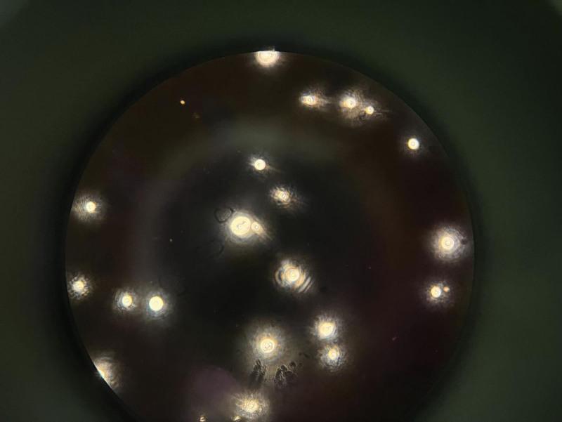
This diagnostic micrograph shows Cryptococcus neoformans in a cerebrospinal fluid (CSF) specimen using an India ink preparation at 400x magnification. The image displays classic negative staining where the dark carbon particles of the ink are excluded by the thick polysaccharide capsules of the yeast. This creates a highly characteristic 'halo' or clear zone surrounding the individual round-to-oval fungal cells against a dark, opaque background. Several yeast cells demonstrate size variation and internal morphology, including central dark cores or concentric ring-like structures. This visual finding is pathognomonic for cryptococcal meningitis in the appropriate clinical context, demonstrating the encapsulated nature of the fungal spores. The distribution pattern shows scattered individual cells and occasional budding forms, which are essential for identifying the yeast’s reproductive activity. This imaging modality is a critical rapid diagnostic tool in medical microbiology for identifying fungal infections in immunocompromised patients.
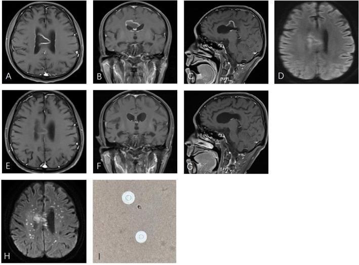
This composite figure illustrates the radiological and microbiological findings of a cerebral cryptococcal infection. Panels A-C show axial, coronal, and sagittal postcontrast T1-weighted MR images revealing a large, centrally located mass in the corpus callosum exhibiting prominent rim enhancement and surrounding vasogenic edema. Panel D is an initial diffusion-weighted image (DWI) showing rim hyperintensity without internal restricted diffusion, a finding that can distinguish this from a typical pyogenic brain abscess. Panels E-G demonstrate significant lesion regression and reduced enhancement 42 days after corticosteroid therapy, illustrating the potentially misleading radiological response of infectious pseudotumors to anti-inflammatory treatment. Panel H shows a subsequent DWI (70 days post-initial imaging) revealing new, multiple punctate hyperintense foci in the centrum semiovale, indicating disease progression and dissemination. Panel I is a cerebrospinal fluid (CSF) microscopy image using India ink stain, identifying the presence of encapsulated Cryptococcus neoformans yeast cells characterized by clear halos against the dark background. This sequence highlights the diagnostic challenge of differentiating fungal masses from neoplastic or demyelinating lesions.
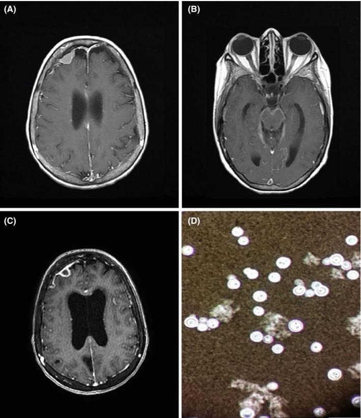
A multi-panel figure illustrating diagnostic findings for neurocryptococcosis. Panels A, B, and C are axial T1-weighted post-contrast brain MRI scans. Panel A shows a right frontal, extra-axial, pseudo-meningiomatous lesion with homogeneous contrast enhancement. Panel B demonstrates diffuse, linear meningeal enhancement consistent with meningitis. Panel C, a follow-up MRI, reveals the progression of the right frontal lesion into an annular (ring-enhancing) structure, accompanied by significant ventricular enlargement indicating secondary hydrocephalus. Panel D is a cerebrospinal fluid (CSF) microscopy image using an India ink preparation. It shows characteristic encapsulated yeasts, identified as Cryptococcus neoformans, which appear as round, budding cells surrounded by a distinct clear halo (the capsule) that excludes the dark ink particles in the background. This composite image serves as an educational resource for identifying the neuroimaging manifestations and laboratory diagnosis of fungal central nervous system infections.
Exercise M-3: Answers
Q1. Identify the Etiological Agent
-
Indian Ink Preparation (CSF smear): The left image shows encapsulated yeast cells with a characteristic clear halo around them against the dark ink background. This is the classic "negative staining" appearance of C. neoformans - the thick polysaccharide capsule excludes the ink particles, producing a clear zone around each round yeast cell. This is pathognomonic.
-
Growth on SDA (Sabouraud Dextrose Agar): The right image shows cream/mucoid colonies typical of C. neoformans. It grows at 37°C (an important virulence feature - differentiates it from non-pathogenic Cryptococcus species).
- Encapsulated, spherical yeast (2-20 µm)
- Single narrow-based budding
- No pseudohyphae
- Thick polysaccharide capsule (the main virulence factor)
- Urease positive
- Produces melanin via laccase enzyme (relevant on Niger seed/birdseed agar)
Q2. Clinical Diagnosis
- The patient is a 45-year-old HIV-infected male - this is a classic opportunistic infection in immunocompromised hosts (CD4+ count typically <100 cells/µL)
- Presenting features: on-and-off fever + severe headache for 1 month + signs of meningeal irritation (neck stiffness, Kernig's/Brudzinski's signs)
- C. neoformans is the most common fungal cause of meningitis in HIV/AIDS patients and accounts for 40-60% of AIDS-defining illnesses (Cummings Otolaryngology)
- The organism is acquired by inhalation from the environment (pigeon/bird droppings, soil), then disseminates hematogenously to the CNS
- Elevated opening pressure
- Mildly elevated protein
- Low glucose
- Lymphocytic pleocytosis
- India ink positive in ~50% of cases (Roberts and Hedges' Clinical Procedures in Emergency)
- Cryptococcal antigen (CrAg) - nearly 100% sensitive
Q3. Treatment of Choice
| Phase | Duration | Drug(s) |
|---|---|---|
| Induction | 2 weeks (minimum) | Liposomal Amphotericin B + Flucytosine (5-FC) |
| Consolidation | 8 weeks | Fluconazole 400 mg/day |
| Maintenance | ≥1 year | Fluconazole 200 mg/day |
- Induction: Liposomal Amphotericin B (3-4 mg/kg/day IV) + Flucytosine (100 mg/kg/day in 4 divided doses) is the gold standard. Recent trials also support Fluconazole + Flucytosine as an alternative induction regimen when amphotericin B is unavailable (Goodman & Gilman's)
- Fluconazole crosses the blood-brain barrier well - used for consolidation and long-term suppression
- Intracranial pressure management is critical - serial lumbar punctures or lumbar drainage to reduce ICP
- Antiretroviral therapy (ART): Should be deferred ~2-10 weeks after starting antifungal therapy to prevent Immune Reconstitution Inflammatory Syndrome (IRIS)
- Maintenance therapy is continued until CD4 count is >200 cells/µL sustained on ART