
A female pt 22 year old complains about 2 times blood in stool but no pain in anal region so tell me the causes and diagnosis treatment
rectal bleeding young adults causes diagnosis
PMID: 37712988
hemorrhoids rectal bleeding causes diagnosis
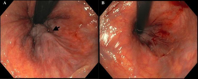
This endoscopic clinical photograph demonstrates Grade III internal hemorrhoids within the rectum before and after sclerotherapy treatment. Image A (pre-treatment) shows a retroflexed view of the anal canal where a prominent, swollen, pale-gray mass is visible, indicated by a black arrow. The hemorrhoid exhibits an irregular, nodular surface texture with dark vertical vascular striations, protruding into the lumen from the surrounding reddish-pink rectal mucosa. Image B (post-treatment) displays the clinical response following sclerotherapy. There is a marked reduction in the size and protrusion of the hemorrhoidal tissue. The previously bulky mass appears flattened and less congested, although some minor surface blood and residual dark vascular markings remain visible. This comparison illustrates the efficacy of sclerotherapy in reducing the volume and vascularity of prolapsing internal hemorrhoidal disease, which is a common minimally invasive intervention for symptomatic, non-bleeding Grade II and III hemorrhoids.
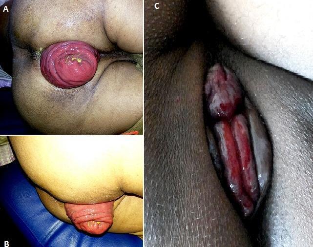
This composite of three clinical photographs provides a visual comparison between full-thickness rectal prolapse and prolapsed internal hemorrhoids, serving as an educational tool for proctological diagnosis. Panel A (posterior view) and Panel B (lateral view) demonstrate a complete rectal prolapse. The images show a large, reddish-pink, cylindrical mass protruding from the anal canal. A pathognomonic feature is the presence of concentric, circular mucosal folds, which distinguish it from other pathologies. Panel C depicts prolapsed internal hemorrhoids. This view shows three distinct, elongated, reddish-purple masses arranged radially within the anal region. Unlike the rectal prolapse in A and B, these masses demonstrate longitudinal or radial folds and are separated by grooves, corresponding to the left lateral, right anterolateral, and right posterolateral anatomical positions. Some surface moisture and vascular congestion are visible. This clinical imagery is essential for differentiating pelvic floor disorders and anorectal conditions based on fold orientation and morphology.
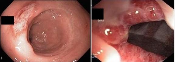
This composite of two endoscopic images demonstrates common rectal pathologies. The top image shows a Solitary Rectal Ulcer (SRU), characterized by a shallow, irregular-shaped lesion with a pale, whitish-grey fibrin base and surrounding erythematous, hyperemic mucosa. The lesion appears flat rather than excavated. The bottom image shows internal hemorrhoids, appearing as multiple, prominent, bulging columns of tissue protruding into the rectal lumen. These hemorrhoids exhibit significant vascular congestion, visible as a deep red or purplish discoloration with a smooth, glistening mucosal surface. The images illustrate the clinical distinction between ulcerative inflammatory processes and vascular-based structural abnormalities in the anorectal region. These findings are clinically significant in the diagnostic workup for symptoms such as bleeding per rectum, tenesmus, and chronic constipation. The content is suitable for medical students and gastroenterology trainees learning to identify anorectal disorders via lower gastrointestinal endoscopy.
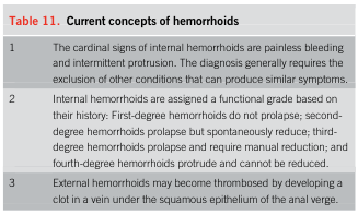
<table> <tr> <td colspan="2"><b>Table 11.</b> Current concepts of hemorrhoids</td> </tr> <tr> <td>1</td> <td>The cardinal signs of internal hemorrhoids are painless bleeding and intermittent protrusion. The diagnosis generally requires the exclusion of other conditions that can produce similar symptoms.</td> </tr> <tr> <td>2</td> <td>Internal hemorrhoids are assigned a functional grade based on their history: First-degree hemorrhoids do not prolapse; second-degree hemorrhoids prolapse but spontaneously reduce; third-degree hemorrhoids prolapse and require manual reduction; and fourth-degree hemorrhoids protrude and cannot be reduced.</td> </tr> <tr> <td>3</td> <td>External hemorrhoids may become thrombosed by developing a clot in a vein under the squamous epithelium of the anal verge.</td> </tr> </table>
Blood in Stool (Hematochezia) - 22-Year-Old Female, Painless
Causes (Differential Diagnosis)
Most Likely (Common in Young Adults)
| Cause | Characteristics |
|---|---|
| Internal Hemorrhoids (Grade I-III) | Most common cause of painless fresh red blood in stool. Blood on surface of stool or on toilet paper. NO perianal pain unless thrombosed. The cardinal signs are painless bleeding and intermittent protrusion. |
| Anal Fissure | Usually painful, but shallow or early fissures may be painless. Bright red streak on stool. |
| Infectious/Infective Colitis | Bloody diarrhea with mucus - Campylobacter, Salmonella, E. coli O157:H7, Shigella, Entamoeba histolytica |
| Inflammatory Bowel Disease (IBD) | Crohn's disease or Ulcerative Colitis - peak onset age 15-35. May have blood + mucus, urgency, diarrhea |
| Juvenile/Colonic Polyp | Painless bright red bleeding; juvenile retention polyps common in young people |
Less Common but Important
| Cause | Note |
|---|---|
| Solitary Rectal Ulcer Syndrome (SRUS) | Painless bleeding + mucus, associated with straining/constipation |
| Meckel's Diverticulum | Can bleed into adulthood; typically massive painless bleed |
| Angiodysplasia | Vascular malformation; more common in elderly but possible in young |
| Colorectal cancer | Rare in 22-year-old but possible if strong family history or IBD background; a 2023 meta-analysis found ~7% neoplastic lesion rate in patients under 40 with rectal bleeding undergoing colonoscopy (Colles et al., 2023) |
| Endometriosis of the bowel | In females - cyclical rectal bleeding correlating with menstrual cycle |
| Diverticular disease | Uncommon at 22 but possible |
As noted in Pye's Surgical Handicraft: "Passage of bright red blood after defaecation may be due to haemorrhoids or an anal fissure. Discovery of haemorrhoids does not obviate the need for further investigations to exclude other pathology. Bleeding may also be due to a polyp, cancer or diverticular disease."
Diagnosis
History to Take
- Character of blood: Bright red on toilet paper vs. mixed in stool vs. dark/tarry
- Frequency and volume: Two episodes so far - amount?
- Associated symptoms: Change in bowel habits, mucus, urgency, weight loss, fever, abdominal cramps
- Menstrual correlation: Cyclical bleeding? (suggests bowel endometriosis)
- Family history: CRC, polyposis, IBD
- Diet / medications: NSAIDs, anticoagulants
- Recent travel: Infectious colitis
Examination
- Perianal inspection: Fissures, external hemorrhoids, skin tags, fistulae
- Digital rectal examination (DRE): Mucosal integrity, masses, sphincter tone
- Proctoscopy / anoscopy: Best to visualize internal hemorrhoids (grades I-III are NOT visible on DRE)
Investigations
- Full Blood Count (FBC) - check for anemia, eosinophilia (IBD/parasites)
- ESR, CRP - inflammation markers
- Stool culture & microscopy - rule out infective cause
- Stool for occult blood (if bleeding intermittent)
- Proctoscopy / sigmoidoscopy - visualize hemorrhoids, fissures, rectal mucosa
- Colonoscopy - gold standard for lower GI evaluation; recommended if: IBD suspected, polyps, no clear benign cause found, family history, or persistent bleeding. In young patients under 40 with rectal bleeding, 7% have neoplastic lesions on colonoscopy.
- MRI pelvis - if endometriosis suspected
- CT colonography - if colonoscopy not feasible
- Technetium-99m scan / Meckel's scan - if Meckel's diverticulum suspected
Treatment (by cause)
Hemorrhoids (most likely)
- Conservative: High-fiber diet, increased fluid intake (>2L/day), avoid straining, sitz baths, stool softeners (lactulose, ispaghula husk)
- Medical: Topical agents (lignocaine, hydrocortisone cream), flavonoids (Daflon)
- Office procedures: Rubber band ligation (Grade I-III) - most effective outpatient treatment; Sclerotherapy (injection); Infrared coagulation
- Surgical: Hemorrhoidectomy for Grade IV or refractory Grade III (Milligan-Morgan or stapled hemorrhoidopexy)
Anal Fissure
- Topical GTN 0.2-0.4% or diltiazem 2% cream (to relax internal sphincter)
- Botulinum toxin injection
- Lateral internal sphincterotomy (surgical)
Infective Colitis
- Oral rehydration, appropriate antibiotics based on culture (e.g., metronidazole for amoeba, ciprofloxacin/azithromycin for Campylobacter/Salmonella if severe)
Inflammatory Bowel Disease (UC / Crohn's)
- Induction: 5-ASA (mesalazine suppositories/enemas for distal UC), corticosteroids for moderate-severe flares
- Maintenance: Mesalazine, azathioprine, biologics (infliximab, adalimumab) for moderate-severe disease
Colonic Polyp
- Endoscopic polypectomy; surveillance colonoscopy
Bowel Endometriosis
- Hormonal therapy (combined OCP, progestogens, GnRH agonists), laparoscopic surgical excision in refractory cases
Summary Table
| Priority | Diagnosis | Key Test | Treatment |
|---|---|---|---|
| Most likely | Internal hemorrhoids | Proctoscopy | Fiber, banding, sclerotherapy |
| Rule out | IBD (UC/Crohn's) | Colonoscopy + biopsy, CRP | 5-ASA, steroids, biologics |
| Rule out | Infective colitis | Stool culture | Antibiotics/supportive |
| Consider | Colonic polyp | Colonoscopy | Polypectomy |
| Consider (female-specific) | Bowel endometriosis | MRI pelvis | Hormonal/surgical |
| Rare | Colorectal cancer | Colonoscopy | Surgery ± adjuvant |
- 22nd Edition Pye's Surgical Handicraft, p. 8152
- Sleisenger and Fordtran's GI and Liver Disease, p. 2917
- Colonoscopy in young patients with rectal bleeding (Colles et al., 2023) - PMID 37712988
If it is dur to menstrual cycle so will it recurrent
bowel endometriosis rectal bleeding cyclical
intestinal endometriosis symptoms diagnosis treatment
Will Blood in Stool Recur if Due to Menstrual Cycle (Bowel Endometriosis)?
Short Answer: YES - but with an important nuance
Why Does It Recur?
- Grows under the influence of estrogen during the proliferative phase
- Bleeds during the secretory/menstrual phase when progesterone drops
- Repeats this cycle every month
- Serosal irritation
- Intramural hemorrhage
- Progressive fibrosis of the intestinal muscle wall
"Once implanted, endometrial tissue still appears to be regulated by hormonal influences so that estrogen promotes and progesterone inhibits growth. These repetitive cycles of growth and sloughing of tissue can lead to serosal irritation, muscle hypertrophy and progressive fibrosis of intestinal muscle." - Sleisenger and Fordtran's GI and Liver Disease
How Rectal Bleeding Happens in Bowel Endometriosis
| Depth of Bowel Involvement | Mechanism of Bleeding |
|---|---|
| Serosal implants (outside bowel) | Usually NO rectal bleeding; causes pain, tenderness |
| Intramural involvement (bowel wall) | Constipation, diarrhea, occasionally bleeding |
| Mucosal penetration (rare - inside bowel lumen) | Hematochezia (visible blood in stool) - this is the stage that causes frank rectal bleeding |
The Pattern of Recurrence
- Bleeding occurs just before or during menstruation
- Resolves after the period ends
- Returns with the next cycle - monthly recurrence
- Patient may also have dysmenorrhea (painful periods), dyspareunia (pain during intercourse), or infertility
- Symptoms may not always correlate perfectly with the cycle
- Bleeding can be irregular or persistent if fibrosis is advanced
- GI symptoms are not necessarily associated with gynecologic symptoms
"Contrary to popular belief, symptoms are not always cyclical and may not fluctuate with hormonal levels; nor are GI symptoms necessarily associated with gynecologic symptoms." - Sleisenger and Fordtran's GI and Liver Disease
What Happens if Left Untreated?
- Fibrosis worsens with each cycle
- Bowel wall thickens - can cause partial obstruction, constipation, narrowed stool caliber
- Risk of bowel obstruction (intussusception, volvulus, stricture)
- Risk of malignant transformation (mainly in deep infiltrating endometriosis)
- Worsening infertility
Diagnosis Confirmation
| Investigation | Finding |
|---|---|
| Ask about menstrual history | Bleeding timing - does it coincide with menstruation? |
| Pelvic examination (rectovaginal) | Tender nodules in cul-de-sac / pouch of Douglas - highly suggestive |
| Pelvic MRI | Best non-invasive test; shows deep infiltrating lesions, bowel wall involvement |
| Transvaginal + transrectal ultrasound | Can detect rectal wall nodules |
| Colonoscopy | Usually normal mucosa unless mucosal penetration has occurred; useful to rule out other causes |
| CA-125 serum | Elevated in some cases (not specific) |
| Laparoscopy | Gold standard for diagnosis; allows direct visualization and biopsy |
Treatment to Stop Recurrence
- Combined oral contraceptive pill (OCP) - taken continuously (no pill-free break) to suppress cycling
- Progestogens (norethisterone, medroxyprogesterone acetate, dienogest)
- GnRH agonists (goserelin, leuprolide) - induce a temporary medical menopause; most effective medically but causes side effects (hot flushes, bone loss)
- Levonorgestrel IUD (Mirena) - for symptom control
- Laparoscopic excision of endometriotic nodules from bowel wall
- Segmental bowel resection (for severe deep infiltrating rectal endometriosis)
- Definitive treatment: Hysterectomy + bilateral salpingo-oophorectomy (reserved for severe, refractory cases in women who have completed family)
Key Takeaway for This Patient
- Detailed menstrual history - timing of bleeding vs. period
- Rectovaginal pelvic examination
- Pelvic MRI
- Gynecology referral
- Sleisenger and Fordtran's GI and Liver Disease, pp. 3991-4014
- Robbins & Kumar Basic Pathology, pp. 553-577
Is any lab test available to find piles fissure and fistula
proctoscopy anoscopy hemorrhoids piles examination
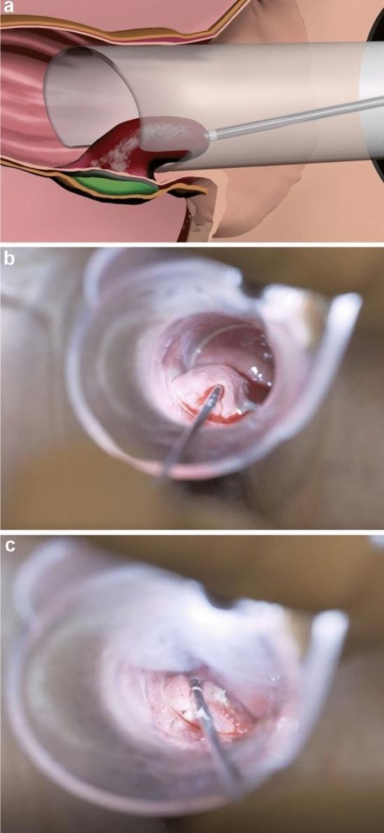
This composite visual represents the Rafaelo® procedure for the treatment of internal hemorrhoids using radiofrequency ablation. Panel (a) is a schematic diagram illustrating the anatomical positioning: a proctoscope is inserted into the anal canal, through which a radiofrequency probe is guided into the hemorrhoidal plexus above the dentate line. A protective layer of local anesthetic is shown injected between the submucosa and the internal sphincter muscle to prevent thermal injury. Panel (b) is a clinical photograph taken via proctoscopy showing the real-time insertion of the fine-needle probe tip into the protruding hemorrhoidal tissue. Panel (c) demonstrates the post-ablation state of the tissue. Visible hallmarks of successful treatment include mucosal blanching (lightening of tissue color) and localized coagulation points on the hemorrhoid surface, indicating thermal denaturation of proteins. The procedure aims to induce tissue fibrosis and subsequent retraction of the hemorrhoidal prolapse, minimizing post-operative pain and avoiding open surgical wounds.
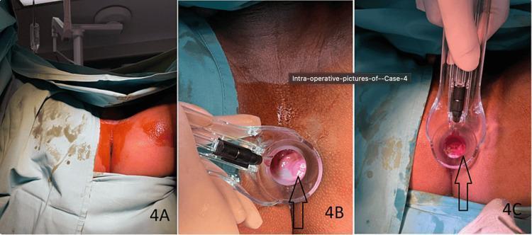
This composite of three clinical photographs documents an intraoperative RAFAELO procedure for internal hemorrhoids. Panel 4A shows the preoperative surgical field with the patient in a prone or lithotomy position, draped with green surgical sheets. The perianal skin is prepped with an orange-tinted antiseptic solution. Panel 4B displays an intraoperative proctoscopy view using a clear plastic proctoscope. A black arrow identifies a prominent, erythematous, fleshy Grade II internal hemorrhoid cushion protruding into the rectal lumen at the 5 o'clock position. Panel 4C shows the post-procedure proctoscopy view following radiofrequency ablation. The targeted hemorrhoidal tissue (indicated by the arrow) appears visibly reduced in volume, less edematous, and significantly more retracted compared to the preoperative state. These images serve as educational material for colorectal surgery, illustrating the visual markers of successful radiofrequency treatment for symptomatic internal hemorrhoids.
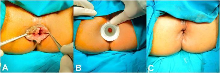
This composite of three clinical photographs documents a surgical hemorrhoidectomy procedure for Grade III or IV hemorrhoids. Panel A shows the preoperative state with significant prolapsed, irregular, reddish-brown hemorrhoidal piles protruding from the anal canal; surgical forceps are used to retract and demonstrate the extent of the prolapse. Panel B illustrates the intraoperative phase, showing the use of a circular white anal dilator to manage internal anal sphincter hypertonicity and provide surgical access to the hemorrhoidal columns. Panel C displays the immediate postoperative result, demonstrating a restored anal anatomy with no visible protruding tissue. The surgical site shows radial suture lines where the hemorrhoidal masses were excised and the mucosa was closed with simple interrupted sutures. This sequence highlights the surgical transition from symptomatic prolapse through intraoperative dilation to postoperative anatomical correction, serving as an educational resource for colorectal surgery and proctology.
anal fistula MRI imaging diagnosis classification
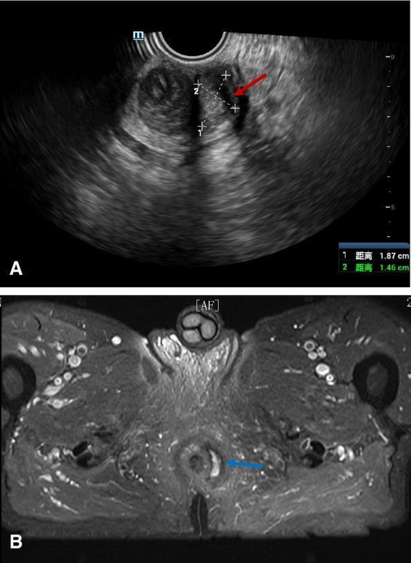
This composite diagnostic image illustrates a Type I anal fistula using two different modalities: 3D pelvic ultrasound (US) and magnetic resonance imaging (MRI). Image A shows a transrectal or pelvic 3D US presenting a heterogeneous perianal region. A red arrow indicates a hypoechoic, cavity-like tract representing the fistula, with calipers measuring its dimensions (1.87 cm and 1.46 cm). Image B is an axial T2-weighted or fat-suppressed MRI of the pelvic floor and anal canal. A blue arrow highlights a hyperintense (bright) signal adjacent to the anal canal, indicating an active inflammatory fistula tract or fluid collection. The comparison demonstrates the clinical utility of both US and MRI in identifying perianal fistulous disease, highlighting the fistula's spatial relationship to pelvic musculature and the anal canal. The images are aimed at an intermediate to advanced medical level, focusing on radiologic diagnosis and classification of anorectal pathologies.
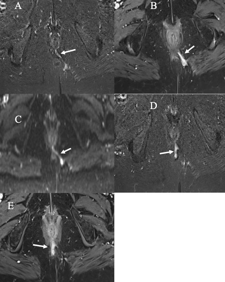
This composite of five axial pelvic MRI scans (labeled A-E) demonstrates a Grade 4 trans-sphincteric perianal fistula with secondary ramification, categorized according to the St. James’s University Hospital classification system. Images A and D utilize STIR (Short Tau Inversion Recovery) sequences, showing the fistula as a hyperintense, fluid-filled tract originating near the 6 o'clock position of the anal canal and crossing the external sphincter. Image B and E are post-contrast fat-suppressed T1-weighted images (T1-WI) showing prominent enhancement of the tract walls, indicating active inflammation. Image C is a Diffusion-Weighted Image (DWI) showing restricted diffusion, which appears as a high-signal (bright) intensity, highlighting the active fistula components. White arrows in all panels identify the primary tract, while panels D and E specifically reveal a secondary ramification branching posteriorly. This diagnostic imaging series is designed for radiology and surgical residents to understand the multi-parametric MRI appearance of complex fistulous disease, including the identification of internal openings and secondary tracts essential for surgical planning.
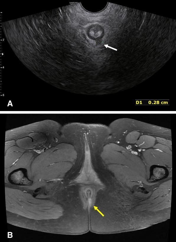
A comparative diagnostic image display showing a Type II anal fistula through two different imaging modalities: (A) a transverse pelvic 3D Ultrasound (US) and (B) an axial Magnetic Resonance Image (MRI). In image A, the 3D ultrasound demonstrates the fistula as a hypoechoic (dark) focal area within the heterogeneous pelvic floor tissue, indicated by a white arrow; a caliper measurement 'D1' shows a width of 0.28 cm. In image B, the MRI (likely T2-weighted or STIR) reveals the fistula as a hyperintense (bright) linear structure, indicated by a yellow arrow, situated within the midline anatomical structures near the anal canal. The surrounding pelvic floor musculature, including the levator ani and external sphincter regions, are visible as darker, well-defined tissues. This comparison illustrates the diagnostic visualization of perianal inflammatory tracks and is used in colorectal surgery planning for classification and localization of fistulous tracts relative to the sphincter complex.
Lab Tests & Investigations for Piles, Fissure, and Fistula
The Most Important Point First
- Confirm the diagnosis
- Grade severity
- Map anatomy (especially fistulas)
- Rule out underlying causes (IBD, Crohn's, cancer, infection)
- Check for complications (anemia, sepsis)
Investigations Broken Down by Condition
1. PILES (Hemorrhoids)
Primary Diagnosis - Clinical Examination
| Examination | What It Shows |
|---|---|
| Perianal inspection | External hemorrhoids, skin tags, prolapsed internal hemorrhoids |
| Digital Rectal Examination (DRE) | Rules out masses, rectal tumors - internal hemorrhoids are NOT felt on DRE as they are soft |
| Proctoscopy / Anoscopy | The key diagnostic tool - directly visualizes internal hemorrhoids above the dentate line, grades them |
| Rigid / Flexible Sigmoidoscopy | Evaluates rectum and sigmoid colon |
| Grade | Description |
|---|---|
| Grade I | Bulge into lumen, do NOT prolapse |
| Grade II | Prolapse on straining, reduce spontaneously |
| Grade III | Prolapse, require manual reduction |
| Grade IV | Permanently prolapsed, cannot be reduced |
Supporting Lab Tests (not diagnostic, but important)
| Test | Purpose |
|---|---|
| Complete Blood Count (CBC/FBC) | Check for anemia due to chronic blood loss (low Hb, low MCV - iron deficiency) |
| Serum Iron / Ferritin / TIBC | Confirm iron deficiency anemia from chronic rectal bleeding |
| Coagulation profile (PT, aPTT) | If bleeding is heavy or unusual - rule out bleeding disorder |
| Stool for occult blood (FOBT) | Detects blood if not visible to naked eye |
| Colonoscopy | If cause of bleeding is unclear, age >40, or red flags present |
2. ANAL FISSURE
Primary Diagnosis - Clinical Examination
| Examination | What It Shows |
|---|---|
| Perianal inspection | Visible tear/split in anal skin (typically at 6 o'clock / posterior midline, or 12 o'clock / anterior midline in females) |
| Gentle DRE | Often too painful to perform acutely; reveals tight hypertonic sphincter |
| Proctoscopy | Usually deferred in acute fissure due to pain; done after healing or under anesthesia |
- Posterior midline (6 o'clock): 90% of cases - most common
- Anterior midline (12 o'clock): More common in females
- Lateral/multiple fissures: Suspect Crohn's disease, TB, STI, HIV - must investigate further
Supporting Lab Tests
| Test | Purpose |
|---|---|
| CBC | Anemia from chronic loss |
| ESR / CRP | Elevated if underlying Crohn's or infection |
| Stool culture | Rule out infective cause |
| TB culture / biopsy | If atypical/lateral fissure - suspect tubercular or Crohn's |
| HIV test | Atypical fissures in lateral position may indicate immunosuppression |
| Colonoscopy + biopsy | If Crohn's disease suspected (multiple, atypical fissures) |
| Anorectal manometry | Measures internal anal sphincter resting pressure - elevated in chronic fissure (hypertonia); guides treatment (e.g., whether to use Botox or sphincterotomy) |
3. ANAL FISTULA
Primary Diagnosis - Clinical Examination
| Examination | What It Shows |
|---|---|
| Perianal inspection | External opening (secondary/outer opening) - discharging pus or blood |
| Digital Rectal Examination | Internal opening felt as tender nodule or induration on rectal wall; identifies abscess |
| Proctoscopy / Sigmoidoscopy | Visualizes internal opening inside the anal canal |
| Probe test (Goodsall's rule) | A probe is gently passed through external opening to trace the fistula tract - determines if it follows anterior or posterior path |
- External opening posterior to a transverse anal line → tract curves posteriorly and opens in the midline internally
- External opening anterior → tract goes straight to the nearest point of the anal canal
Imaging Investigations (CRITICAL for fistula)
| Investigation | Use | Best For |
|---|---|---|
| MRI Pelvis (STIR/T2) | Gold standard - maps fistula tract, secondary extensions, relation to sphincters, detects abscesses | Complex/high fistulas, recurrent fistulas, pre-surgical planning |
| Endoanal Ultrasound (EAUS) | Visualizes fistula tract and sphincter integrity; done bedside | Simple low fistulas, assessing sphincter damage |
| 3D Transrectal Ultrasound | High-resolution sphincter mapping | Surgical planning |
| Fistulogram (contrast X-ray) | Inject dye into external opening + X-ray shows tract path | Low-tech, not commonly used now - replaced by MRI |
| CT with contrast | For pelvic abscess, Crohn's, complex supralevator fistulas | When MRI not available or IBD complications |
Lab Tests for Fistula
| Test | Purpose |
|---|---|
| CBC | Leukocytosis if active sepsis/abscess |
| CRP / ESR | Active inflammation - also elevated in Crohn's-related fistula |
| Fasting blood glucose / HbA1c | Diabetes is a major risk factor for fistula and impairs healing |
| HIV test | Unusual or recurrent fistulas suggest immunosuppression |
| Crohn's workup (ASCA, calprotectin) | Fecal calprotectin elevated in IBD; ASCA positive in Crohn's |
| Stool culture | If infectious (TB, Actinomyces) cause suspected |
| Biopsy of fistula tract | If Crohn's, TB, or malignancy suspected - histopathology |
| Anorectal manometry | Assesses sphincter function before surgical planning |
Summary Table
| Condition | CLINICAL exam (primary) | Key Investigation | Lab Tests |
|---|---|---|---|
| Piles | Proctoscopy / Anoscopy | Proctoscopy (grades I-IV) | CBC (anemia), colonoscopy if needed |
| Anal Fissure | Perianal inspection | Anorectal manometry | CBC, CRP, colonoscopy if atypical |
| Anal Fistula | DRE + Probe test | MRI pelvis (gold standard) | CBC, CRP, glucose, HIV, biopsy if Crohn's |
Practical Approach for Your 22-Year-Old Patient
- Step 1: Perianal inspection + DRE + Proctoscopy - this single outpatient examination will diagnose piles and fissure on the spot
- Step 2: CBC + serum ferritin - check if chronic blood loss has caused anemia
- Step 3: If fistula suspected (external opening, discharge, recurrent perianal abscess) - add MRI pelvis
- Step 4: If atypical or no clear diagnosis - colonoscopy
Key principle: Proctoscopy/anoscopy is the single most important investigation for all three conditions (piles, fissure, fistula). No blood test replaces it.