
Brachial cleft cyst radiology ans
Reading File
Finding Sources
Searching PubMed
branchial cleft cyst imaging
Searching the Web
branchial cleft cyst radiology ultrasound CT MRI imaging features
Finding Sources
Reading Web Page
https://pmc.ncbi.nlm.nih.gov/articles/PMC6323601/
Branchial cleft cyst - Radiology
A branchial cleft cyst is a congenital epithelial cyst, usually presenting as a painless lateral neck swelling. Second branchial cleft cysts are the commonest, typically located near the angle of the mandible along the anterior border of the sternocleidomastoid muscle (SCM). Their tract characteristically passes between the internal and external carotid arteries toward the tonsillar fossa. K J Lee’s Essential Otolaryngology, p. 677.
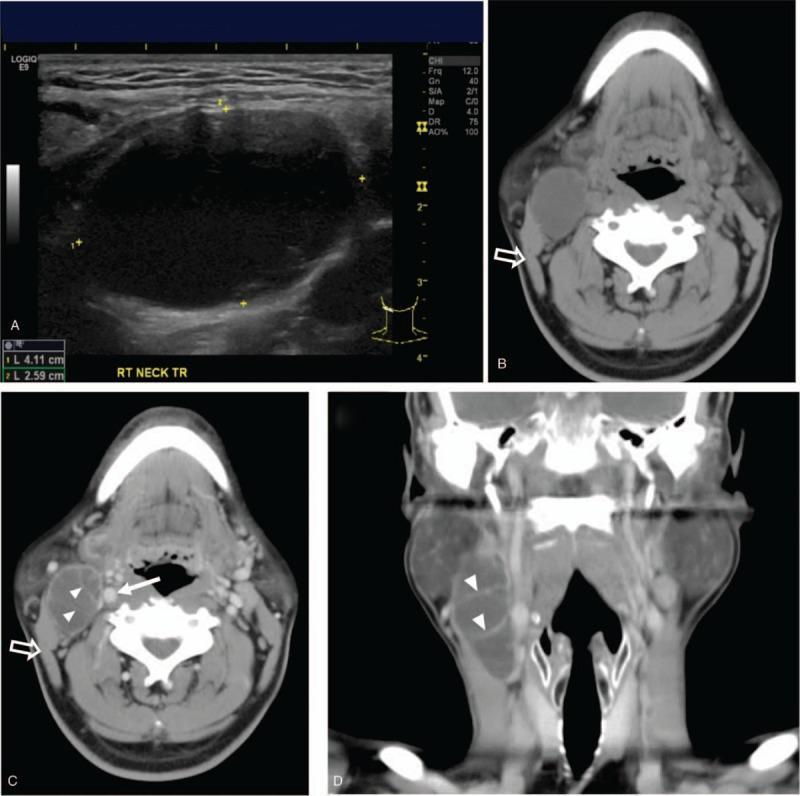
Ultrasound
- Well-defined, thin-walled cystic lesion.
- Usually anechoic or homogeneously hypoechoic.
- May contain fine internal debris, giving pseudo-solid appearance.
- Posterior acoustic enhancement is common.
- No internal vascularity on Doppler.
- Infection causes internal echoes, septations, wall thickening, and peripheral hyperemia.
CT neck with contrast
- Well-circumscribed, round/ovoid, non-enhancing, low-attenuation lesion.
- Usually lies anterior to SCM and lateral to carotid space, commonly at the mandibular angle.
- In a second cleft cyst, extension or a tapered “beak/notch” between the internal and external carotid arteries is highly suggestive.
- Infected cyst: thick irregular rim enhancement, surrounding fat stranding, possibly fluid-fluid level or abscess formation.
MRI
- T1: usually low signal, but may be high with proteinaceous fluid, hemorrhage, or infection.
- T2/STIR: high signal intensity.
- No internal enhancement in an uncomplicated cyst. Thin rim enhancement may occur.
- MRI is useful for defining a sinus/fistulous tract and relation to carotid vessels, parotid gland, and facial nerve.
Typical locations by type
| Type | Usual location |
|---|---|
| First cleft cyst | Periauricular, external auditory canal, or within/superficial/deep to parotid |
| Second cleft cyst | Anterior SCM, level II neck, near angle of mandible, lateral to carotid space |
| Third/Fourth cleft anomaly | Lower neck, pyriform sinus region, often left-sided |
Differential diagnosis
- Cystic metastatic cervical lymph node, especially HPV-associated oropharyngeal squamous cell carcinoma in adults
- Necrotic lymphadenopathy, including tuberculous nodes
- Lymphatic malformation
- Thyroglossal duct cyst, usually midline
- Dermoid/epidermoid cyst
- Abscess, plunging ranula
Important exam point
In an adult with a new cystic level II neck mass, do not assume branchial cyst without excluding cystic nodal metastasis from an oropharyngeal primary. Suspicious features include a mural nodule, solid enhancing component, irregular thick wall, extracapsular spread, or additional abnormal lymph nodes.
The typical ultrasound, CT, and MRI appearances are summarized in this radiology review; CT is particularly useful for mapping extent and vascular relationships, while MRI better depicts soft tissues and tracts, as described by NCBI StatPearls.
Classify various orbital tumours. Describe radiological features of tumours affecting the eyeball (globe).
Reading File
Finding Sources
Finding Sources
Finding Sources
Searching Images
retinoblastoma CT MRI intraocular tumor calcification radiology image
Searching Images
uveal melanoma MRI intraocular tumour radiology image
Searching PubMed
intraocular tumors imaging
Searching the Web
radiology orbital tumors classification imaging features intraocular retinoblastoma uveal melanoma
Finding Sources
Finding Sources
Reading Web Page
https://pmc.ncbi.nlm.nih.gov/articles/PMC9600631/
Reading File
Reading File
Classification of orbital tumours
Orbital tumours may be classified by site of origin, which is the most useful radiological classification.
| Compartment/site | Common tumours |
|---|---|
| Globe / intraocular | Retinoblastoma, uveal melanoma, choroidal metastasis, medulloepithelioma, choroidal haemangioma, intraocular lymphoma |
| Optic nerve and sheath | Optic nerve glioma, optic nerve sheath meningioma, lymphoma, metastasis |
| Intraconal orbit | Cavernous venous malformation, schwannoma, optic nerve glioma, lymphoma |
| Extraconal orbit | Dermoid/epidermoid, lacrimal gland tumours, capillary haemangioma, lymphoma, metastasis |
| Lacrimal gland | Pleomorphic adenoma, adenoid cystic carcinoma, lymphoma, inflammatory dacryoadenitis |
| Diffuse/infiltrative orbital disease | Lymphoma, leukemia/granulocytic sarcoma, idiopathic orbital inflammation, metastases |
| Bony orbit / adjacent sinus extension | Osteoma, fibrous dysplasia, Ewing sarcoma, rhabdomyosarcoma, sinonasal carcinoma with orbital invasion |
| Vascular lesions and tumour mimics | Venous malformation, lymphatic malformation, varix, carotid-cavernous fistula |
Another practical classification is by age:
- Children: retinoblastoma, rhabdomyosarcoma, optic nerve glioma, dermoid, capillary haemangioma, leukemic infiltrate.
- Adults: cavernous venous malformation, lymphoma, meningioma, lacrimal gland tumours, metastasis, and uveal melanoma.
Radiological features of tumours affecting the globe
1. Retinoblastoma
The commonest primary intraocular malignancy of childhood, usually presenting before 5 years with leukocoria. It arises from the retina. Grainger & Allison's Diagnostic Radiology, p. 1572.
Ultrasound
- Intraocular echogenic retinal mass.
- Calcification causes acoustic shadowing, a highly useful finding.
- Internal vascularity on colour Doppler.
- May show retinal detachment, vitreous seeding, hemorrhage, or pseudohypopyon.
CT
- Hyperattenuating soft-tissue mass, usually in the posterior globe.
- Nodular or stippled calcification is characteristic and is seen in most cases.
- Contrast enhancement is present.
- Assesses bony orbit and gross extraocular extension, but CT is now avoided where feasible in children because of ionizing radiation and the risk of radiation-induced second malignancy in hereditary disease.
MRI
- Best modality for local staging and intracranial assessment.
- Tumour is typically:
- T1: intermediate to hyperintense relative to vitreous.
- T2: relatively hypointense compared with vitreous.
- Post-contrast: moderate to marked enhancement.
- DWI: restricted diffusion.
- Look specifically for:
- Optic nerve invasion, especially post-laminar involvement.
- Scleral/extrascleral extension.
- Choroidal invasion.
- Orbital extension.
- Pineal or suprasellar primitive neuroectodermal tumour in hereditary disease, termed trilateral retinoblastoma.
Key point: An enhancing intraocular mass with calcification in a child should be regarded as retinoblastoma until proved otherwise. The diagnostic text describes calcification in about 95% of cases and emphasizes dedicated orbital and whole-brain MRI for intracranial spread. Grainger & Allison's Diagnostic Radiology, p. 1572.
2. Uveal melanoma
This is the commonest primary intraocular malignancy in adults. Most arise in the choroid, with less frequent involvement of the ciliary body or iris. Grainger & Allison's Diagnostic Radiology, p. 1571.
Ultrasound
- Solid, dome-shaped or mushroom-shaped choroidal mass.
- Low-to-medium internal echoes on A-scan.
- Homogeneous echogenic lesion on B-scan.
- Associated exudative retinal detachment is common.
- Colour Doppler shows internal vascularity.
CT
- Hyperdense enhancing intraocular mass.
- Usually non-calcified.
- May demonstrate globe deformation, retinal detachment, scleral thickening, or extrascleral extension.
- CT is less specific than MRI for melanoma characterization.
MRI
The classical MRI appearance is due to paramagnetic melanin:
- T1: hyperintense relative to vitreous.
- T2: hypointense relative to vitreous.
- Moderate to marked homogeneous enhancement after gadolinium.
- May be dome-shaped, lentiform, or have a mushroom configuration when the tumour breaks through Bruch membrane.
- MRI is valuable for detecting extrascleral spread, optic nerve involvement, and associated retinal detachment.
Amelanotic melanoma may not show the classic T1-high/T2-low signal pattern.
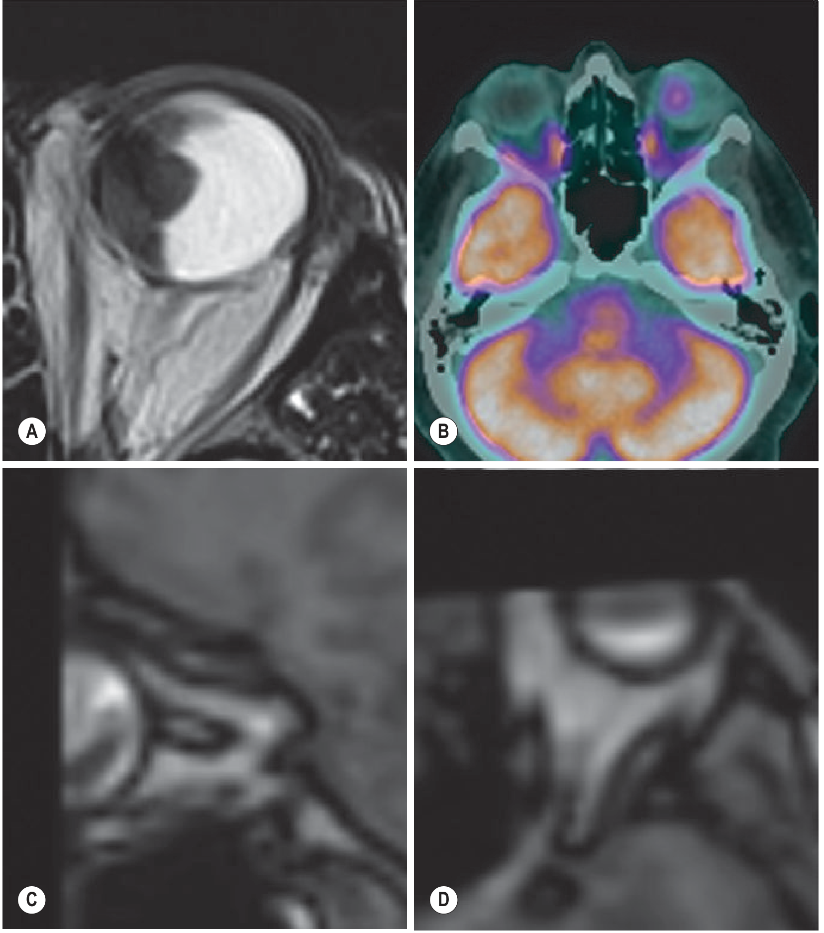
Choroidal melanoma: nodular lesion at the globe with associated retinal hemorrhage. Grainger & Allison's Diagnostic Radiology, p. 1571.
3. Choroidal metastases
- Most common intraocular malignancy overall in adults.
- Usually from breast carcinoma in women and lung carcinoma in men.
- Often bilateral and multifocal.
- Preferentially involve the posterior choroid.
Imaging
- Ultrasound: plateau-shaped, relatively echogenic choroidal mass with subretinal fluid.
- CT/MRI: enhancing choroidal or subretinal thickening/mass.
- MRI signal is variable and lacks the characteristic melanotic MRI pattern.
- Associated retinal detachment is frequent.
- Search for systemic primary malignancy and other metastatic deposits.
4. Medulloepithelioma
A rare, malignant embryonal tumour of the non-pigmented ciliary epithelium, usually in young children.
Imaging
- Arises from the ciliary body, therefore located anteriorly in the globe.
- Multiloculated cystic and solid intraocular mass.
- Ultrasound: echogenic solid portions with cystic spaces.
- CT: enhancing ciliary-body mass; dystrophic calcification may occur.
- MRI: heterogeneous lesion, commonly T2 hyperintense cystic areas with enhancing solid components.
- Can mimic retinoblastoma, but ciliary-body origin and multiple cystic spaces favour medulloepithelioma.
5. Choroidal haemangioma
Circumscribed choroidal haemangioma
- Solitary, benign vascular tumour.
- Usually a well-defined orange-red posterior choroidal lesion.
Imaging
- Ultrasound: acoustically solid echogenic choroidal mass.
- MRI: typically hyperintense on T2 and enhances strongly.
- Associated exudative retinal detachment may occur.
- Unlike melanoma, it is generally T2 hyperintense, whereas melanotic melanoma is usually T2 hypointense.
Diffuse choroidal haemangioma
- Associated with Sturge-Weber syndrome.
- Diffuse thickening of the choroid and may produce glaucoma or retinal detachment.
6. Intraocular lymphoma/leukemic infiltration
- May involve the retina, choroid, vitreous, optic nerve, or uvea.
- MRI may show diffuse or focal enhancing intraocular/uveal thickening with restricted diffusion.
- Often associated with CNS lymphoma or systemic hematologic malignancy.
- Leukemic infiltration may be diffuse; retinal hemorrhages are common in leukemia.
Imaging differentiation: retinoblastoma vs melanoma
| Feature | Retinoblastoma | Uveal melanoma |
|---|---|---|
| Typical patient | Child under 5 years | Adult, usually middle or older age |
| Origin | Retina | Choroid/uveal tract |
| Calcification | Common and characteristic | Uncommon |
| Shape | Irregular retinal mass | Dome, lentiform, or mushroom-shaped mass |
| T1 MRI | Intermediate relative to vitreous | Typically hyperintense if melanotic |
| T2 MRI | Relatively hypointense | Typically hypointense if melanotic |
| Important extension | Optic nerve, choroid, sclera, orbit, brain | Extrascleral spread and hematogenous liver metastases |
General radiological approach to an intraocular mass
- Ultrasound with Doppler: first-line for lesion location, calcification, internal vascularity, and retinal detachment.
- MRI orbit with contrast and DWI: best for lesion characterization, optic nerve/scleral/extraocular spread, and intracranial extension.
- CT: best for calcification and bone, but use selectively, especially in children.
- Assess for complications: retinal detachment, vitreous seeding, hemorrhage, glaucoma, optic nerve invasion, extrascleral extension, and distant metastasis.
This is a shared conversation. Sign in to Orris to start your own chat.