
Explain detail about Wilson’s disease
Kayser-Fleischer ring Wilson disease cornea
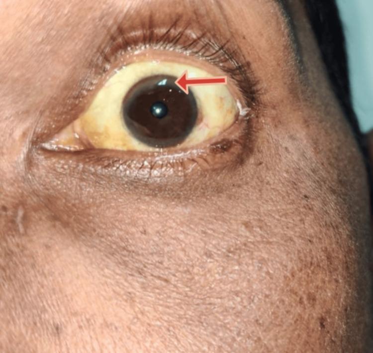
A close-up clinical photograph of a left eye and periorbital region demonstrating characteristic signs of hepatolenticular degeneration (Wilson disease). The sclera exhibits marked icterus (yellowish discoloration), consistent with hyperbilirubinemia or underlying chronic liver disease. At the peripheral margin of the cornea, a brownish-yellow Kayser-Fleischer (KF) ring is visible, particularly highlighted by a red arrow in the superior-temporal quadrant. This ring represents copper deposition in Descemet's membrane. The iris is dark brown, and the surrounding skin shows a slightly uneven pigmentation. The image serves as an educational example of ophthalmologic manifestations associated with systemic metabolic disorders and copper metabolism dysfunction.
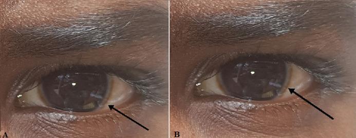
This clinical photograph consists of two panels (A and B) showing the right and left eyes of a patient with dark-pigmented skin. The primary pathological finding is a Kayser-Fleischer ring, visible as a distinct brownish-golden or greenish-brown discoloration at the periphery of the cornea near the limbus. The ring is most prominent in the superior and inferior poles, appearing as a circumferential band that encircles the dark brown iris. The sclera shows mild conjunctival vascularity but is otherwise white. The pupils are round and reactive to light. This ocular sign is a hallmark clinical manifestation of Wilson disease, resulting from copper deposition in Descemet's membrane. The image serves as a classic educational example for hepatology, neurology, and ophthalmology, illustrating the diagnostic physical exam findings associated with impaired copper metabolism.
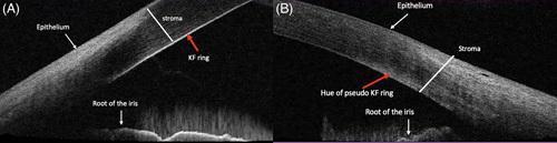
This diagnostic image consists of two panels (A and B) featuring Anterior Segment Optical Coherence Tomography (AS-OCT) cross-sections of the peripheral cornea and anterior chamber angle. The image compares the appearance of corneal deposits in different clinical conditions. Panel A illustrates a classic Kayser-Fleischer (KF) ring, characteristic of Wilson disease, visualized as a discrete hyperintense line located specifically on the Descemet membrane at the posterior corneal surface. In contrast, Panel B demonstrates a 'pseudo-KF ring,' often associated with non-Wilsonian hyperbilirubinemia, which appears as a diffuse, scattered hyper-reflective hue localized within the posterior stroma rather than a distinct line on the Descemet membrane. Both panels are labeled with key anatomical structures including the epithelium, stroma, and the root of the iris. This comparison serves as an educational tool for ophthalmologists and hepatologists to differentiate between true copper deposits and pigmentary changes due to bile pigments using high-resolution imaging.
"Wilson disease" AND treatment
Wilson's Disease (Hepatolenticular Degeneration)
Overview
Pathophysiology
- In the trans-Golgi network: incorporates copper into apoceruloplasmin to form functional ceruloplasmin, which is secreted into blood.
- In lysosomes: transports non-ceruloplasmin-bound copper into bile canaliculi for excretion in bile (the major route of copper elimination, since biliary copper does not recirculate enterohepatically).
- Copper cannot be excreted into bile, so it accumulates in hepatocyte cytoplasm and lysosomes, generating reactive oxygen species that damage hepatocytes.
- Copper cannot be loaded into apoceruloplasmin, so serum ceruloplasmin is low (though low ceruloplasmin itself does not cause the toxicity - it's a downstream marker, not the pathogenic mechanism).
Clinical Features
- ~20% purely hepatic, ~20% neurologic + hepatic, ~50% neurologic/psychiatric presentations in adults (some degree of liver involvement is essentially always present).
- Hepatic: ranges from asymptomatic transaminase elevation to acute hepatitis, chronic hepatitis, cirrhosis, or fulminant acute liver failure (about 5% of patients present this way and need urgent transplant). Fatigue, jaundice, hepatomegaly, ascites, and secondary endocrine effects (delayed puberty, amenorrhea) may occur.
- Neurologic: presents later (typically after age 20) as a movement disorder mimicking Parkinsonism - dysarthria, dystonia, rigidity, tremor, chorea, abnormal gait, poor handwriting, occasionally peripheral neuropathy.
- Psychiatric: personality change, irritability, depression, anxiety, psychosis, or bipolar-like symptoms, often in late teens/early 20s - frequently mistaken for primary psychiatric disease.
- Ocular - Kayser-Fleischer rings: golden-brown to green copper deposits in Descemet's membrane at the corneal limbus. Present in ~95% of patients with neuropsychiatric disease but only 50-65% of purely hepatic presentations. Sunflower cataracts can also occur.
- Other: Coombs-negative hemolytic anemia, renal tubular (Fanconi-like) dysfunction, skeletal abnormalities (osteopenia, arthropathy) (Bradley and Daroff's Neurology in Clinical Practice; Goldman-Cecil Medicine).
Diagnosis
| Test | Utility |
|---|---|
| Serum ceruloplasmin | Low in most patients; most useful as a screening test, but can be falsely normal/elevated (e.g., inflammation, pregnancy, estrogen use) or falsely low in other conditions |
| 24-hour urinary copper | Elevated; the most specific widely used test |
| Hepatic copper content (liver biopsy) | >250 μg/g dry weight is diagnostic; the most sensitive test but invasive |
| Slit-lamp exam | Detects Kayser-Fleischer rings |
| Serum copper | Not diagnostically useful - can be low, normal, or high depending on disease stage |
| Genetic testing (ATP7B sequencing) | Confirmatory but complicated by the huge number of possible mutations; most useful for family/first-degree relative screening |
Treatment
- First-line chelators: D-penicillamine or trientine (triethylenetetramine dihydrochloride). Trientine lacks the sulfhydryl groups of penicillamine and is often preferred as second-line or in penicillamine-intolerant patients due to a better side-effect profile; some centers now use it first-line.
- Zinc salts (e.g., zinc acetate): block intestinal copper absorption by inducing metallothionein in enterocytes; used as maintenance therapy or in milder/presymptomatic disease, and often combined with a chelator initially. Zinc is also considered relatively safer in pregnancy.
- Tetrathiomolybdate: an experimental chelator, close to clinical use.
- Liver transplantation: curative for the metabolic defect; indicated in fulminant hepatic failure or decompensated cirrhosis unresponsive to medical therapy.
- Gene therapy for Wilson's disease is under active investigation but not yet standard of care (Goldman-Cecil Medicine; Sleisenger and Fordtran's Gastrointestinal and Liver Disease, Ch. 76; Yamada's Textbook of Gastroenterology).
Recent Evidence (supplementary)
- A 2024 systematic review/meta-analysis on pregnancy outcomes in Wilson's disease found continued anti-copper treatment during pregnancy reduces adverse outcomes (PMID: 39206599).
- A 2025 systematic review describes cardiac ventricular dysfunction as an underrecognized extrahepatic manifestation of Wilson's disease (PMID: 40904321).
- A 2025 systematic review characterized the mutation spectrum in Arab populations, reinforcing how genetically heterogeneous ATP7B mutations are across regions (PMID: 39922954).