
Explain D-dimer test
The D-dimer test measures a fibrin degradation product released when a blood clot breaks down. It is primarily used as a rule-out test for venous thromboembolism (VTE) - including deep vein thrombosis (DVT) and pulmonary embolism (PE). A negative result (low D-dimer) makes VTE very unlikely; a positive result is not specific and requires further imaging. It is also used in DIC (disseminated intravascular coagulation) workup.
Searching sources...
Reading File
Now let me search the medical library and PubMed in parallel for comprehensive D-dimer information.
Finding Sources
Searching PubMed
"D-dimer" AND "venous thromboembolism" AND diagnosis
Good results. Now let me read the richest sources in parallel - the Tietz Lab Medicine section (most detailed for lab tests) and the Fishman's Pulmonary/Murray & Nadel sections.
Reading File
Reading File
Reading File
Excellent. Let me now read more content from Tietz (the dedicated D-dimer for exclusion of VTE section) and Murray & Nadel (D-dimer testing section), plus the age-adjusted D-dimer info.
Reading File
Reading File
Finding Sources
Now let me get the Harrison's content on age-adjusted D-dimer and the Rosen's Emergency Medicine D-dimer testing section, plus the D-dimer/DIC paragraph from Tietz.
Reading File
Reading File
I now have comprehensive information from multiple authoritative sources. Let me also fetch the fibrinogen/D-dimer formation diagram which is highly relevant.
Loading Image
Loading Image
I now have comprehensive information from multiple authoritative textbook sources along with excellent diagrams. Here is the complete, well-sourced answer:
D-Dimer Test
What Is D-Dimer?
D-dimer is a fibrin degradation product (FDP) - a small protein fragment released into the bloodstream when a blood clot (thrombus) is broken down by fibrinolysis. Its name comes from the two cross-linked "D" domains of fibrin that remain joined after plasmin cleaves the molecule.
Formation - How D-Dimer Is Generated
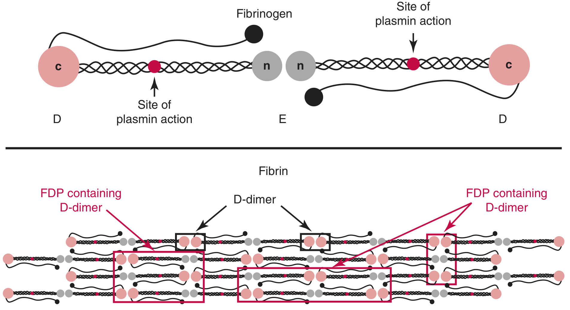
The key steps are:
- Thrombin cleaves fibrinogen (340 kDa) into fibrin monomer
- Fibrin monomers polymerize to form a fibrin network
- Factor XIIIa cross-links the D domains of adjacent fibers, forming the D-D epitope
- Plasmin (the key fibrinolytic enzyme) cleaves the fibrin between D and E domains
- Soluble fibrin degradation products (FDPs) containing the D-dimer moiety are released into plasma
D-dimer is specific to cross-linked fibrin - it is not generated by cleavage of fibrinogen, fibrin monomers, or non-cross-linked fibrin. - Tietz Textbook of Laboratory Medicine, 7th Ed.
Clinical Uses
1. Rule Out Venous Thromboembolism (DVT / PE) - PRIMARY USE
D-dimer is the most extensively studied and best-performing blood test for VTE. It is used as an exclusionary tool, not a confirmatory one.
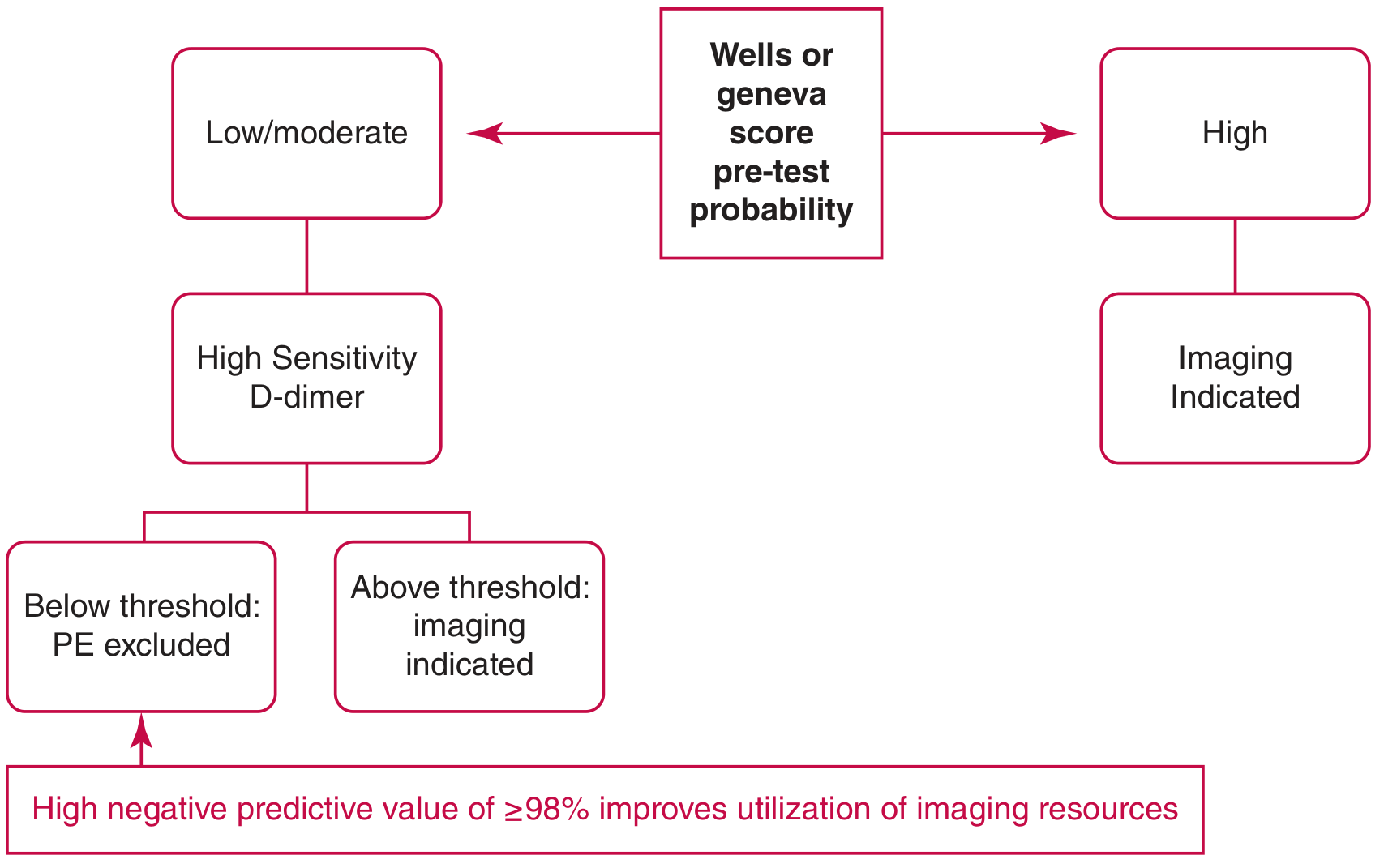
The logic:
- D-dimer below threshold + low/moderate pre-test probability → PE/DVT effectively ruled out; no imaging needed
- D-dimer elevated → Non-specific; imaging (CT pulmonary angiography or ultrasound) is required
- High pre-test probability → Do NOT measure D-dimer; proceed directly to imaging
D-dimer assays for VTE exclusion must meet minimum performance criteria: ≥98% sensitivity and ≥98% negative predictive value (lower CI at 95% and 90% respectively). - Tietz Textbook of Laboratory Medicine, 7th Ed.
2. Disseminated Intravascular Coagulation (DIC)
Quantitative D-dimer is useful in the evaluation of DIC, where simultaneous widespread clot formation and fibrinolysis produce markedly elevated D-dimer levels. - Tietz Textbook of Laboratory Medicine, 7th Ed.
3. Other Contexts
- Aortic dissection (elevated in most cases)
- COVID-19 and sepsis monitoring
- Pregnancy complications (pre-eclampsia, placental abruption)
Test Performance
| Parameter | Value |
|---|---|
| Sensitivity for PE/DVT | 95-98% |
| Specificity | 40-55% (low) |
| Negative predictive value | 99-100% |
D-dimer testing is highly sensitive but not specific. Elevated levels occur in nearly all patients with thromboembolism, but also in a wide range of other conditions. - Rosen's Emergency Medicine; Murray & Nadel's Textbook of Respiratory Medicine
Causes of Elevated D-Dimer (False Positives)
Because specificity is low, D-dimer can be elevated in many conditions besides VTE:
- Advanced age
- Pregnancy
- Surgery / trauma (postoperative period)
- Active malignancy / cancer
- Infection and sepsis
- Inflammatory states (rheumatic diseases, COVID-19)
- Liver disease
- Stroke
- Atrial fibrillation
- Renal failure
Causes of False-Negative D-Dimer
- Subacute or chronic PE (D-dimer has a half-life of < 8 hours in circulation)
- Severe lipemia
- Ongoing warfarin therapy
- Isolated subsegmental PE (small clot burden)
Assay Types and Reporting Units
Several assay formats exist with differing performance:
| Assay Type | Performance |
|---|---|
| ELISA / immunoturbidimetric / LIA | Highly sensitive; suitable for VTE exclusion |
| Latex agglutination (semiquantitative) | Lower sensitivity; NOT appropriate for VTE exclusion |
A critical source of confusion is the reporting units:
- D-Dimer Units (DDU): based on a D-dimer fragment (~185 kDa)
- Fibrinogen Equivalent Units (FEU): based on intact fibrinogen (~340 kDa)
- FEU is approximately twice DDU for the same sample
- Example: 500 ng/mL FEU = 250 ng/mL DDU
Using the wrong units can result in incorrect thresholds and missed diagnoses. Clinicians must know which unit system their lab uses. - Tietz Textbook of Laboratory Medicine, 7th Ed.; Murray & Nadel's Textbook of Respiratory Medicine
Standard Cutoff and Age-Adjusted D-Dimer
Standard cutoff: 500 ng/mL (FEU) is the traditional upper limit of normal.
Problem with elderly patients: D-dimer sensitivity is preserved across age groups, but specificity decreases markedly with age - leading to many false positives and unnecessary CT scans in older patients.
Age-adjusted D-dimer (for patients >50 years with low/intermediate pre-test probability):
Threshold = Age × 10 ng/mL (FEU)
For example, a 70-year-old patient's D-dimer threshold would be 700 ng/mL rather than 500 ng/mL.
This was prospectively validated in the ADJUST-PE trial (2014, Righini et al.), a multicenter multinational study showing:
- Increased percentage of patients in whom PE could be ruled out
- Failure rate of only 0.3% using the age-adjusted threshold
- 10-20% reduction in CT pulmonary angiograms in patients over 70
Note: The age-adjusted D-dimer applies to PE exclusion, not to suspected DVT. - Harrison's Principles of Internal Medicine 22E; Rosen's Emergency Medicine; Murray & Nadel's
Advanced Strategies: Variable Thresholds
YEARS Algorithm
Uses three clinical questions (signs of DVT, hemoptysis, PE as most likely diagnosis) to stratify patients:
- If none of the three are present: D-dimer threshold raised to 1000 ng/mL
- If any are present: standard 500 ng/mL threshold
- Validated in a large multicenter study; reduced need for CTPA by 14%
High Pre-Test Probability (PTP) Caveat
In patients with a HIGH pre-test probability (Wells score ≥6), do not order D-dimer - proceed directly to imaging. D-dimer is only useful when PTP is low or intermediate. A negative D-dimer in a high-PTP patient is unreliable and should not be used to exclude VTE. - Rosen's Emergency Medicine
Wells Score for DVT (Used Alongside D-Dimer)
| Clinical Characteristic | Score |
|---|---|
| Active cancer | +1 |
| Paralysis/paresis or plaster immobilization of legs | +1 |
| Bedridden ≥3 days or major surgery within 12 weeks | +1 |
| Localized tenderness along deep venous distribution | +1 |
| Entire leg swollen | +1 |
| Calf swelling ≥3 cm larger than asymptomatic side | +1 |
| Pitting edema in symptomatic leg only | +1 |
| Collateral superficial veins (non-varicose) | +1 |
| Previously documented DVT | +1 |
| Alternative diagnosis at least as likely as DVT | -2 |
Score <2: DVT unlikely → D-dimer can exclude DVT if negative
Score ≥2: DVT likely → proceed to imaging
- Murray & Nadel's Textbook of Respiratory Medicine, 2-Volume
Summary
| Feature | Details |
|---|---|
| What it detects | Cross-linked fibrin degradation products |
| Primary use | Rule OUT VTE (DVT/PE) |
| Standard threshold | 500 ng/mL FEU |
| Age-adjusted threshold | Age × 10 ng/mL (for >50 years) |
| Sensitivity | 95-98% |
| Specificity | 40-55% (low) |
| NPV when negative | 99-100% |
| Used alone? | Only in LOW or INTERMEDIATE pre-test probability |
| High pre-test probability | Skip D-dimer; go directly to imaging |
The key clinical rule: A normal D-dimer in a patient with low/moderate pre-test probability effectively rules out VTE with very high confidence (~99-100% NPV) and avoids radiation exposure from CT scanning. A positive D-dimer is non-specific and always requires confirmatory imaging.
Sources: Tietz Textbook of Laboratory Medicine 7th Ed. | Harrison's Principles of Internal Medicine 22E | Murray & Nadel's Textbook of Respiratory Medicine | Fishman's Pulmonary Diseases and Disorders | Rosen's Emergency Medicine | Recent evidence: ADJUST-PE validated age-adjusted D-dimer (PMID: 37452732 - van Es N et al., Eur Heart J 2023)
This is a shared conversation. Sign in to Orris to start your own chat.