
Give the the devleopment of placenta in the figure format and explain in simple language
Reading File
Now let me search the medical library for placenta development content and images in parallel.
Finding Sources
Finding Sources
Excellent! Found the key source - "The Developing Human". Let me read both sections in parallel.
Reading File
Reading File
Great content and images found. Let me also get the detailed content from The Developing Human on placental structure, and fetch the key images in parallel.
Reading File
Loading Image
Loading Image
Loading Image
I now have excellent textbook diagrams and content. Let me fetch the remaining key images (the full-term placenta cross-section and the chorion villi detail).
Loading Image
Loading Image
I now have all the textbook diagrams and rich content needed. Let me compile the complete, well-illustrated explanation.
Development of the Placenta
The placenta is the lifeline between the mother and baby - it delivers oxygen and nutrients to the fetus and removes waste. Here is the full development explained stage by stage, with textbook diagrams.
🔵 Stage 1: Implantation (Day 6-7 after fertilization)
When the fertilized egg (blastocyst) arrives in the uterus, it burrows into the uterine lining (endometrium). The outer layer of the blastocyst - called the trophoblast - starts this process.
The trophoblast splits into two layers:
- Cytotrophoblast - inner layer of individual cells
- Syncytiotrophoblast - outer layer where cells fuse together to invade the uterine wall
Think of it like a seed planting itself into the soil - the trophoblast is the "roots" that anchor the embryo.
🔵 Stage 2: Formation of Chorionic Villi (Weeks 2-3)
The trophoblast grows finger-like projections called chorionic villi into the uterine wall. These go through three stages:
| Stage | What it contains |
|---|---|
| Primary villi | Just trophoblast cells |
| Secondary villi | Trophoblast + mesoderm (connective tissue) grows in |
| Tertiary villi | Trophoblast + mesoderm + blood vessels form inside |
By Week 3, the villi have their own fetal blood vessels, and the placenta can now exchange gases and nutrients with the mother's blood.
🔵 Stage 3: Formation of Two Chorion Types (Weeks 5-8)
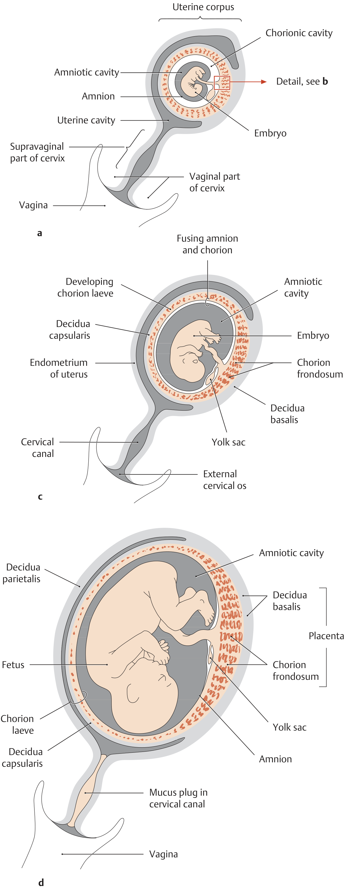
This is a key step. Look at the diagram below - at 5 weeks, villi cover the entire surface of the chorionic sac. By 8 weeks, two distinct regions form:
| Region | Name | What happens |
|---|---|---|
| Villi facing the uterine wall (decidua basalis) | Chorion frondosum ("bushy chorion") | Villi grow and branch rapidly - becomes the fetal part of placenta |
| Villi facing away from the wall (decidua capsularis) | Chorion laeve ("smooth chorion") | Blood supply reduces, villi shrink and disappear |
Simple analogy: Imagine ivy growing on a wall. The side touching the wall grows thick and lush (chorion frondosum); the side facing away from the wall withers (chorion laeve).
🔵 Stage 4: Fetal Membranes at 16 Weeks
By 16 weeks, the fetus is clearly inside an organized system:
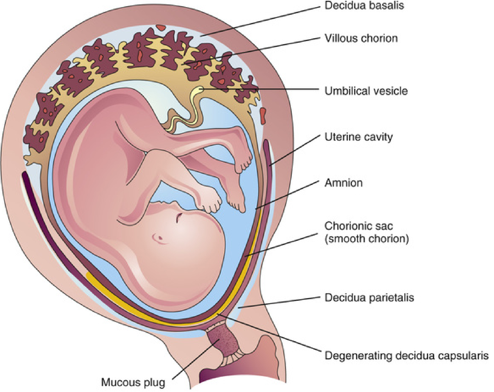
The uterine lining (decidua) is divided into 3 parts:
- Decidua basalis - under the placenta (becomes the maternal part of the placenta)
- Decidua capsularis - over the embryo (degenerates by 22 weeks)
- Decidua parietalis - lines the rest of the uterus
🔵 Stage 5: The Fully Formed Placenta (From ~20 Weeks)
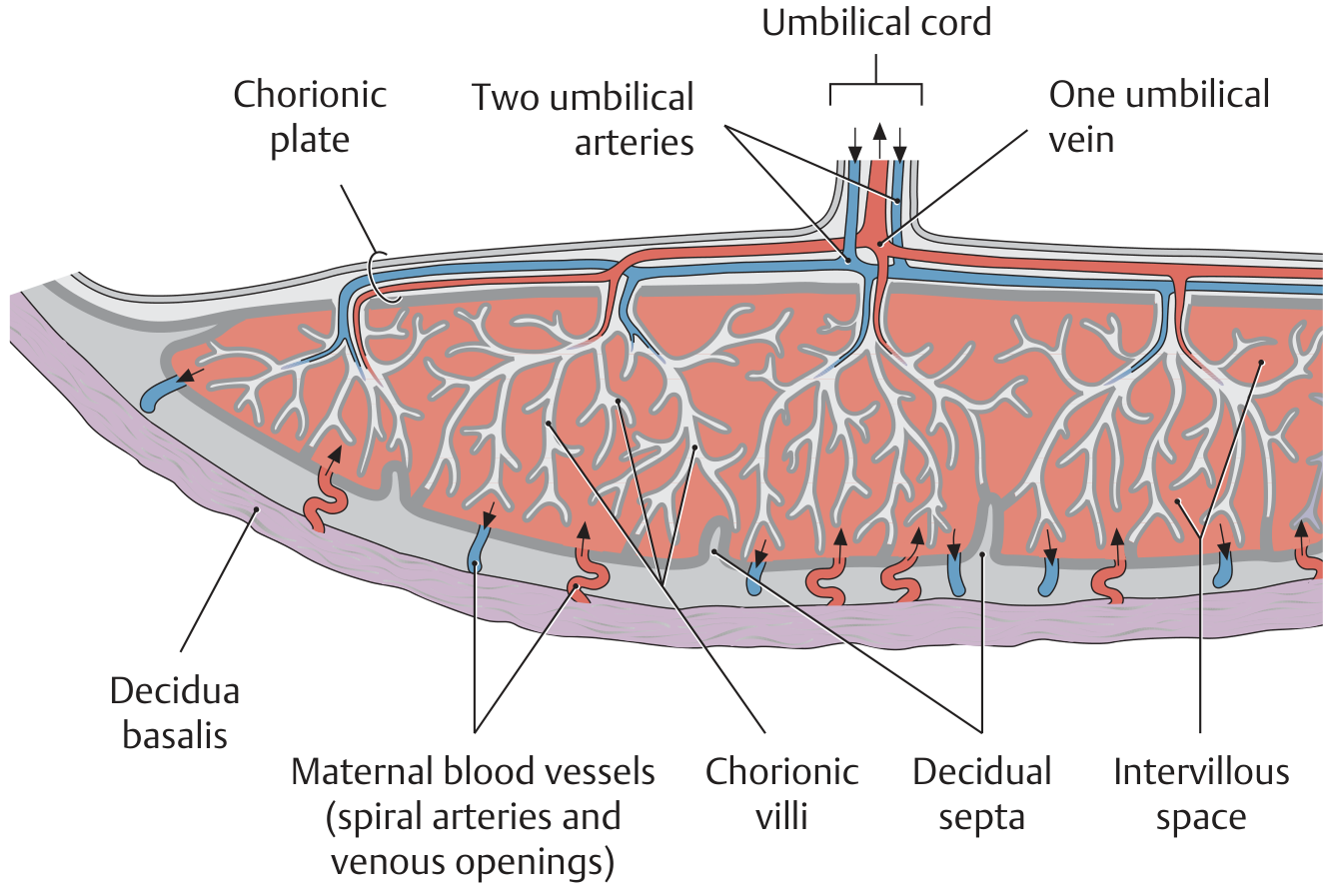
The mature placenta has two parts:
1. Fetal Part = Villous Chorion (Chorion Frondosum)
- The fetal blood vessels run through branching villi
- The umbilical cord connects the fetus to this plate
2. Maternal Part = Decidua Basalis
- The uterine wall that the villi dig into
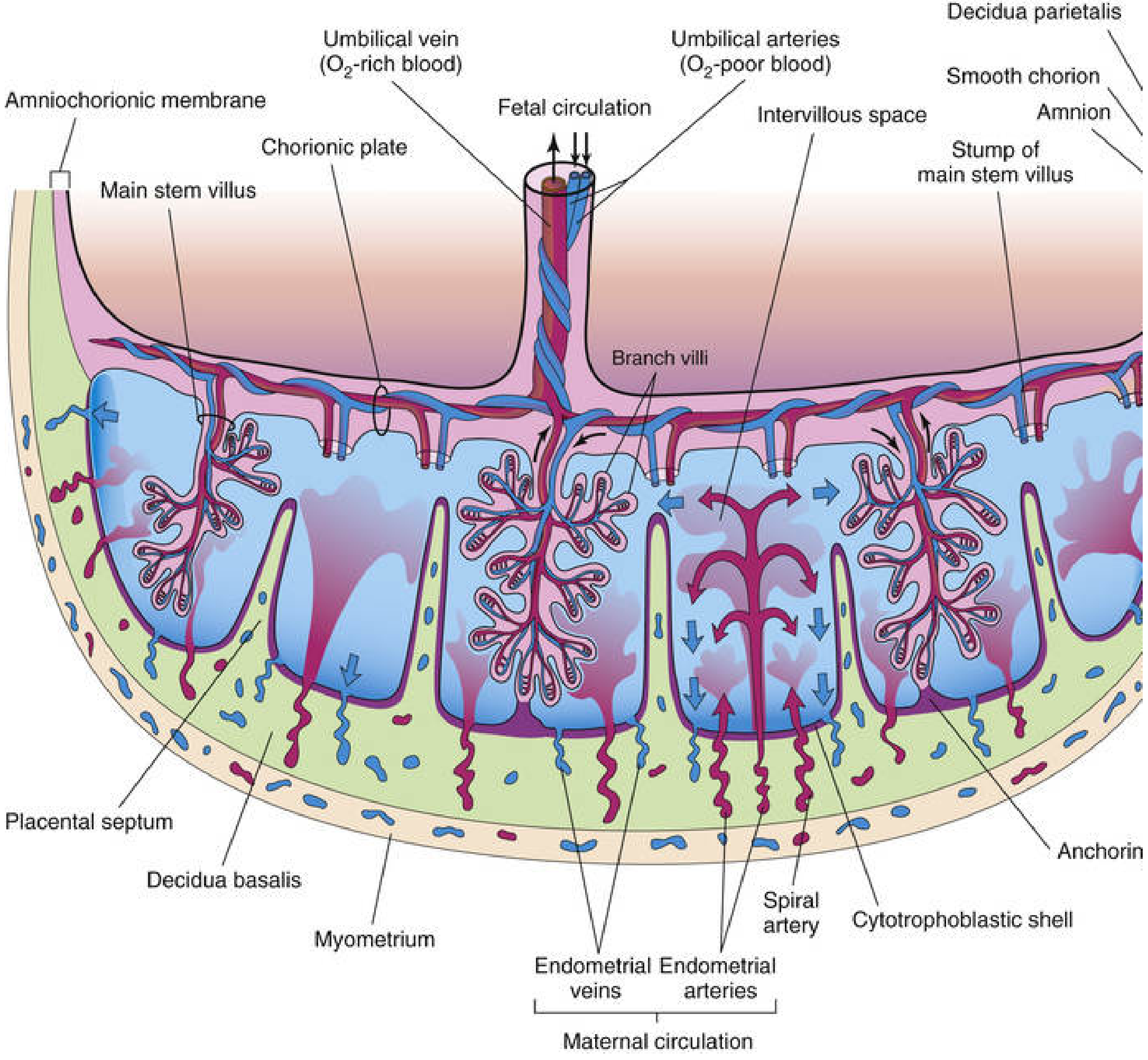
- Mother's spiral arteries open into spaces between the villi
Here is the cross-section of the mature placenta showing blood flow:
And the schematic overview of the mature placenta's structure:
🔵 How Exchange Happens (The Placental Barrier)
The mother's blood and the baby's blood never actually mix. Instead:
Mother's spiral arteries
        ↓
Intervillous space (pool of maternal blood)
        ↓
Nutrients + O₂ diffuse ACROSS the villus wall
        ↓
Fetal capillaries inside villi → umbilical vein → baby
The wall of the villus acts like a selective filter - letting through what the baby needs and blocking most harmful substances (though not everything - alcohol, some drugs, and certain viruses can cross).
🔵 Summary Timeline
| Week | Event |
|---|---|
| Week 1 | Blastocyst implants; trophoblast invades endometrium |
| Week 2 | Primary villi form; lacunae (spaces) fill with maternal blood |
| Week 3 | Secondary → Tertiary villi; fetal blood vessels form; exchange begins |
| Week 5 | Villi cover entire chorionic sac |
| Week 8 | Chorion frondosum and chorion laeve distinct |
| Week 10 | Placenta fully functional |
| Week 20 | Placenta fully formed; covers 15-30% of uterine wall |
| Term (40 wks) | Placenta weighs ~500g (1/6 of fetal weight) |
🔵 Key Facts to Remember
- The placenta has a fetal side (villous chorion) and a maternal side (decidua basalis)
- The umbilical cord has 2 arteries (carry deoxygenated blood from fetus) and 1 vein (carries oxygenated blood to fetus)
- The placenta is divided into cotyledons (about 15-30) by decidual septa
- The placenta uses 40-60% of oxygen reaching the uterus just for its own metabolism
- Homeobox genes (HLX, MSX2, DLX3) control trophoblast invasion and regulate all of this development
Sources: The Developing Human: Clinically Oriented Embryology (Moore & Persaud); THIEME Atlas of Anatomy - General Anatomy and Musculoskeletal System
This is a shared conversation. Sign in to Orris to start your own chat.