
How to differentiate between Bell's palsy and facial palsy if a patient came how to differentiate between this two What are the investigation and examination for it
Reading File
Now let me search the medical library for relevant content on Bell's palsy and facial palsy differentiation.
Finding Sources
Finding Sources
Finding Sources
Good results. Now let me read the most relevant sections in parallel.
Reading File
Reading File
Reading File
Excellent content. Let me fetch more from Adams & Victor's (the neurology textbook) for the UMN vs LMN differentiation and investigations, and also look for the differential diagnosis table in Cummings.
Reading File
Reading File
Now let me read more from Adams & Victor's for the UMN vs LMN clinical features and examination findings, and also the differential diagnosis section from Cummings.
Reading File
Reading File
Now let me fetch the facial nerve diagram from Adams & Victor's and also search for the UMN vs LMN examination table.
Loading Image
Finding Sources
Let me read the Rosen's Emergency Medicine table and also search for the House-Brackmann grading scale and examination approach.
Reading File
Finding Sources
Now let me read the Scott-Brown's assessment section and the House-Brackmann grading table.
Reading File
Reading File
I now have comprehensive information to write a thorough answer. Let me compile everything.
Bell's Palsy vs Facial Palsy: Differentiation, Examination & Investigations
The key to answering this question lies in understanding that Bell's palsy IS a type of facial palsy - specifically, it is the idiopathic (no identifiable cause) lower motor neuron (LMN) facial nerve palsy. The clinical question is therefore: "Is this facial palsy Bell's palsy, or does it have another cause?"
The first and most fundamental step is distinguishing upper motor neuron (UMN) from lower motor neuron (LMN) facial palsy, and then among LMN causes, deciding if it fits Bell's palsy or another diagnosis.
Step 1: UMN vs LMN Facial Palsy (Most Critical Distinction)
This is the single most important bedside differentiation.
| Feature | UMN (Central) Facial Palsy | LMN (Peripheral) Facial Palsy |
|---|---|---|
| Forehead involvement | Spared - forehead wrinkles normally | Affected - forehead cannot be raised (entire side) |
| Eye closure | Preserved or near-normal | Incomplete/absent - Bell's phenomenon present |
| Which muscles | Lower face only (contralateral) | All ipsilateral facial muscles |
| Laterality | Contralateral to lesion | Ipsilateral to lesion |
| Taste | Preserved | May be lost (anterior 2/3 tongue) |
| Hyperacusis | Absent | May be present (stapedius affected) |
| Lacrimation | Normal | May be affected |
| Associated deficits | Hemiplegia, other CN signs (e.g., CN VI) | Usually isolated; may have ear/parotid signs |
Why is the forehead spared in UMN lesions? Because the upper part of the facial motor nucleus receives bilateral cortical input (from both hemispheres). So a unilateral cortical/subcortical lesion (stroke, tumor) only knocks out the contralateral lower face. A brainstem or peripheral nerve lesion affects the entire ipsilateral face. - Gray's Anatomy for Students
Bell's phenomenon: On attempted eye closure, the eyeball rolls upward on the paralyzed side - the eyelid cannot close, leaving the white sclera visible. This is a classic sign of LMN facial palsy. - Adams and Victor's Principles of Neurology, 12th Edition, p.1370
Step 2: Diagnosing Bell's Palsy (Among LMN Causes)
Bell's palsy is a diagnosis of exclusion. Taverner's minimum diagnostic criteria are:
- Paralysis or paresis of all muscle groups of one side of the face
- Sudden onset
- Absence of CNS disease signs
- Absence of ear or cerebellopontine angle disease
As Cummings Otolaryngology states: "The term Bell palsy should be reserved for cases of facial paralysis that have signs and symptoms consistent with the disease and in which a diligent search for another cause is negative... The dictum that 'all that palsies are not Bell' cannot be overemphasized." - Cummings Otolaryngology Head and Neck Surgery
Step 3: Differential Diagnosis of LMN Facial Palsy (Red Flags for non-Bell's)
| Cause | Distinguishing Features |
|---|---|
| Bell's palsy (HSV reactivation) | Sudden onset, complete LMN palsy, no vesicles, resolves in weeks |
| Ramsay Hunt syndrome (VZV) | Vesicular rash on pinna/ear canal/palate, more severe, worse prognosis, associated hearing loss, tinnitus, vertigo |
| Lyme disease | Tick bite history, erythema migrans, can be bilateral palsy, CSF pleocytosis |
| Parotid malignancy | Slow onset, partial palsy (only 1-2 branches), palpable parotid mass, NO sudden onset |
| Acoustic neuroma / CPA tumor | Hearing loss, tinnitus, CN VI involvement, gradual onset |
| Otitis media / Cholesteatoma | Ear pain, discharge, conductive hearing loss |
| Sarcoidosis | Bilateral palsy, uveitis, enlarged parotid (Heerfordt syndrome), elevated ACE |
| HIV | CSF pleocytosis, risk factors, other systemic features |
| Stroke (UMN) | Forehead sparing, hemiplegia, other CNS signs |
| MS | Young patient, other demyelinating episodes, white matter lesions on MRI |
| Trauma | History, hemotympanum, base of skull fracture |
| Leprosy | Endemic area, thickened nerves, skin patches |
Sources: Adams and Victor's Principles of Neurology, Cummings Otolaryngology, Scott-Brown's Otorhinolaryngology
Step 4: Clinical Examination
History
- Duration and onset (sudden = Bell's; gradual = tumor)
- Progression: worsening over weeks is a red flag against Bell's
- Associated ear symptoms (pain, discharge, vesicles, hearing loss)
- Tick bite, travel history, skin rash
- Family history, diabetes, hypertension, pregnancy
- Pain behind the ear (common in Bell's, precedes paralysis by 1-2 days)
Cranial Nerve Examination (Systematic)
Test each branch of the facial nerve:
- Temporal branch: raise eyebrows, wrinkle forehead
- Zygomatic branch: close eyes tight (look for Bell's phenomenon)
- Buccal branch: puff cheeks, smile showing teeth
- Marginal mandibular: depress lower lip
- Cervical branch: platysma contraction
Also examine:
- Taste (anterior 2/3 tongue with sugar/salt) - chorda tympani involvement
- Hearing/tuning fork (Rinne and Weber) - stapedius, inner ear, cochlea
- Lacrimation (Schirmer's test) - greater petrosal nerve
- Otoscopy - vesicles (Ramsay Hunt), cholesteatoma, effusion, perforation
- Parotid gland palpation - mass?
- Neck/lymph nodes - malignancy, lymphoma
- Skin exam - vesicles (ear, palate), leprosy patches
- Eye exam - uveitis (sarcoidosis), papilledema (raised ICP)
- Full neurological exam - assess for other CN deficits, cerebellar signs, pyramidal signs
Grading Severity - House-Brackmann Scale
Use this to document severity and monitor recovery:
| Grade | Description |
|---|---|
| I | Normal function |
| II | Mild dysfunction - slight weakness, complete eye closure with minimal effort |
| III | Moderate dysfunction - obvious asymmetry, complete eye closure with effort |
| IV | Moderately severe - forehead absent, incomplete eye closure |
| V | Severe - barely perceptible motion, asymmetry at rest |
| VI | Total paralysis |
Source: Scott-Brown's Otorhinolaryngology Head & Neck Surgery, Table 95.2
Step 5: Investigations
First-Line (All patients with new facial palsy)
- MRI brain and temporal bones with gadolinium - gadolinium-enhanced MRI shows abnormal signal in the facial nerve in Bell's palsy; also excludes CPA tumor, brainstem pathology, MS, parotid lesion
- Otoscopy - mandatory (rule out otitis media, cholesteatoma, vesicles of Ramsay Hunt)
- Blood glucose / HbA1c - diabetes increases risk and worsens prognosis
- Blood pressure - hypertension association
Targeted Based on Clinical Suspicion
- Lyme serology (ELISA, Western blot) - tick exposure, endemic area, bilateral palsy
- HIV test - risk factors, young patient
- VZV serology - if Ramsay Hunt suspected without vesicles (zoster sine herpete)
- ACE level, CXR - sarcoidosis suspected (bilateral palsy, uveitis, lymphadenopathy)
- FBC, ESR, CRP - infection, inflammatory causes
- LP (lumbar puncture) - if Lyme or HIV suspected (both cause CSF pleocytosis)
- CT temporal bone - trauma, cholesteatoma, bony erosion
- CT/MRI parotid - if parotid mass palpated
- Chest X-ray - sarcoidosis, malignancy
Electrodiagnostic Tests (For prognosis in Bell's palsy, NOT for diagnosis)
- Electroneurography (ENoG) - compares peak-to-peak amplitude of compound action potential (CAP) bilaterally. If degeneration >90%, prognosis worsens significantly. Best done days 3-14.
- Electromyography (EMG) - denervation potentials appear 10+ days after onset. Loss of voluntary motor units in first 3-4 days = poor prognosis. Retention of voluntary activity past day 7 = good prognosis.
- Nerve Excitability Test (NET) - threshold difference >3.5 mA between sides suggests degeneration
- Nerve Conduction Velocity - normal 37-58 m/s; velocity <10 m/s = poor outcome
Sources: Cummings Otolaryngology Head and Neck Surgery, Adams and Victor's Principles of Neurology 12th Ed
Facial Nerve Anatomy (Helps Localize the Lesion)
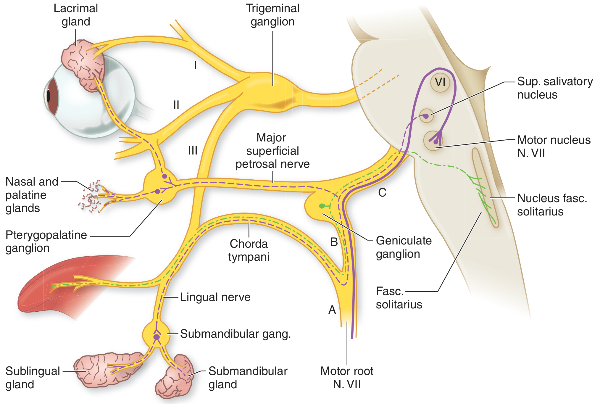
Figure: The VII (facial) nerve showing lesion sites A (stylomastoid foramen), B (distal to geniculate ganglion), C (proximal to geniculate ganglion) - Adams and Victor's Principles of Neurology
| Lesion Site | Motor Weakness | Taste | Lacrimation | Hyperacusis | Hearing |
|---|---|---|---|---|---|
| Above nucleus (cortex/subcortex) | Lower face only (UMN) | Intact | Normal | Absent | Normal |
| At nucleus/pons (brainstem) | Whole face + CN VI palsy | Intact | Normal | Absent | Normal |
| Geniculate ganglion (Ramsay Hunt) | Whole face | Lost | Reduced | Present | ± Affected |
| Facial canal (Bell's palsy) | Whole face | Lost | Normal | Present | Normal |
| Stylomastoid foramen | Whole face | Intact (chorda tympani above) | Normal | Absent | Normal |
| Parotid (distal) | Partial (1-2 branches) | Intact | Normal | Absent | Normal |
Quick Clinical Summary
If a patient comes in with facial weakness, ask yourself:
- Is forehead spared? → UMN (stroke/tumor) - refer neurology urgently
- Is forehead involved? → LMN - now look for a cause
- Any ear vesicles/pain? → Ramsay Hunt (VZV) - add antivirals
- Slow onset / parotid mass? → Tumor - urgent imaging
- Tick exposure / bilateral? → Lyme - check serology
- Sudden complete LMN palsy, no red flags? → Bell's palsy (treat with prednisolone within 72h)
This is a shared conversation. Sign in to Orris to start your own chat.