
Small round red bump type acne or pimples on scrotum
scrotal folliculitis small red bumps pimples scrotum
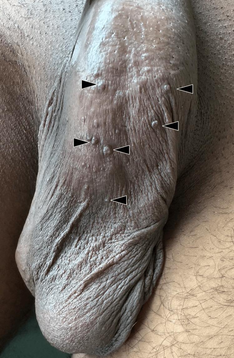
This clinical photograph shows a close-up view of the human scrotum. The scrotal skin exhibits numerous, discrete, small (1-4 mm) dome-shaped papules distributed across its surface, as highlighted by black arrowheads. The lesions present a reddish-brown to dusky-red hue, contrasting against the hyperpigmented scrotal skin. The texture of some papules appears slightly hyperkeratotic or smooth, while the surrounding background skin maintains its characteristic wrinkled and folded appearance. These findings are clinically characteristic of angiokeratoma of Fordyce, a benign vascular dermatosis involving ectatic superficial dermal vessels. The image serves as an educational reference for dermatological examination of male genital lesions, emphasizing morphology, color, and clustered distribution patterns typical of vascular-origin papules in this anatomical region.
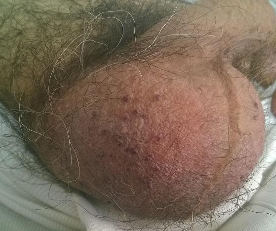
This clinical photograph shows a close-up view of human scrotal skin. The anatomical region is characterized by typical rugosity and coarse pubic hair distribution. Scattered across the surface of the scrotum are multiple, small (approximately 1-3 mm), well-circumscribed, dome-shaped papules. These lesions exhibit a dark red to purple or violaceous hue, characteristic of angiokeratomas of Fordyce. The papules appear as discrete vascular lesions and are distributed randomly across the scrotal wall. The surrounding skin shows mild erythema but is otherwise intact without signs of ulceration or active bleeding. This image is an educational example of benign vascular ectasias common in the urogenital area, often associated with increased venous pressure or aging. It serves as a classic diagnostic reference for distinguishing benign scrotal lesions from more concerning dermatological conditions.
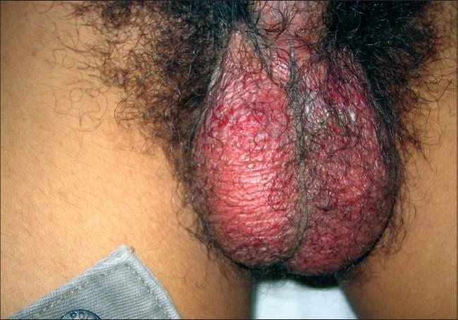
This clinical photograph shows a close-up view of the male genitalia, specifically focusing on the scrotum. The image demonstrates diffuse, bright erythema (redness) across the entire scrotal surface. The skin texture appears markedly irregular with multiple superficial erosions and denuded areas, indicating a loss of the epidermal barrier. Small, pinpoint red spots suggest possible petechiae or active inflammatory foci. There is an absence of discrete nodules or large ulcerative craters, with the primary finding being generalized inflammation and superficial skin breakdown. Clinically, this presentation is consistent with severe scrotal dermatitis, an inflammatory reaction, or a manifestation of a systemic cutaneous syndrome. The educational focus is on identifying scrotal skin integrity loss and recognizing diffuse genital erythema as a sign of acute dermatological or infectious pathology.
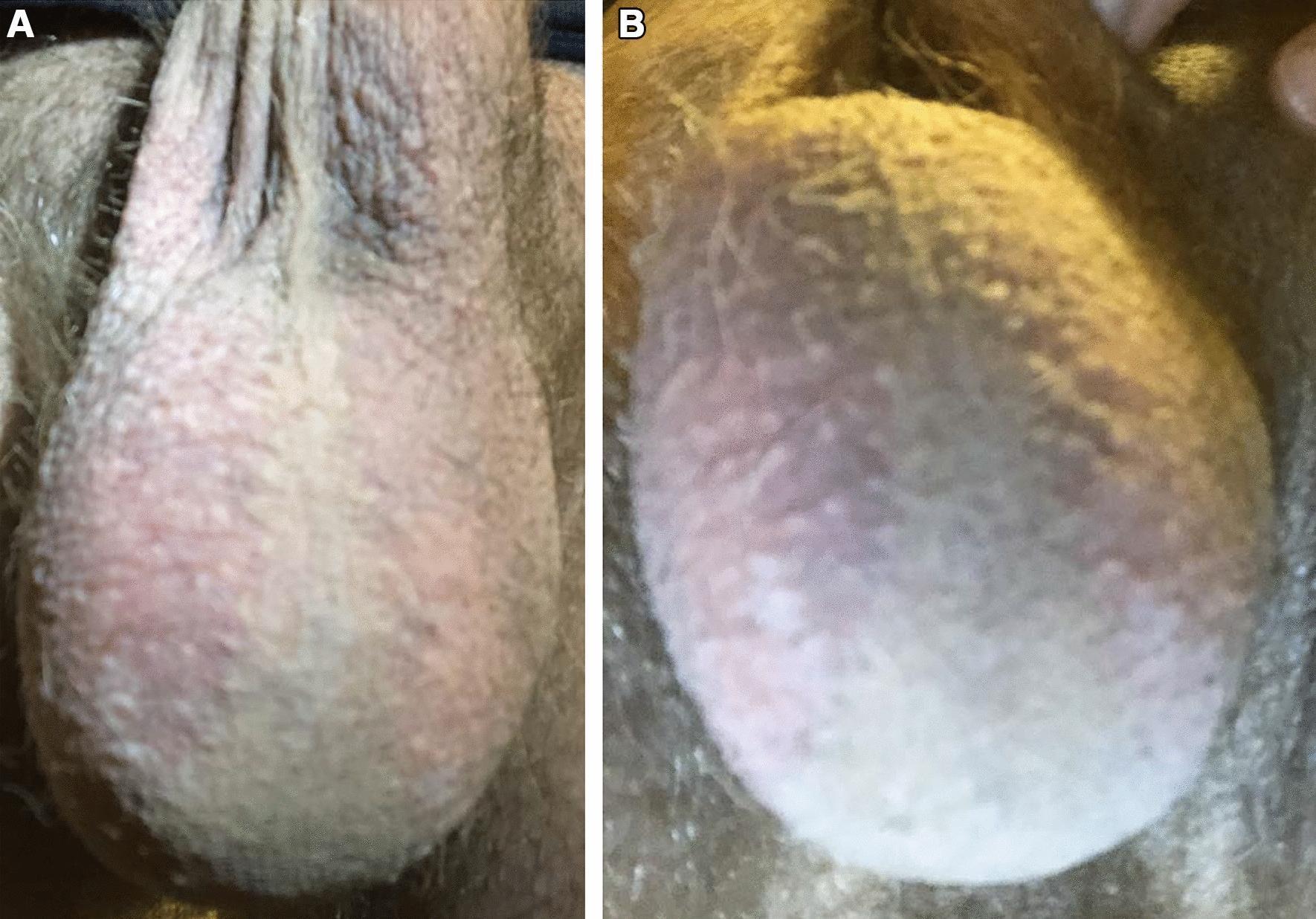
Two-panel clinical photograph (A and B) of the male external genitalia illustrating localized cutaneous erythema of the scrotum. Image A depicts the anterior scrotum and ventral penile shaft, showing well-demarcated, bilateral erythema. A key clinical finding is the distinct sparing of the vertical scrotal midline (raphe) where hair follicles are absent, while the erythematous area corresponds precisely to the distribution of scrotal hair follicles. Image B shows the underside of the scrotum from an inferior angle, demonstrating that the skin in this region is unaffected, retaining a normal skin tone and texture. There are no visible signs of ulceration, induration, edema, or pustules. The presentation is characteristic of a follicle-associated dermatosis or an atypical inflammatory response, such as 'Red Scrotum Syndrome' or a localized folliculitis, emphasizing the diagnostic importance of topographical distribution and the sparing of hairless anatomical regions in dermatology.
small red bumps pimples on scrotum causes treatment differential diagnosis
Small Red Bumps / Pimple-Like Lesions on the Scrotum
1. Folliculitis (Most Common "Pimple" Type)
- Cause: Usually Staphylococcus aureus; also triggered by ingrown hairs, sweating, friction, or shaving
- Appearance: Red, round bumps - sometimes with a white/yellow pus center
- Symptoms: Mildly tender or itchy; not usually painful at rest
- Treatment: Warm compresses, gentle antibacterial wash (chlorhexidine or benzoyl peroxide), topical mupirocin; oral antibiotics (e.g., cefalexin) if widespread
2. Angiokeratoma of Fordyce
- Appearance: 1-2 mm red or purple papules on the scrotum - can look exactly like red pimples (Campbell-Walsh Urology)
- Symptoms: Usually completely asymptomatic; may rarely bleed if scratched
- Treatment: Not needed unless they bleed; laser (Nd:YAG, KTP) can be used cosmetically
3. Epidermal (Sebaceous) Cysts
- Appearance: Flesh-colored to red, dome-shaped; may have a central pore
- Symptoms: Usually painless unless infected (then red, tender, fluctuant)
- Treatment: Leave alone if asymptomatic; incision and drainage or excision if infected/bothersome (Campbell-Walsh Urology, Smith & Tanagho's General Urology)
4. Molluscum Contagiosum
- Appearance: Small, dome-shaped papules with a characteristic central dimple (umbilication); flesh-colored or slightly red
- Symptoms: Usually painless; may itch
- Treatment: Often self-limiting; cryotherapy, curettage, or topical agents (imiquimod, cantharidin) can speed resolution - RACGP reference
5. Genital Herpes (HSV)
- Appearance: Starts as red bumps or blisters that then break open into painful ulcers/erosions
- Symptoms: Pain, burning, itching; may have flu-like symptoms on first episode
- Key difference: Herpes lesions cluster, blister, ulcerate, and heal - then recur. A regular pimple does not blister.
- Diagnosis: Swab of active lesion for HSV PCR
6. Idiopathic Scrotal Calcinosis
When to See a Doctor
- Bumps persist beyond 2-3 weeks without improvement
- They blister, ulcerate, or weep fluid
- They are painful (especially with flu-like symptoms - think herpes)
- They are multiplying rapidly
- You have had unprotected sex recently (rule out STI)
- There is a firm lump inside the scrotum (rule out testicular pathology)
Practical Self-Care (for likely folliculitis/blocked pores)
- Keep the area clean and dry; use a gentle, non-soap wash
- Avoid tight underwear that traps sweat
- Do not squeeze or pop the bumps - risks spreading infection
- Wear breathable, cotton underwear
- Avoid shaving the area if it triggers breakouts
Disclaimer: This is educational information only, not a substitute for examination by a healthcare provider. Only a clinician can distinguish these conditions on physical exam.
Scrotum and penins acne type
acne folliculitis red papules penis scrotum genital skin
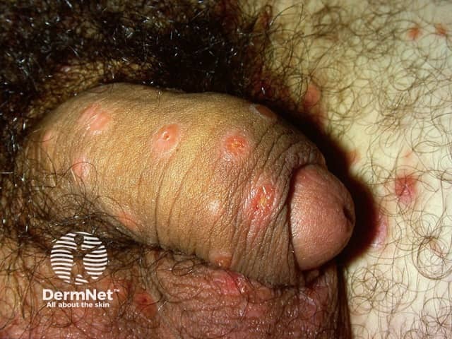
Clinical photography of external genital skin; close-up macro view of the penis, including the glans and adjacent shaft. Location: external genitalia with scattered small erythematous papules along the dorsal and ventral shaft and near the corona. Lesions are discrete, 2–4 mm in diameter, slightly raised, and vary from pink to red with mild surface erythema; surrounding skin shows normal folds and mild friction lines. The image captures a non-ulcerated, non-pustular eruption without obvious vesicles or crusts. Visual impression favors inflammatory or infectious papules rather than a single dominant lesion. Differential diagnoses include genital warts (condylomata acuminata, HPV types 6/11), folliculitis, irritant contact dermatitis or allergic dermatitis, candidal dermatitis, and less likely psoriasis or lichen planus. The appearance differs from pearly penile papules, which are arranged circularly around the corona and are typically flesh-colored. Diagnostic significance rests on clinical correlation: sexual history, symptoms, timing, lesion evolution, and potential exposure. Further workup may include dermoscopy, HPV typing, fungal culture, or skin biopsy if uncertainty persists. Clinical relevance spans dermatology, sexual health, infectious disease, and primary care; appropriate patient education and STI screening may be indicated. This image serves as educational material for recognition and differential diagnosis of penile dermatoses in clinical practice.
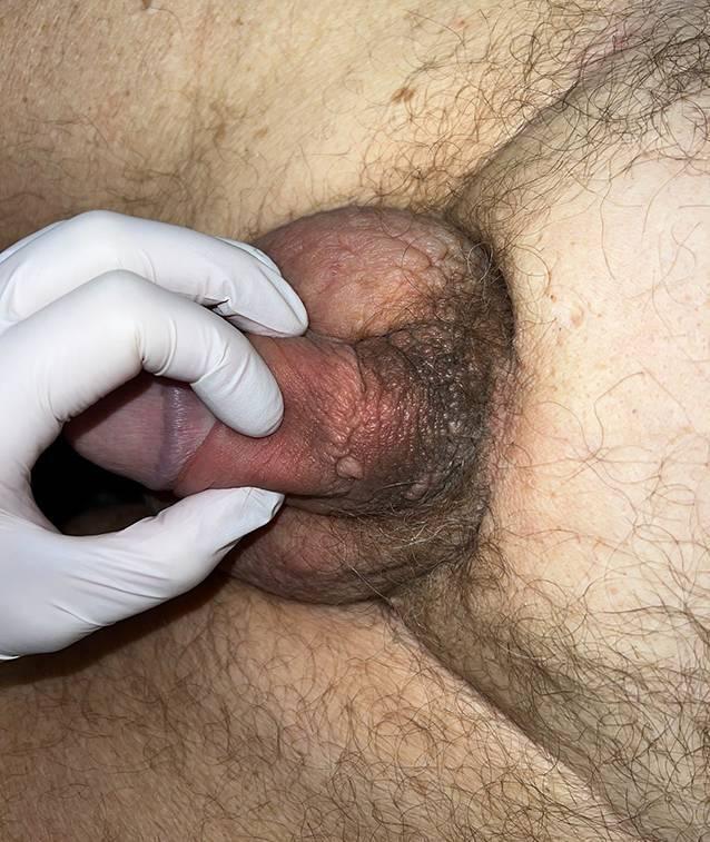
Clinical photograph showing a ventral close-up of the male genital region. The image demonstrates significant scrotal edema, characterized by diffuse swelling and a tense, shiny appearance of the scrotal skin. At the base (radix) of the penis, there are multiple skin-colored to brownish verrucous papules. These papules are arranged in a circular distribution and show a tendency to coalesce into larger plaques. The surrounding skin of the mons pubis and scrotum exhibits induration and texture changes resembling peau d'orange (orange-peel appearance), suggestive of lymphatic obstruction or cutaneous infiltration. A gloved hand is visible for clinical manipulation, highlighting the firmness of the underlying tissue. This presentation is consistent with localized lymphedema and secondary skin changes associated with malignant infiltration or chronic inflammatory processes in the urogenital area.
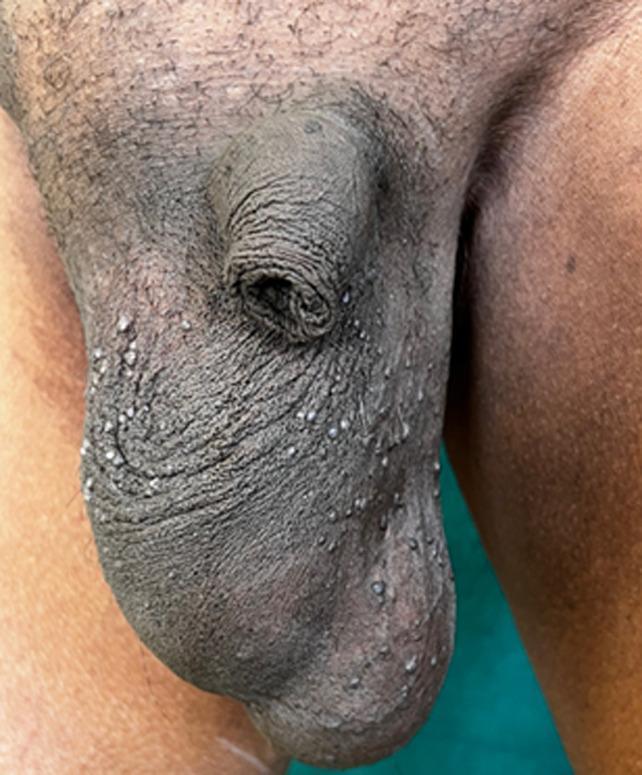
This clinical photograph shows the scrotum and base of the penis of an individual with dark skin. The primary findings are multiple, small, discrete papules scattered across the scrotal surface, consistent with Angiokeratomas of Fordyce. These lesions are approximately 1-3 mm in diameter, dome-shaped, and appear slightly lighter or more reflective than the surrounding hyperpigmented, rugose scrotal skin. The distribution is widespread but relatively uniform. The scrotal skin exhibits a typical rough, wrinkled texture (rugosity), and there is a prominent fold of redundant skin near the superior aspect of the scrotum. This image serves as a classic educational representation of benign vascular lesions of the genital skin, often presenting in older men, and helps in the differential diagnosis of common genital papules.
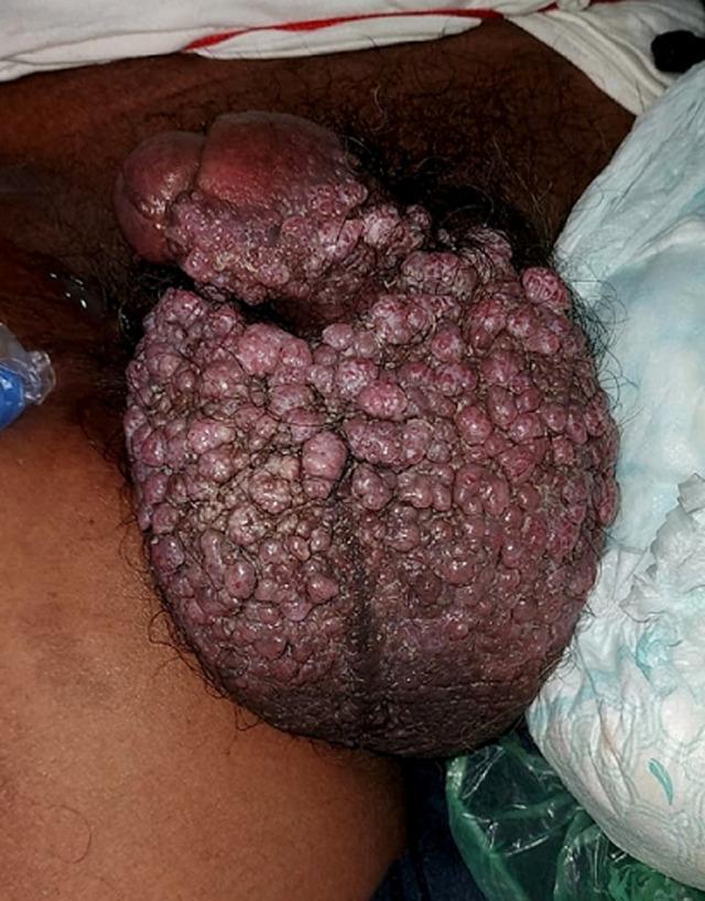
This clinical photograph displays a human male genital region exhibiting extensive multinodular lesions. The anatomical focus is the scrotum and the proximal shaft of the penis. The skin texture is significantly altered by the presence of numerous, densely packed, exophytic nodules. These individual lesions range from small papules to medium-sized nodules, many of which have a rounded or irregular morphology. Their color is predominantly a dark, purplish-red or violaceous hue, characteristic of vascular or infiltrative skin pathologies. The distribution is widespread and confluent, nearly entirely replacing the normal scrotal skin surface and extending onto the penile shaft. The clinical presentation is highly suggestive of advanced dermatological or systemic conditions such as Kaposi sarcoma, cutaneous lymphoma, or severe idiopathic scrotal calcinosis. This image serves as a teaching tool for medical students and clinicians to recognize atypical dermatological manifestations of internal or infectious diseases in the urogenital region.
acne type bumps on penis and scrotum causes differential diagnosis treatment dermatology
https://pmc.ncbi.nlm.nih.gov/articles/PMC6620174
Acne-Type Bumps on Both Penis and Scrotum
1. Folliculitis (Most Common)
- Cause: Staphylococcus aureus most often; also friction, sweating, shaving, tight clothing
- Appearance: Small red bumps, sometimes with a white/yellow head; may be tender
- Key feature: Centered on hair follicles; you can often see a hair at the center
- Treatment:
- Warm compresses 2-3x daily
- Wash with antibacterial/benzoyl peroxide soap
- Topical mupirocin 2% if localized
- Oral cefalexin or flucloxacillin if widespread or recurring
2. Fordyce Spots (Sebaceous Glands)
- Appearance: Tiny (1-3 mm), whitish-yellow or pale bumps on the penile shaft (especially ventral) and scrotum; not red but can look "pimple-like" when inflamed
- Symptoms: Painless, non-itchy; more visible when skin is stretched
- Important: These are ectopic sebaceous glands - a normal anatomical variant
- Treatment: None needed; RACGP guidelines confirm no treatment required
3. Angiokeratoma of Fordyce (Vascular)
- Appearance: 1-2 mm red or blue-purple papules; look like small blood blisters or red pimples (Campbell-Walsh Urology)
- Symptoms: Asymptomatic; rarely bleed if scratched
- Treatment: Not needed; laser if cosmetically bothersome
4. Molluscum Contagiosum
- Appearance: Smooth, dome-shaped papules 2-5 mm; central dimple (umbilication) is the hallmark - can look like a round pimple but firmer, pearl-like
- Symptoms: Usually painless; may itch slightly
- Treatment: Often self-resolves in months; cryotherapy, curettage, imiquimod 5% cream, or cantharidin can be used (PMC review)
5. Genital Herpes (HSV-1 or HSV-2)
- Appearance: Starts as clustered red papules/bumps → progresses to blisters → painful ulcers → crusting and healing
- Symptoms: Burning, tingling, pain; first episode may have fever and swollen lymph nodes
- Key difference from a pimple: Herpes blisters and ulcerates; regular folliculitis does not
- Diagnosis: HSV PCR swab of active lesion
- Treatment: Aciclovir 400 mg TDS x 5-10 days (or valaciclovir 500 mg BD)
6. Genital Warts (HPV - Condyloma Acuminata)
- Appearance: Flesh-colored, cauliflower-like or flat papules; can be pink-red on moist skin; appear on shaft and scrotum
- Symptoms: Painless usually; may itch
- Treatment: Podophyllotoxin 0.5% solution, imiquimod cream, cryotherapy, or laser
7. Scabies
- Key symptom: Severe itching, worse at night - also affects finger webs, wrists, waist
- Appearance: Small red papules, burrows (fine wavy lines); partner/household contacts often affected too
- Treatment: Permethrin 5% cream applied neck to toe, left 8-14 hours; repeat at 1 week (PMC)
8. Seborrheic Dermatitis
- Appearance: Red, slightly scaly plaques on the penile shaft and glans; less "pimple-like" and more of a patch
- Cause: Overgrowth of Malassezia yeast
- Treatment: Topical ketoconazole or mild corticosteroid
9. Pearly Penile Papules (Normal Variant)
- Located specifically at the corona of the glans penis in a ring pattern - not the shaft or scrotum
- Completely normal, no treatment needed
Visual Reference
Comparison Table
| Feature | Folliculitis | Angiokeratoma | Molluscum | Herpes | Scabies |
|---|---|---|---|---|---|
| Color | Red, may have pus | Red/purple | Flesh/pearl | Red → ulcer | Red |
| Pain | Mild tenderness | None | None | Painful | Itchy |
| Central feature | Hair/pus | Vascular | Dimple | Blister | Burrow |
| Contagious | No | No | Yes (STI) | Yes (STI) | Yes |
| Night itch | No | No | No | No | Yes |
| Both penis + scrotum | Yes | Yes | Yes | Yes | Yes |
When to See a Doctor Urgently
- Bumps blister, ulcerate, or produce open sores
- Burning/pain during or after sexual contact
- Discharge from the urethra
- Swollen groin lymph nodes
- Partner has a known STI
- Bumps spread rapidly over days
- Night-time itching affecting you and your partner/household
Self-Care for Likely Folliculitis/Blocked Pores
- Wash gently once daily with a mild antibacterial cleanser
- Wear loose, breathable cotton underwear
- Avoid shaving the area - or use a clean single-blade razor with shaving gel
- Do not squeeze or pop - introduces more bacteria
- Change and wash underwear daily
Disclaimer: This information is educational and not a substitute for in-person examination. Genital skin conditions often look similar to each other - a clinician (GP or dermatologist/GUM specialist) can examine, swab, and diagnose accurately. If there is any chance of an STI, get tested.