
Hypertrophic gaestropathy for md pathology exam
hypertrophic gastropathy Menetrier disease gastric rugae histology
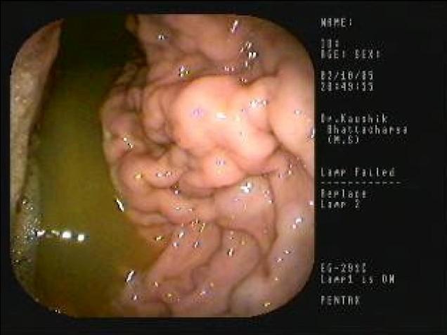
This clinical photograph displays a human upper gastrointestinal endoscopy focused on the gastric body. The image demonstrates a classic presentation of Menetrier's disease, a rare hypertrophic gastropathy. The primary visual finding is the presence of massively enlarged, thickened gastric rugae that exhibit a convoluted, cerebriform, or 'cobblestone' appearance. These hypertrophic folds appear congested and edematous, significantly obstructing the gastric lumen compared to normal mucosal architecture. A pool of greenish-yellow bile-stained fluid is visible in the dependent portion of the stomach (lower-left), suggesting an altered gastric environment or reflux. This imaging modality is essential for identifying the characteristic mucosal hyperplasia and protein-losing gastropathy associated with the condition. The educational focus is on the endoscopic identification of giant rugal folds and the differential diagnosis of hypertrophic gastropathies, including Zollinger-Ellison syndrome and infiltrative malignancies.
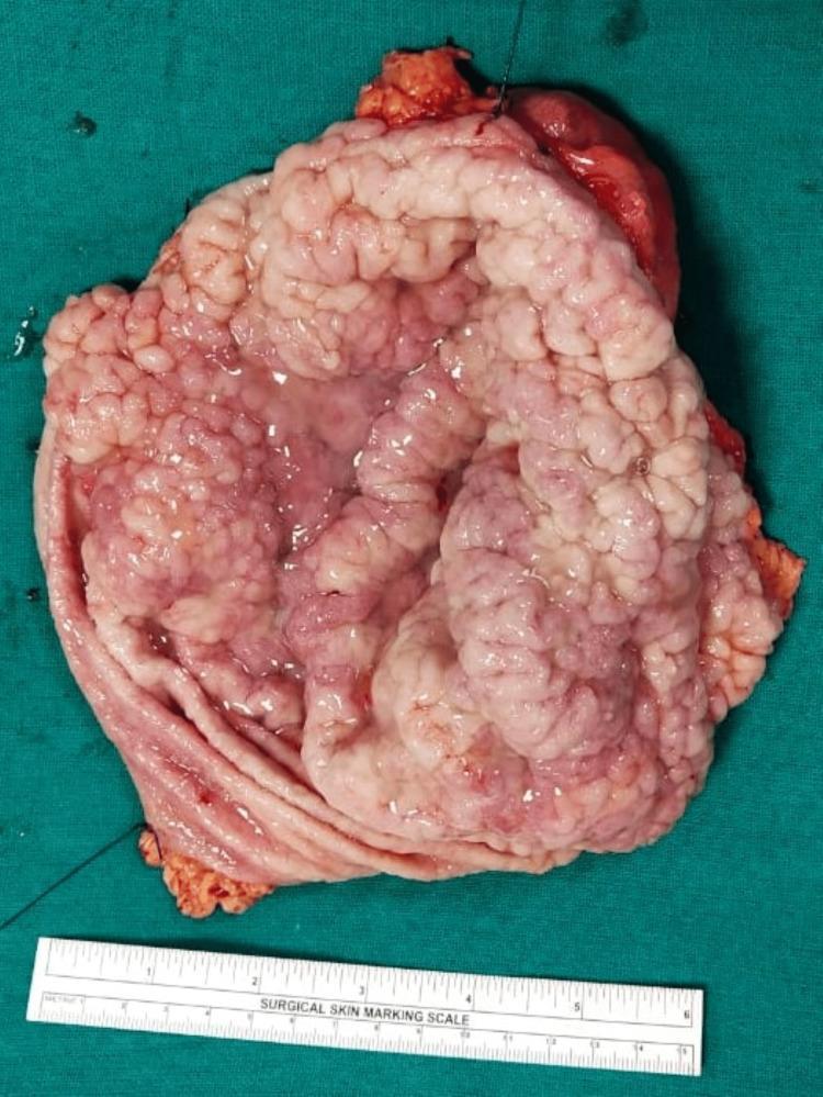
This clinical photograph displays a gross pathological specimen of a cut-open total gastrectomy. The image captures the internal mucosal surface of the stomach, which exhibits marked, diffuse hypertrophy of the gastric rugae. These folds are grossly thickened, firm, and convoluted, resulting in a distinct 'cerebriform' or brain-like appearance, characteristic of Menetrier's disease (hypoproteinemic hypertrophic gastropathy). The mucosal surface appears glistening and lobulated with varied coloration ranging from pale pink to deeper red, suggesting focal areas of congestion or vascular changes. A surgical skin marking scale at the bottom provides a size reference, indicating the specimen's longitudinal dimension is approximately 15 cm. This visual demonstrates severe foveolar hyperplasia and mucosal thickening without an obvious localized mass, contrasting with the typical appearance of gastric adenocarcinoma or lymphoma. The specimen serves as a classic educational example of hypertrophic gastropathy in a surgical pathology context.
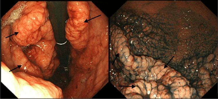
This dual-panel diagnostic image presents endoscopic views of the stomach illustrating classic features of Ménétrier disease. The left-hand panel shows a standard white-light endoscopy of the gastric body, revealing massive, convoluted, and thickened gastric rugae (marked by black arrows) that resemble cerebral gyri. These folds are hyperplastic with a reddish-pink mucosal surface. The right-hand panel demonstrates the same anatomical region using blue dye chromoendoscopy. The application of the dye enhances the surface architecture by pooling in the mucosal crevices, highlighting a reticular or 'net-like' pattern and emphasizing the irregular, bosselated texture of the hypertrophied folds. This side-by-side comparison illustrates the utility of chromoendoscopy in delineating mucosal topography for the evaluation of hypertrophic gastropathy and foveolar hyperplasia. Target audience: medical students and gastroenterology specialists.
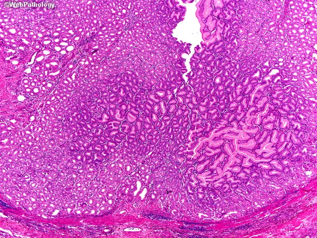
This image shows gastric body mucosa obtained by biopsy, examined under light microscopy with standard Hematoxylin and Eosin staining. The histologic hallmark is marked foveolar hyperplasia with elongated, tortuous glands and frequent cystic dilatation of mucous glands, a pattern strongly suggestive of hypertrophic gastropathy. The surface epithelium is mucin-rich, with tall, columnar foveolar cells forming crowded, branching pits; deep oxyntic glands are variably atrophic in affected foci. Lamina propria is mildly edematous and may contain modest chronic inflammatory infiltrates. The overall architecture is diffuse rather than polypoid, consistent with a non-neoplastic, hyperplastic process. No definite dysplasia is observed in this field; however, Menetrier-type hypertrophic gastritis (Menetrier disease) carries a recognized risk of gastric adenocarcinoma with time. The pattern correlates clinically with adult-onset, often severe disease characterized by diffuse thickening of the gastric folds and protein-losing enteropathy, whereas pediatric forms are typically self-limited. On this section, the combination of foveolar hyperplasia, gland tortuosity, and cystic dilation constitutes the diagnostic clue. This histology supports a diagnosis of Menetrier-type hypertrophic gastropathy and has implications for management, including monitoring for nutritional deficiency, edema, and potential neoplastic transformation, and consideration of EGFR-targeted therapies in selected patients.
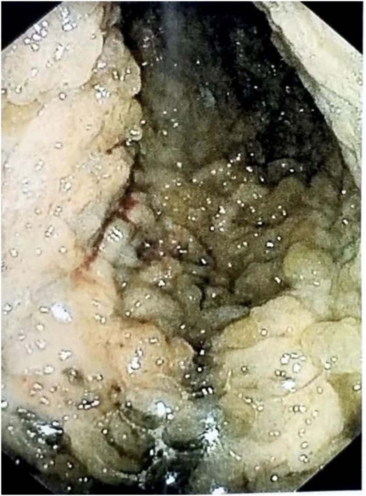
This clinical photograph displays a preoperative upper endoscopy of the stomach, demonstrating hallmark features of Menetrier's disease. The image reveals massive, cerebriform (brain-like) enlargement of the gastric rugae, specifically involving the fundus and body of the stomach. These hypertrophied folds appear thick, convoluted, and edematous, significantly narrowing the gastric lumen. The mucosa exhibits a variegated color palette, ranging from pale beige to brownish-green, with a moist, reflective surface texture likely caused by excessive mucus production (foveolar hyperplasia). Small, bright reflective spots across the tissue surface indicate the presence of fluid or mucous film. A localized area of linear erythema is visible, suggesting capillary congestion or minor mucosal erosion. The visual evidence of extreme mucosal thickening correlates with the clinical presentation of protein-losing gastropathy, hypoalbuminemia, and cachexia, providing a classic endoscopic example of hypertrophic gastropathy.
Hypertrophic gaestropathy for md pathology exam